Heath JA, Frederick PC. Relationship among .... 2006;26:536-539. Oskarsson A, Palminger Hallén I, Sundberg J. Exposure to toxic elements via breast milk.
13 Environmentally Induced Oxidative Stress and Disruption of Brain Thyroid Hormone Homeostasis in Autism Spectrum Disorders Elizabeth M. Sajdel-Sulkowska Department of Psychiatry, Harvard Medical School and Brigham and Women’s Hospital, Boston, MA, USA In memory of my son and a budding neuroscientist, Zachary L. Sulkowski, BS 1. Introduction Autism Spectrum Disorder (ASD) is a group of neuropsychiatric disorders characterized by impairments of language, social interactions, and movements and involves neurochemical, morphological, and neuroanatomical changes in specific brain regions including several cortical regions: the cerebellum, corpus callosum, basal ganglia and the limbic system (Sajdel-Sulkowska et al., 2010). It affects 1% of the births and its incidence is on the rise. Although its causation is assumed to have a strong genetic component, most of the known genetic risks have been associated with copy number variants (CNVs). Even an international genome-wide scan (AGP; Anney et al., 2010) failed to discover “the critical autism loci”. Furthermore, ASD concordance for monozygotic twins aged 18 years and younger, is less than 90 percent (Rosenberg et al., 2009). Thus nongenetic, environmental triggers of ASD pathology are gaining recognition as likely causal factors although the mechanisms involved in the environmental impact are not fully understood. This chapter focuses on the developmental impact of environmental pollutants that interfere with the thyroid hormone (TH), a key hormone involved in the regulation of brain development (Oppenheimer and Schwartz, 1997), as a possible factor contributing to autistic pathology. Many environmental toxicants, such as herbicides, polychlorinated biphenyls (PCBs), bisphenol A (BPA) and organic mercury compounds are potent disruptors of the endocrine system including TH. TH plays a critical role in brain development by virtue of regulating cellular metabolism, growth, differentiation and maturation and is indispensable for the proper development of the central nervous system (CNS). TH deficiency during CNS development results in disorders such as cretinism and a spectrum of psychoneurological disorders including both neurological and cognitive deficiencies (Vermiglio et al., 1995). It has been suggested that maternal hypothyroxinemia during critical periods may disrupt the developmental processes and produce morphological brain changes leading to autism (Roman, 2007). Hypothyroidism during pregnancy has been proposed as one of the twelve
252
Autism – A Neurodevelopmental Journey from Genes to Behaviour
autism risk factors (King, 2011). Yet studies addressing TH plasma levels, both 3’,3,5triiodothyronine (T3) and 3,5,3’,5’-tetraiodothyronine (thyroxine,T4), and thyroid stimulating hormone (TSH) failed to show major abnormalities in autism; thus TH involvement in autistic pathology has been ruled out. What has been overlooked in dismissing a TH-autism relationship is the fact that the majority of active TH hormone (T3) in the brain does not come from circulation, but is converted from the prohormone (T4) locally in the brain by deiodinase activity through the removal of iodine. Thus, while plasma TH levels may be within the normal range, its levels in the brain may be inadequate to support normal developmental processes. In vivo animal studies suggest that environmental toxicants can affect brain deiodinase activity and are supported by in vitro studies suggesting a direct inhibition of deiodinase enzymes by environmental triggers of oxidative stress (Mori et al., 1996; Lamirand et al., 2008). This chapter focuses on the developmental impact of environmental pollutants that trigger oxidative stress and disrupt brain homeostasis of TH, a key hormone involved in the regulation of brain development (Oppenheimer and Schwartz, 1997), as a plausible factor contributing to autistic pathology.
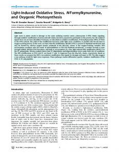
Fig. 1. Relative contribution of plasma-originated vs. tissue originated T3 in the CNS and the peripheral tissues.
Environmentally Induced Oxidative Stress and Disruption of Brain Thyroid Hormone Homeostasis in Autism Spectrum Disorders
253
2. Brain TH homeostasis: contribution of circulating vs. locally produced active forms of TH, T3 THs, both T3 and T4, are produced in the thyroid gland. The ratio of T3 to T4 released into the blood is 1:20. Both T3 and T4 then reach the individual body organs, where the prohormone T4 is converted to the biologically active hormone T3. The organ/tissue levels of T3 are regulated locally primarily by the activity of two different selenoenzymes, deiodinases type 2 (D2) and type 3 (D3), (Leonard, 1992; Silva et al., 1982; Bianco et al., 2002), although deiodinase type 1 is also involved (D1; Bates et al., 1999). In the CNS, approximately 70-80% of T3 originates from intracerebral T4 to T3 conversion, while the plasma contribution amounts to 20-30 % (Leonard, 1992); Bianco et al., 2002), and D2 is responsible for most of the T3 supply within the brain (Crantz et al., 1982). Mice with a globally targeted disruption of the Dio2 gene (D2KO mice) have ~50% less T3 content in their cerebral cortex, cerebellum, and hypothalamus (Galton et al., 2007). The extent of the local brain T4 to T3 conversion is in contrast to the peripheral tissues where T3 comes mostly from plasma. In the brain T3 exerts its major effect by binding to the nuclear TH receptor (TR), a ligand-regulated transcription factor, and regulates T3-dependent gene transcription. TR-mediated transcription may be modulated by various substances. The nuclear hormone receptor superfamily contains more than 40 transcriptional factors and most of these receptors are present in the brain. (Koibuchi et al., 2003). Excess T4 and T3 are then converted to inactive metabolites rT3 and 3,3’-diiodothyronine (T2) by D3. D2 is localized mainly in the glial cells (Guadano-Ferraz et al., 1997), but the Purkinje cell localization has been observed during specific developmental periods (Verhoelst et al., 2005). D3 is localized mainly in neurons including the Purkinje cells (Verhoelst et al., 2002). D3 activity increases in hyperthyroidism, and decreases in hypothyroidism (Dratman et al., 1983) and is thought to protect neurons from excessive T3 levels. D2 and D3 activity balance has been shown to be critical for the regulation of the intraneuronal level of the active form of T3 (Leonard, 1982; Bianco et al., 2002). Both D2 and D3 activities have been demonstrated in the human brain (Campos-Barros et al., 1996). While the majority of brain T3 is derived through the conversion of T4 to T3 by D2 coded by the Dio2 gene, some T3 is transported from plasma through the blood-brain barrier, a process mediated in part by the monocarboxylate transporter 8 (Mct8/MCT8). Using mice with inactivated Mct8 (Slc16a2) and Dio2 genes it has been shown that T3 from plasma and intracerebrally generated T3 play a distinct role in the brain and specifically in the regulation of TH-dependent gene expression. Inactivation of the Mct8 gene (Mct8KO) was without effect on the expression of 31 of these genes, but Dio2 inactivation selectively affected the expression of negatively regulated genes (Morte et al., 2010). In our recent study, thimerosal (TM) exposure resulted in decreased cerebellar D2 activity and overexpression of genes negatively regulated by TH (Sulkowski et al., accepted).
3. Systemic changes in TH in autism Several clinical studies, to date, have shown no evidence of TH abnormalities in autism. The study of a small group of patients between the ages of 7 and 21 years showed no clinical evidence of hypothyroidism with reported levels of plasma T3, T4, and TSH all within the normal range (Abbassi et al., 1978). Similarly, a study of a larger population of autistic children showed normal levels of T3, T4 and TSH (Cohen et al., 1980). On the other hand,
254
Autism – A Neurodevelopmental Journey from Genes to Behaviour
others reported significantly lower TSH levels in autistic boys as compared to mentally retarded or control groups (Hashimoto et al., 1991) and marginal changes in diurnal rhythms of serum TSH (Nir et al., 1995). Thus, while the evidence for the involvement of TH in autistic pathology is not compelling, there appears to be a tendency for TSH abnormalities in autism. Based on these findings further research for TH involvement in autism has been abandoned. However, others have tested the theory of a mild neonatal hypothyroidism in autism in animal models (Sadamatsu et al., 2006)
4. Altered deiodinase activities and brain TH homeostasis in other pathologies Considering that most of the brain T3 is generated by the activity of D2, it is surprising that no studies of the deiodinase activity in autism have been reported. Interestingly, in Alzheimer’s disease, where there is also no evidence of systemic TH abnormalities is also missing, as assessed by serum TH levels (McKhann et al., 1984), there is evidence of localized intra-brain hypothyroidism. Direct measures of T3 and T4 in the postmortem AD brains indicated no changes in T4 levels, but significantly lower T3 levels in advanced stages of the disease (Davis et al., 2008), suggesting decreased conversion of T4 to T3, possibly due to decreased D2 activity. Furthermore, both the level of rT3 and the rT3:T4 ratio in the cerebrospinal fluid (CSF) are significantly increased, suggesting an abnormal intracerebellar TH metabolism most likely due to an increase in D3 activity (Sampaolo et al., 2005). An increase in the CSF rT3 concentration has been found in other disorders involving the CNS. The CSF levels of T4 and free rT3 were increased during endogenous depression as compared to levels after recovery suggesting increased production of rT3 from T4 in the brain (Kirkegaard and Faber, 1991). These observations lend further support to the concept of local intra-brain regulation of TH homeostasis and its relevance to various pathological conditions.
5. Disruption of brain TH homeostasis by environmental toxicants Considering the absence of systemic TH abnormalities in autism and postulating the impact of environmental toxicants on brain TH homeostasis, we will examine some of their neurotoxic properties. Many environmental pollutants, including BPA, PCBs, organochlorine (dicofol, endosulfan) and organophosphate (Diazinon) pesticides, as well as metals such as lead, mercury and cadmium (Schantz and Windholm (2001) are considered to be endocrine disruptors. While most of them have been classified as endocrine disruptors, some of them, like PCBs (Venkataraman et al., 2007) and PBDE (Messer et al., 2010) and perchlorates (Brar et al., 2010), are also classified as TH disruptors. PCBs and PBDEs compete with T3 by virtue of having a similar chemical structure (Koibuchi et al, 2003). Table 1 summarizes the data on environmental toxicants implicated in ASD pathology. TH plays a critical role in brain development, and thus toxicants that affect TH homeostasis are most likely to interfere with brain development. It has been proposed that transient maternal hypothyroxinemia induced by environmental antithyroid agents such as PCBs, perchlorates, mercury and coal derivatives, could contribute to autistic pathology (Roman, 2009). This hypothesis was based on a leading ecological study in Texas that correlated higher levels of autism with the environmental release of mercury from industrial sources (Palmer et al., 2006). A potential association between autism and metal concentrations,
Environmentally Induced Oxidative Stress and Disruption of Brain Thyroid Hormone Homeostasis in Autism Spectrum Disorders
255
including mercury, has been reported in the San Francisco Bay area (Windham et al., 2006). A similar relationship has been postulated between autism and polybrominated diphenyl esters (PBDEs), potent thyroid hormone mimetics, used in home furnishings and electronics (Messer et al., 2010). Some of the toxicants interfere directly with TH synthesis and alter plasma TH levels, others bind plasma to the TH transport protein, transthyretin (TTR), resulting in a lower rate of T4 transport to the fetal brain (Schroder-van der Elst et al., 1998). However, others like mercury do not produce changes in the circulating TH (Watanabe et al., 1999), and yet they disrupt TH actions. TOXICANT SOURCE
ASSOCIATION ENDOCRINE TH WITH AUTISM DISRUPTOR DISRUPTOR
PLASMA TH (T3,T4, TSH)
BPA
Plastics
Brown, 2009
: TH Zoeller ? et al., 2005; no change: Nieminen et al., 2002; Kobayashi et al., 2002
Jain et al., 2011
DICOFOL
Pesticides
Roberts et al., 2007
: T4:Van den ? Berg et al., 1991
?
ENDOSUL- Pesticides FAN
Roberts et al., 2007
?
?
Hinkal et al., 1995
No change: Watanabe et al., 1999; lower: Tan et al., 2009
(Sulko Stringari et wski et al., 2008 al., submitt ed)
Aydogan et al., 2008
Schoeters et al., 2008
BRAIN OXIDATIVE T3/T4 STRESS IN D2, D3 BRAIN
METHYLMERCURY/ ETHYLMERCURY
Industrial Tan et al., 2009; Sringari et al., byproducts/ Windham et al., 2008 Pharmaceutical 2006 Air, food
PCBs
Industrial byproducts, food
Roman, 2009; Venkataraman Brar et al., 2008 Total and Kimura-Kuroda et al., 2007 free T4: Morse et al., 2007 et al., 1996
Vendkatara man et al., 2007; Hassoun et al., 2010
PBDE
Flame retardants
Messer et al., 2010
Messer et al, 2010
TH mimetic: Messer et al., 2010
Giordano et al., 2008; Zhang et al., 2010
PERCHLOR Drinking water Roman et al, ATES 2009
Roman, 2009
Bekkedal et al., Liuet al., 2008 2004
Liu et al., 2008
Table 1. Environmental toxicants associated with autistic pathology. As discussed above, the major source of the biologically active hormone T3 in the brain is the local intra-brain conversion of T4 to T3, while a small fraction comes from circulating T3. Thus it is possible that a direct action on some of the endocrine disruptors on brain deiodinases affects brain TH homeostasis. Indeed, we have observed the inhibition of the brain deiodinase D2 following perinatal exposure to TM (Sulkowski et al., accepted). Most of the toxicants implicated in ASD pathology are also potent triggers of oxidative stress (Table 1). As evidence derived from in vitro studies suggests, in response to oxidative stress D3 increases while D2 decreases (Lamirand et al., 2008; Freitas et al., 2010). Thus it is likely that the effect of many of these toxicants on brain deiodinases is mediated via mechanisms involving oxidative stress (Sulkowski et al., accepted).
256
Autism – A Neurodevelopmental Journey from Genes to Behaviour
Many of the toxicants, including heavy metals, (Bokara et al., 2008) and specifically mercury (Hg; Windham et al., 2006; Palmer et al., 2009), have been identified as factors exerting a range of harmful neurological and cognitive effects in humans and experimental animals, and have been implicated in the etiology of a number of neuropsychiatric disorders including Alzheimer’s disease (Gerhardsson et al., 2008), Parkinson’s disease (MonnetTschudi et al., 2006) and autism (Windham et al., 2006; Palmer et al., 2009). A specifically strong association has been observed between Hg exposure and autism; we will thus consider the Hg effect in relation to brain TH homeostasis in greater detail. The major environmental organic compounds of mercury include methylmercury (Met-Hg) and ethylmercury (Et-Hg). Met-Hg can be found in contaminated fish; Et-Hg is a metabolite of TM used in the United States in some maternal flu vaccines and in infant vaccines in the developing countries (Sulkowski et al., accepted). Hg compounds accumulate significantly in the pituitary and thyroid glands in both animals (Nishida et al., 1986) and humans (Kosta et al., 1975), and interfere with the hypothalamic-pituitary-thyroid (HTP) axis. Exposure to Met-Hg can produce hypothyroid conditions (Nishida et al., 1989), although changes in TH plasma levels based on both animal and human studies are inconsistent (Tan et al., 2009). Met-Hg has been shown to cross the placenta (Nordenhall et al., 1995) and Hg also enters the milk (Morgan et al., 2006) and is taken up by suckling pups (Oskarsson et al., 1995). Hg accumulates in both fetal and neonatal brains (Linares et al., 2004; Orct et al., 2006; Zareba et al., 2007) potentially affecting neurodevelopment (Orct et al., 2006). In rats, postnatal exposure (P1-P30) results in impairments in motor coordination and learning (Sakamoto et al., 2004). Perinatal TM exposure in rats results in the impairment of auditory functions and motor learning (Sulkowski et al., accepted). In humans, Met-Hg exposure in expectant mothers due to fish consumption is associated with `increased mercury accumulation in the infant brains accompanied by behavioral abnormalities, which include deficits in motor, attention, and verbal performance that are more pronounced in males (Gao et al., 2007), while the postnatal Met-Hg exposure in humans appears to have no recognizable effects (Debes et al., 2006). Hg compounds in general are potent endocrine disruptors (Heath et al., 2005;Windham et al., 2006; Palmer et al., 2009; Tan et al., 2009) and are also specifically TH disruptors (Stingari et al., 2008). Organic Hg compounds are also potent triggers of oxidative stress. Exposure to Met-Hg or Et-Hg in vivo or in vitro (Linares et al., 2004; Kaur et al., 2006; Rush et al., 2009; Glaser et al., 2010; Yin et al., 2011), induces oxidative stress that leads to a cascade of other changes including decreased neurogenesis, increased neuronal apoptosis and impaired synaptic plasticity in the neonatal brain. Results of one of our recently completed studies indicate that perinatal TM exposure increases cerebellar 3-nitrotyrosine (3-NT; Sulkowski et al., accepted), a well accepted marker of oxidative stress found in over fifty different pathologies including autism (Sajdel-Sulkowska, 2010). Further, Met-Hg is not only a potent trigger of oxidative stress, but also a disruptor of antioxidant defense systems (Chang and Tsai, 2008; Barcelos et al., 2011). Gestational exposure to Met-Hg in mice results in increased lipid peroxidation via interference in brain GSH levels (Stringari et al., 2008), while gestational exposure (G12-G14) in rats to Met-Hg (5 mg/kg) induces oxidative stress and reduces the antioxidant enzyme superoxide dismutase (SOD) in the hippocampus (Vincente et al., 2004). Hg compounds have been shown to target tissue deiodinases (Sulkowski et al., accepted). Our data derived from in vivo experiments in rats, supports results of earlier in vitro studies (Lamirand et al., 2008). Other in vitro studies indicated that the exposure of neuronal cells to
Environmentally Induced Oxidative Stress and Disruption of Brain Thyroid Hormone Homeostasis in Autism Spectrum Disorders
257
Met-Hg (Kim et al., 2005) or neuroblastoma cells to TM (James et al., 2005) results in a depletion of GSH which is both an antioxidant and a cofactor of deiodinases (Goswani and Rosenberg, 1988; Bhat et al., 1989; Croteau et al., 1998; Goemann et al., 2010). Thus, cerebellar D2 activity might be impaired due to a lack of the reducing cofactor. In primary astrocyte culture, GSH counteracts the impact of oxidative stress, and decreases D3 activity but increases D2 activity (Lamirannd et al., 2008). It is of interest that T3 regulates GSH levels in the developing brain and treatment of astrocyte cultures with TH results in increased GSH levels and improved antioxidative defense, suggesting that TH plays a positive role in maintaining GSH homeostasis and protecting the brain from oxidative stress (Dasgupta et al., 2007). Thus is it is also possible that a decrease in D2 activity could further amplify the effects of oxidative stress. As discussed above, tissue levels of T3 are regulated by D2 and D3, which are selenoproteins and are consequently sensitive to selenium availability. Selenium is not only a cofactor of deiodinases but also a potent antioxidant. Thus, environmental contaminants that sequester selenium or induce oxidative stress are likely to affect deiodinase activity. Met-Hg has been shown to interact with selenium (Soldin et al., 2008) and can inhibit the function of selenoproteins such as the deiodinases (Watanabe et al., 2001). We have also shown that TM exposure increases levels of oxidative stress (Sulkowski et al., accepted), which has been found previously to decrease expression of the Dio2 gene (Lamirannd et al., 2008).
6. Sexually dimorphic responses to environmental endocrine disruptors and sex ratio in autism When discussing the impact of environmental factors on CNS, it is critical to recognize the sexual dimorphism of their effects (Nguon et al., 2005a). Sex-dependent responses to a number of environmental pollutants including organophosphate pesticides (Dam et al., 2000), have been previously reported. Our earlier studies on the perinatal exposure to PCBs in rats demonstrated sex-dependent effects on cerebellar and motor functions with males being more sensitive (Nguon et al., 2005b). Even at low concentrations, different PCB congeners interfere with TH status in a sex-dependent manner (Abdelouahab et al., 2008). Our recently completed study on the perinatal exposure to TM revealed not only sex- but also strain-dependent effects on motor learning and cerebellar oxidative stress and D2 activity (Sulkowski et al., accepted). Specifically, in the Spontaneously Hypertensive Rats (SHR), a strain more sensitive to inflammation (Ballerio et al., 2007), perinatal exposure to TM resulted in decreased cerebellar D2 activity in male, but not in female neonates, and this decrease was correlated with a disruption of T3-dependent gene expression (Sulkowski et al, accepted). Our findings are in agreement with earlier observations both in humans (Gao et al., 2007) and in animals (Sobutskii et al., 2007) showing that the developing males appear to be more sensitive to Hg exposure. Furthermore, gene profiling studies in the rat cerebellum following perinatal exposure to a number of toxicants including PCBs, pesticides and methylmercury, showed differential sex-dependent effects of on gene expression (Padhi et al., 2008). Although the precise mechanism involved in this dimorphism is not known, in the cerebellum, developmentally-timed progesterone synthesis in the Purkinje cells (Sakamoto et al., 2003), differential regulation of progesterone-receptors by estradiol (Quadros et al., 2002; Guerra-Araiza et al., 2002), and the formation of estradiol from testosterone in the Purkinje cells (Sakamoto et al., 2003), have been implicated in these
258
Autism – A Neurodevelopmental Journey from Genes to Behaviour
differential effects. It is thus interesting that the Purkinje cells express D2 at specific times during development (Verhoelst et al., 2005). Therefore it is possible that environmental toxicants interfere with TH homeostasis by acting on the Purkinje cells.
7. Could localized, intra-brain TH deficiency contribute to the pathology of ASD and present new venues for the diagnosis and treatment of autism It is clear from the preceding discussion that there are no systemic TH changes in autism, that some environmental factors disrupt TH regulation without any effect on systemic TH status, and that it is the local intra-brain T4 to T3 conversion rather than circulating T3 levels that are responsible for the majority of brain T3. Furthermore, the T3 generated locally in the brain by D2 controls the expression of genes negatively regulated by TH, while plasma T3 controls the expression of the positively regulated genes (Morte et al., 2010). Thus, systemic hypothyroidism that is known to interfere with normal brain development may regulate the expression of genes distinct from those that are regulated by the locally generated T3, and is thus likely to result in a different set of morphological and functional abnormalities. Animal studies have indicated that in the developing rat cerebellum, systemic TH deficiency affects cerebellar granule cell migration. Also, Purkinje cell migration requires reelin (Miyata et al., 2010). Reelin is one of the genes whose abnormal expression is implicated in autism (Fatemi et al., 2005) and is also regulated by T3 produced locally in the fetal brain from T4 by deiodinase activity mostly in astrocytes but also in Purkinje cells (Verhoelst et al., 2005). It is possible that the aberrant Purkinje cell migration in ASD contributes to the decrease in Purkinje cells in ASD (Courchesne, 1991). Furthermore, in ASD, the lower intra-brain T3 levels occur in the absence of a systemic T3 deficiency (Davis et al., 2008), most likely due to the increased activity of D3 (Sampaolo et al., 2005). Similar studies involving postmortem ASD brains are now being initiated in our laboratory. Although none of the studies so far provide direct evidence for the disruption of brain TH metabolism in autism, there is a sufficient amount of indirect data to warrant pursuing the hypothesis that environmentally induced oxidative stress and local brain hypothyroidism contributes to ASD pathology. According to this hypothesis, brain region-specific oxidative stress in autism may be associated with increased D3 and decreased D2 activity resulting in a region-specific T3 deficiency in the brain. Future human studies utilizing the CSF of living ASD individuals or postmortem brain tissue of ASD donors will support its validity. Such findings would have several significant implications. They may result in methods of early ASD diagnosis; detection of high brain D3 levels in postmortem human brains may suggest the benefits of measuring the levels of its product (rT3) in the CSF of living patients to assess the risks, monitor the disease progression and efficacy of ongoing treatment. Furthermore, several tissue-specific and TH receptor (TR)-specific thyromimetics have been developed as potential treatment for atherosclerosis, obesity and Type 2 diabetes and might be able to correct local brain TH deficiency without systemic thyrotoxicity (Baxter and Webb, 2009) and may thus be considered as potential therapeutic agents. Finally, confirmation that autism may be associated with increased D3 and decreased D2 activity resulting in a regionspecific T3 deficiency in the brain could lead to or reinforce dietary treatments, because D2 activity can be modulated not only by selenium but also by xenobiotic compounds (da-Silva et al., 2007). In conclusion, TH abnormalities in autism warrant a second look.
Environmentally Induced Oxidative Stress and Disruption of Brain Thyroid Hormone Homeostasis in Autism Spectrum Disorders
259
8. Acknowledgments The author is indebted to Dr. Ann Marie Zavacki for introducing her to the concept of local brain TH regulation. The author also expresses her gratitude to her friend, Patricia McCann, and son, Professor Adam J. Sulkowski, for their expert editorial work.
9. References Abbassi V, Linscheid T, Coleman M. Triiodothyronine (T3) concentration and therapy in autistic children. J Autism Child Schizophr. 1978;8:383-7. Abdelouahab N, Mergler D, Takser L, Vanier C, St-jean M, Baldwin M, Spear PA, Chan HM. Gender differences in the effects of organochlorines, mercury and lead on thyroid hormone levels in lakeside communities of Quebeck (Canada). Environ Res. 2008;107:380-92. Anney R, Klei L, Pinto D, Regan R, Conroy J, Magalhaes TR. A genome-wide scan for common alleles affecting risk for autism. Hum Mol Genet. 2010;19:4072-82. Aydoğan M, Korkmaz A, Barlas N, Kolankaya D. The effect of vitamin C on bisphenol A, nonylphenol and octylphenol induced brain damages of male rats. Toxicology. 2008;249:35-9. Ballerio R, Gianazza E, Mussoni L, Miller I, Gelosa P, Guerrini U, Eberini I, Gemeiner M, Belcredito S, Tremoli E, Sironi L. Gender differences in endothelial function and inflammatory markers along the occurrence of pathological events in stroke-prone rats. Exp Mol Pathol. 2007;82:33-41. Barcelos GR, Grotto D, Serpeloni JM, Angeli JP, Rocha BA, de Oliveira Souza VC, Vicentini JT, Emanuelli T, Bastos JK, Antunes LM, Knasmuller S, Barbosa F Jr. Protective properties of quercetin against DNA damage and oxidative stress induced by methylmercury in rats. Arch Toxicol. 2011; PMID:21286687. Bates JM, St. Germain DL, Galton VA. Expression profiles of the three iodothyronine deiodinases, D1, D2, and D3, in the developing brain. Endocrinology 140:844-851, 1999. Baxter JD, Webb P. Thyroid hormone mimetics: potential applications in atherosclerosis, obesity and type 2 diabetes. Nat Rev Drug Discov. 2009;8:308-20. Bekkedal MY, Arfsten D, Mattie D. An evaluation of neurobehavioral tests used to assess the neurodevelopmental effects of early ammonium perchlorate exposure. J Toxicol Environ Health 2004;67:835-44. Bellinger FP, Bedi KS, Wilson P, Wilce PA. Ethanol exposure during the third trimester equivalent results in long-lasting decreased synaptic efficacy but not plasticity in the CA1 region of the rat hippocampus. Synapse 1999;31:51-58. Bhat GB, Iwase K, Hummel BC, Walfish PG. Kinetic characteristics of a thioredoxinactivated rat hepatic and renal low-Km iodothyronine 5'-deiodinase. Biochem J. 1989;258:785-92. Bianco AC, Salvatore D, Gereben B, Berry MJ, Larsen PR. Biochemistry, cellular and molecular biology, and physiological roles of the iodothyronine selenodeiodinases. Endocr Rev. 2002;23:38-89. Bokara KK, Brown E, McCormick R, Yallapragada PR, Rajanna S, Bettaiya R. Leadinduced increase in antioxidant enzymes and lipid peroxidation products in developing rat brain. Biometals 2008; 21:9-16.
260
Autism – A Neurodevelopmental Journey from Genes to Behaviour
Brar NK, Waggoner C, Reyes JA, Fairey R, Kelley KM. Evidence for thyroid endocrine disruption in wild fish in San Francisco Bay, California, USA. Relationships to contaminant exposures. Aquat Toxicol. 2010;96:203-15. Brown JS Jr. Effects of bisphenol-A and other endocrine disruptors compared with abnormalities of schizophrenia: an endocrine-disruption theory of schizophrenia. Schizophr Bull. 2009;35:256-78. Campos-Barros A, Hoell T, Musa A, Sampaolo S, Stoltenburg G, Pinna G, Eravci M, Meinhold H, Baumgartner A. Phenolic and tyrosyl ring iodothyronine deiodination and thyroid hormone concentrations in the human central nervous system. J Clin Endocrinol Metab. 1996;81:2179-85. Chang JY, Tsai PF. Prevention of methylmercury-induced mitochondrial depolarization, glutathione depletion and cell death by 15-deoxy-delta-12,14-prostaglandin J(2). Neurotoxicology 2008;29:1054-1061. Chen J, Berry MJ. Selenium and selenoproteins in the brain and brain diseases. J Neurochem. 2003;86:1-12. Cohen DJ, Young JG, Lowe TL, Harcherik D. Thyroid hormone in autistic children. J Autism Dev Disord. 1980;10:445-50. Crantz FR, Silva JE, Larsen PR. An analysis of the sources and quantity of 3,5,3'triiodothyronine specifically bound to nuclear receptors in rat cerebral cortex and cerebellum. Endocrinology 1982;110:367-375. Croteau W, Bodwell JE, Richardson JM, St Germain DL. Conserved cysteines in the type 1 deiodinase selenoprotein are not essential for catalytic activity. J Biol Chem. 1998 ;273:25230-25236. Dam K, Seidler FJ, Slotkin TA. Chlorpyrifos exposure during a critical neonatal period elicits gender-selective deficits in the development of coordination skills and locomotor activity. Brain Res Dev Brain Res. 2000;121:179-87. Dasgupta A, Das S, Sarkar PK. Thyroid hormone promotes glutathione synthesis in astrocytes by up regulation of glutamate cysteine ligase through differential stimulation of its catalytic and modulator subunit mRNAs. Free Radic Biol Med. 2007;42:617-626. da-Silva WS, Harney JW, Kim BW, Li J, Bianco SD, Crescenzi A, Christoffolete MA, Huang SA, Bianco AC. The small polyphenolic molecule kaempferol increases cellular energy expenditure and thyroid hormone activation. Diabetes. 2007;56:767-76. Davis JD, Podolanczuk A, Donahue JE, Stopa E, Hennessey JV, Luo LG, Lim YP, Stern RA. Thyroid hormone levels in the prefrontal cortex of post-mortem brains of Alzheimer’s disease patients. Curr Aging Sci. 2008;1:175-81. Debes F, Budtz-Jørgensen E, Weihe P, White RF, Grandjean P. Impact of prenatal methylmercury exposure on neurobehavioral function at age 14 years. Neurotoxicol Teratol. 2006;28:363-75. Dratman MB, Crutchfield FL, Gordon JT, Jennings AS. Iodothyronine homeostasis in rat brain during hypo- and hyperthyroidism. Am J Physiol. 1983;245:E185-93. Fatemi SH, Snow AV, Stary JM, Araghi-Niknam M, Reutiman TJ, Lee S, Brooks AI, Pearce DA. Reelin signaling is impaired in autism. Biol Psychiatry. 2005;57:777-87. Freitas BC, Gereben B, Castillo M, Kalló I, Zeöld A, Egri P, Liposits Z, Zavacki AM, Maciel RM, Jo S, Singru P, Sanchez E, Lechan RM, Bianco AC. Paracrine signaling by glial
Environmentally Induced Oxidative Stress and Disruption of Brain Thyroid Hormone Homeostasis in Autism Spectrum Disorders
261
cell-derived triiodothyronine activates neuronal gene expression in the rodent brain and human cells. J Clin Invest. 2010;120:2206-17. Galton VA, Wood ET, St Germain EA, Withrow CA, Aldrich G, St Germain GM, Clark AS, St Germain DL. Thyroid hormone homeostasis and action in the type 2 deiodinasedeficient rodent brain during development. Endocrinology 2007;148:3080-3088. Gao Y, Yan CH, Tian Y, Xie HF, Zhou X, Yu XD, Yu XG, Tong S, Zhou QX, Shen XM. Prenatal exposure to mercury and neurobehavioral development of neonates in Zhoushan City, China. Environ Res. 2007;105:390-399. Gereben B, Zavacki AM, Ribich S, Kim BW, Huang SA, Simonides WS, Zeold A, Bianco AC. Cellular and molecular basis of deiodinase-regulated thyroid hormone signaling. Endocr Rev 2008;29:898-938. Gerhardsson L, Lundh T, Minthon L, Londos E. Metal concentrations in plasma and cerebrospinal fluid in patients with Alzheimer's disease. Dement Geriatr Cogn Disord. 2008;25:508-15. Giordano G, Kavanagh TJ, Costa LG. Neurotoxicity of a polybrominated diphenyl ether mixture (DE-71) in mouse neurons and astrocytes is modulated by intracellular glutathione levels. Toxicol Appl Pharmacol. 2008;232:161-8. Glaser V, Nazari EM, Muller YM, Feksa L, Wannmacher CM, Rocha JB, de Bem AF, Farina M, Latini A. Effects of inorganic selenium administration in methylmercuryinduced neurotoxicity in mouse cerebral cortex. Int J Dev Neurosci. 2010;28:631637. Goemann IM, Gereben B, Harney JW, Zhu B, Maia AL, Larsen PR. Substitution of serine for proline in the active center of type 2 iodothyronine deiodinase substantially alters its in vitro biochemical properties with dithiothreitol but not its function in intact cells. Endocrinology 2010;151:821-829. Goswami A, Rosenberg IN. Effects of glutathione on iodothyronine 5'-deiodinase activity. Endocrinology 1988;123:192-202. Guadaño-Ferraz A, Obregón MJ, St Germain DL, Bernal J. The type 2 iodothyronine deiodinase is expressed primarily in glial cells in the neonatal rat brain. Proc Natl Acad Sci U S A. 1997;94:10391-6. Guerra-Araiza C, Coyoy-Salgado A, Camacho-Arroyo I. Sex differences in the regulation of progesterone receptor isoforms expression in the rat brain. Brain Res Bull. 2002 Oct 30;59(2):105-9. Hashimoto T, Aihara R, Tayama M, Miyazaki M, Shirakawa Y, Kuroda Y. Reduced thyroidstimulating hormone response to thyrotropin-releasing hormone in autistic boys. Dev Med Child Neurol. 1991;33:313-9. Hassoun EA, Periandri-Steinberg S. Assessment of the roles of antioxidant enzymes and glutathione in 3,3',4,4',5-Pentachlorobiphenyl (PCB 126)-induced oxidative stress in the brain tissues of rats after subchronic exposure. Toxicol Environ Chem. 2010;92:301. Heath JA, Frederick PC. Relationship among mercury concentrations, hormones, and nesting effort of white Ibises (Eudocimus albus) in the Florida Everglades. Auk 2005;122:255-267. Hincal F, Gürbay A, Giray B. Induction of lipid peroxidation and alteration of glutathione redox status by endosulfan. Biol Trace Elem Res. 1995;47:321-6.
262
Autism – A Neurodevelopmental Journey from Genes to Behaviour
Jain S, Mahendra Kumar CH, Suranagi UD, Mediratta PK. Protective effect of Nacetylcysteine on bisphenol A-induced cognitive dysfunction and oxidative stress in rats. Food Chem Toxicol. 2011 Mar 30. [Epub ahead of print] James SJ, Slikker W 3rd, Melnyk S, New E, Pogribna M, Jernigan S. Thimerosal neurotoxicity is associated with glutathione depletion: protection with glutathione precursors. Neurotoxicology. 2005;26:1-8. Kaur P, Aschner M, Syversen T. Glutathione modulation influences methyl mercury induced neurotoxicity in primary cell cultures of neurons and astrocytes. Neurotoxicology 2006;27:492-500. Kim CY, Watanabe C, Satoh H. Effects of buthionine sulfoximine (BSO) on mercury distribution after Hg(o) exposure. Toxicology. 1995;98:67-72. Kimura-Kuroda J, Nagata I, Negishi-Kato M, Kuroda Y. Thyroid hormone-dependent development of mouse cerebellar Purkinje cells in vitro. Develop Brain Res. 2002;137:55-65. Kimura-Kuroda J, Nagata I, Kuroda Y. Disrupting effect of hydroxyl-polychlorinated biphenyl (PCB) congeners on neuronal development of cerebellar Purkinje cells: a possible causal factor for developmental brain disorders? Chemosphere. 2007;67:S412-20. King CR. A novel embryological theory of autism causation involving endogenous biochemicals capable of initiating cellular gene transcription: A possible link between twelve autism risk factors and the autism 'epidemic' Med Hypotheses. 2011 Mar 7. (Epub ahead of print) Kirkegaard C, Faber J. Free thyroxine and 3,3’,5’-triiodothyronine levels in cerebrospinal fluid in patients with endogenous depression. Acta Endocrinol (Copenh). 1991;124:166-72. Kobayashi K, Miyagawa M, Wang RS, Suda M, Sekiguchi S, Honma T. Effects of in utero and lactational exposure to bisphenol A on thyroid status in F1 rat offspring. Ind Health. 2005;43:685-90. Koibuchi N, Jingu H, Iwasaki T, Chin WW. Current perspectives on the role of thyroid hormone in growth and development of cerebellum. Cerebellum. 2003;2:279-89. Lamirand A, Pallud-Mothré S, Ramaugé M, Pierre M, Courtin F. Oxidative stress regulates type 3 deiodinase and type 2 deiodinase in cultured rat astrocytes. Endocrinology. 2008;149:3713-3721. Leonard JL. Regulation of T3 production in the brain. Acta Med Austriaca. 1992;19:5-8. Linares AF, Loikkanen J, Jorge MF, Soria RB, Novoa AV. Antioxidant and neuroprotective activity of the extract from the seaweed, Halimeda incrassata (Ellis) Lamouroux, against in vitro and in vivo toxicity induced by methyl-mercury. Vet Hum Toxicol. 2004;46:1-5. Liu F, Gentles A, Theodorakis CW. Arsenate and perchlorate toxicity, growth effects, and thyroid histopathology in hypothyroid zebrafish Danio rerio. Chemosphere. 2008;71:1369-76. Malin JP, Ködding R, Fuhrmann H, von zur Mühlen A. T4, T3 and rT3 levels in serum and cerebrospinal fluid of patients with amyotrophic lateral sclerosis. J Neurol. 1989;236:57-9. McKhann G, Drachman D, Folstein M, Katzman R, Price D, Stadlan EM. Clinical diagnosis of Alzheimer's disease: report of the NINCDS-ADRDA Work Group under the
Environmentally Induced Oxidative Stress and Disruption of Brain Thyroid Hormone Homeostasis in Autism Spectrum Disorders
263
auspices of Department of Health and Human Services Task Force on Alzheimer's Disease. Neurology. 1984;34:939-44. Messer A. Mini-review: polybrominated diphenyl ether (PBDE) flame retardants as potential autism risk factors. Physiol behave. 2010;100:245-9. Miyata T, Ono Y, Okamoto M, Masaoka M, Sakakibara A, Kawaguchi A, Hashimoto M, Ogawa M. Migration, early axonogenesis, and Reelin-dependent layer-forming behavior of early/posterior-born Purkinje cells in the developing mouse lateral cerebellum. Neural Dev. 2010;5:23. Monnet-Tschudi F, Zurich MG, Boschat C, Corbaz A, Honegger P. Involvement of environmental mercury and lead in the etiology of neurodegenerative diseases. Rev Environ Health. 2006;21:105-17. Mori K, Stone S, Braverman LE, Devito WJ. Involvement of tyrosine phosphorylation in the regulation of 5”-deiodinase in FRTL-5 rat thyroid cells and rat astrocytes. Endocrinology. 1996;137:1313-8. Morse DC, Wehler EK, Wesseling W, Koeman JH, Brouwer A. Alterations in rat brain thyroid hormone status following pre- and postnatal exposure to polychlorinated biphenyls (Aroclor 1254).Toxicol Appl Pharmacol. 1996;136:269-79. Morte B, Ceballos A, Diez D, Grijota-Martinez C, Dumitrescu AM, Di Cosmo C, Galton VA, Refetoff S, Bernal J. Thyroid hormone-regulated mouse cerebral cortex genes are differentially dependent on the source of the hormone: a study in monocarboxylate transporter-8- and deiodinase-2-deficient mice. Endocrinology 2010;151:2381-7. Nguon K, Ladd B, Baxter MG, Sajdel-Sulkowska EM. Sexual dimorphism in cerebellar structure, function, and response to environmental perturbations.Prog Brain Res. 2005;148:341-51. Nguon K, Baxter MG, Sajdel-Sulkowska EM. Perinatal exposure to polychlorinated biphenyls differentially affects cerebellar development and motor functions in male and female rat neonates. The Cerebellum 2005b;4:112-122. Nieminen P, Lindström-Seppä P, Mustonen AM, Mussalo-Rauhamaa H, Kukkonen JV. Bisphenol A affects endocrine physiology and biotransformation enzyme activities of the field vole (Microtus agrestis).Gen Comp Endocrinol. 2002;126:183-9. Nir I, Meir D, Zilber N, Knobler H, Hadjez J, Lerner Y. Brief report: circadian melatonin, thyroid-stimulating hormone, prolactin, and cortisol levels in serum of young adults with autism. J Autism Dev Disoerd. 1995;25:641-54. Nordenhäll K, Dock L, Vahter M. Transplacental and lactational exposure to mercury in hamster pups after maternal administration of methyl mercury in late gestation. Pharmacol Toxicol. 1995;77:130-135. Oppenheimer JH, Schwartz HL. Molecular basis of thyroid hormone-dependent brain development. Endocr Rev. 1997;18:462-75. Orct T, Blanusa M, Lazarus M, Varnai VM, Kostial K. Comparison of organic and inorganic mercury distribution in suckling rat. J Appl Toxicol. 2006;26:536-539. Oskarsson A, Palminger Hallén I, Sundberg J. Exposure to toxic elements via breast milk. Analyst. 1995;120:765-770. Padhi BK, Pelletier G, Williams A, Berndt-Weis L, Yauk C, Bowers WJ, Chu I. Gene expression profiling in rat cerebellum following in utero and lactational exposure to mixtures of methylmercury, polychlorinated biphenyls and organochlorine pesticides. Toxicol Lett. 2008;176:93-103.
264
Autism – A Neurodevelopmental Journey from Genes to Behaviour
Palmer RF, Blanchard S, Wood R. Proximity to point sources of environmental mercury release as a predictor of autism prevalence. Health Place 2009; 15:18-24. Quadros PS, Lopez V, De Vries GJ, Chung WC, Wagner CK. Progesterone receptors and the sexual differentiation of the medial preoptic nucleus.J Neurobiol. 2002;51:24-32. Roberts EM, English PB, Grether JK, Windham GC, Somberg L, Wolff C. Maternal residence near agricultural pesticide applications and autism spectrum disorders among children in the California Central Valley. Environ Health Perspect. 2007;115:1482-9. Roman GC. Autism: transient in utero hypothyroxinemia related to maternal flavonoid ingestion during pregnancy and to other environmental antithyroid agents. J Neurol Sci. 2007;262:15-26. Rosenberg RE, Law JK, Yenokyan G, McGready J, Kaufmann WE, Law PA. Characteristics and concordance of autism spectrum disorders among 277 twin pairs. Arch Pediatr Adolesc Med. 2009;163:907-14. Rush T, Hjelmhaug J, Lobner D. Effects of chelators on mercury, iron, and lead neurotoxicity in cortical culture. Neurotoxicology 2009;30:47-51. Sadamatsu M, Kanai H, Xu X, Liu Y, Kato N. Review of animal models for autism: implication of thyroid hormone. Congenit Anom (Kyoto). 2006;46:1-9. Sajdel-Sulkowska EM, Lipinski B, Windom H, Audhya T, McGinnis W. Oxidative stress in autism: cerebellar 3-nitrotyrosine levels. Am. J. Biochem Biotechnol 2008;4:73-84. Sajdel-Sulkowska EM. Oxidative stress and neurotrophin signaling in autism. In: Autism: Oxidative stress, inflammation and immune abnormalities. CRC Press (A Chauhan, V. Chauhan, W.T. Brown, edts) pp. 47-60, 2010. Sajdel-Sulkowska EM, Xu M, McGinnis W, Koibuchi N. Brain region-specific changes in oxidative stress and neurotrophin levels in autism spectrum disorders (ASD). Cerebellum. 2011;10:43-8. Sakamoto M, Kakita A, de Oliveira RB, Sheng Pan H, Takahashi H. Dose-dependent effects of methylmercury administered during neonatal brain spurt in rats. Brain Res Dev Brain Res. 2004;152:171-176. Sampaolo S, Campos-Barros A, Mazziotti G, Carlomagno S, Sannino V, Amato G, Carella C, Di Iorio G. Increased cerebrospinal fluid levels of 3,3’,5’-triiodothyronine in patients with Alzheimer’s disease. J Clin Endocrinol Metab. 2005;90:198-202. Schantz SL, Widholm JJ. Cognitive effects of endocrine-disrupting chemicals in animals. Environ Health Perspect. 2001;109:1197-206. Schoeters G, Den Hond E, Dhooge W, van Larebeke N, Leijs M. Endocrine disruptors and abnormalities of pubertal development. Basic Clin Pharmacol Toxicol. 2008;102:16875. Silva JE, Larsen PR. Comparison of iodothyronine 5'-deiodinase and other thyroid-hormonedependent enzyme activities in the cerebral cortex of hypothyroid neonatal rat. Evidence for adaptation to hypothyroidism. J Clin Invest. 1982;70:1110-23. Sobutskii MP, Kovan‘ko EG, Liutinskii SI, Ivanov SD. Effect of age and gender on genotoxic and biochemical indexes in animal blood after low doses of radiation-mercury exposure. Adv Gerontol. 2007;20:91-96. Soldin OP, O'Mara DM, Aschner M. Thyroid hormones and methylmercury toxicity. Biol Trace Elem Res. 2008;126:1-12. Stringari J, Nunes AK, Franco JL, Bohrer D, Garcia SC, Dafre AL, Milatovic D, Souza DO, Rocha JB, Aschner M, Farina M. Prenatal methylmercury exposure hampers
Environmentally Induced Oxidative Stress and Disruption of Brain Thyroid Hormone Homeostasis in Autism Spectrum Disorders
265
glutathione antioxidant system ontogenesis and causes long-lasting oxidative stress in the mouse brain. Toxicol Appl Pharmacol. 2008;227:147-154. Sulkowski ZL, Chen T, Midha S, Zavacki AM, Sajdel-Sulkowska EM. Perinatal exposure to thimerosal results in a sex- and strain-dependent neurodevelopmental abnormalities. Cerebellum (accepted) Tan SW, Meiller JC, Mahaffey KR. The endocrine effects of mercury in humans and wildlife. Crit Rev Toxicol. 2009; 39:228-269. Van den Berg KJ, van Raaij JA, Bragt PC, Notten WR. Interactions of halogenated industrial chemicals with transthyretin and effects on thyroid hormone levels in vivo. Arch Toxicol. 1991;65:15-9. Venkataraman P, Muthuvel R, Krishnamoorthy G, Arunkumar A, Sridhar M, Srinivasan N, Balasubramanian K, Aruldhas MM, Arunakaran J. PCB (Aroclor 1254) enhances oxidative damage in rat brain regions: protective role of ascorbic acid. Neurotoxicology. 2007;28:490-8. Verhoelst CH, Vandenborne K, Severi T, Bakker O, Zandieh Doulabi B, Leonard JL, Kuhn ER, van der Geyten S, Darras VM. Specific detection of type III iodothyronine deiodinase protein in chicken cerebellar purkinje cells. Endocrinology. 2002;143:2700-7. Verhoelst CH, Roelens SA, Darras VM. Role of spatiotemporal expression of iodothyronine deiodinase proteins in cerebellar cell organization. Brain Res Bull. 2005;67:196-202. Vermiglio F, Lo Presti VP, Scaffidi Argentina G, Finocchiaro MD, Gullo D, Squatrito S, Trimarchi F. Maternal hypothyroxinaemia during the first half of gestation in an iodine deficient area with endemic cretinism and related disorders. Clin Endocrinol (Oxf). 1995;42:409-15. Vicente E, Boer M, Netto C, Fochesatto C, Dalmaz C, Rodrigues Siqueira I, Gonçalves CA. Hippocampal antioxidant system in neonates from methylmercury-intoxicated rats. Neurotoxicol Teratol. 2004;26:817-823. Watanabe C. Selenium deficiency and brain functions: the significance for methylmercury toxicity. Nippon Eiseigaku Zasshi. 2001;55:581-589. Watanabe C, Yoshida K, Kasanuma Y, Kun Y, Satoh H. In utero methylmercury exposure differentially affects the activities of selenoenzymes in the fetal mouse brain. Environ Res. 1999;80:208-14. Windham GC, Zhang L, Gunier R, Croen LA., Grether JK. Autism spectrum disorders in relation to distribution of hazardous air pollutants in the San Francisco bay area. Environ Health Perspect. 2006; 114:1438-1444. Yin ZZ, Lee E, Ni M, Jiang H, Milatovic D, Rongzhu L, Farina M, Rocha JB, Aschner M. Methylmercury-induced alterations in astrocyte function are attenuated by ebselen. Neurotoxicology 2011; PMID:21300091. Zareba G, Cernichiari E, Hojo R, Nitt SM, Weiss B, Mumtaz MM, Jones DE, Clarkson TW. Thimerosal distribution and metabolism in neonatal mice: comparison with methyl mercury. J Appl Toxicol. 2007;27:511-518. Zavacki AM, Ying H, Christoffolete MA, Aerts G, So E, Harney JW, Cheng SY, Larsen PR, Bianco AC. Type 1 iodothyronine deiodinase is a sensitive marker of peripheral thyroid status in the mouse. Endocrinology 2005;146:1568-75.
266 Zhang
Autism – A Neurodevelopmental Journey from Genes to Behaviour
ZX, Wang XH, Zou LW, Ding SS, Zhai JX. [Oxidative stress of decabromodiphenylether in mice brain tissue]. Zhonghua Lao Dong Wei Sheng Zhi Ye Bing Za Zhi. 2010;28:900-3. Zoeller RT, Bansal R, Parris C. Bisphenol-A, an environmental contaminant that acts as a thyroid hormone receptor antagonist in vitro, increases serum thyroxine, and alters RC3/neurogranin expression in the developing rat brain. Endocrinology. 2005;146:607-12.