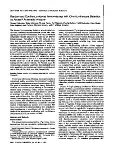
Westwood, Massachusetts). CelluTome Epidermal Micrograft Harvesting Procedure. An automated system for suction blister epidermal harvesting. (CelluTome ...
ORIGINAL INVESTIGATION
Epidermal Micrografts Produced via an Automated and Minimally Invasive Tool Form at the Dermal/ Epidermal Junction and Contain Proliferative Cells That Secrete Wound Healing Growth Factors Sandra N. Osborne, PhD; Marisa A. Schmidt, BS; Kathleen Derrick, MS; and John R. Harper, PhD
ABSTRACT OBJECTIVE: The aim of this scientific study was to assess epidermal micrografts for formation at the dermal-epidermal (DE) junction, cellular outgrowth, and growth factor secretion. Epidermal harvesting is an autologous option that removes only the superficial epidermal layer of the skin, considerably limiting donor site damage and scarring. Use of epidermal grafting in wound healing has been limited because of tedious, time-consuming, and inconsistent methodologies. Recently, a simplified, automated epidermal harvesting tool (CelluTome Epidermal Harvesting System; Kinetic Concepts Inc, San Antonio, Texas) that applies heat and suction concurrently to produce epidermal micrografts has become commercially available. The new technique of epidermal harvesting was shown to create viable micrografts with minimal patient discomfort and no donor-site scarring. DESIGN: This study was a prospective institutional review boardYapproved healthy human study. SETTING: This study was conducted at the multispecialty research facility, Clinical Trials of Texas, Inc, in San Antonio, Texas. PATIENTS: The participants were 15 healthy human volunteers. RESULTS: Epidermal micrografts formed at the DE junction, and migratory basal layer keratinocytes and melanocytes were proliferative in culture. Basement membraneYspecific collagen type IV was also found to be present in the grafts, suggesting that the combination of heat and vacuum might cause partial delamination of the basement membrane. Viable basal cells actively secreted key growth factors important for modulating wound healing responses, including vascular endothelial growth factor, hepatocyte growth factor, granulocyte colony-stimulating factor, platelet-derived growth factor, and transforming growth factor >.
CONCLUSIONS: Harvested epidermal micrografts retained their original keratinocyte structure, which is critical for potential re-epithelialization and repigmentation of a wound environment. KEYWORDS: epidermal micrograft, split-thickness skin graft, cellular outgrowth ADV SKIN WOUND CARE 2015;28:397Y405
Autologous split-thickness skin grafts (STSGs) often succeed as coverage over wounds when conservative therapies fail; however, STSGs generally require anesthesia and hospitalization and result in donor-site trauma that can increase the risk of complications for the patient. Autologous epidermal grafts that have been minced to expand and cover wounds much larger than the donor site provide an immediate wound coverage option. Unlike an STSG, an epidermal graft contains only the superficial epidermal layer of the skin. Advantages of epidermal grafts include minimal scarring to the donor site, and no risk of graft rejection. Epidermal suction blister grafting was first demonstrated by Reverdin, who reported the results of pinch grafting, or ‘‘epidermic grafting,’’ in 1869.1 Epidermal grafting using suction blisters in vivo on normal human skin was pioneered by Kiistala and Mustakallio2 and was later applied by Falabella3 to treat vitiligo. Although epidermal suction blister grafting has been successfully used for many decades in treating pigmentation disorders, reports of its application in wound management are more recent. However, the most commonly used suction blister method of epidermal grafting involves manual use of syringes to raise epidermal blisters and has been described by authors as cumbersome, time consuming, and inconsistent.4Y8 Larger syringes (920 mL) may take hours to create blisters, and smaller syringes (G10 mL) provide blisters that are
Sandra N. Osborne, PhD, is Senior Scientist; Marisa A. Schmidt, BS, and Kathleen Derrick, MS, are both Scientist III; and John R. Harper, PhD, is Senior Vice President, Clinical Affairs and Clinical Sciences; all at Kinetic Concepts Inc (KCI), an Acelity Company, San Antonio, Texas. The authors have disclosed that they are all employees of KCI, an Acelity Company. Acknowledgment: The authors thank Brad Jonietz for managing the project; Andy Ziegler, Sameer Sabir, and Robert LaRoche for thoughtful discussions, guidance, and training; Karen Beach, Anthony Rycerz, Michael Manwaring, Ricardo Martinez, and Julissa Ramos for critical review of the manuscript; Leslie Gutierrez for technical assistance with sample preparation for histology; Jagan Achi and Isabella Pacetti for help with data operations; Zhenmei Liu, David Flores, and Roberta James for statistical analyses; Michele Hilmer, Lorenzo Pina, Staci Poettgen, and Jill Twardowski for assistance with clinical operations; Alex Van Engelenburg for histological processing at HistoTox Laboratories (Boulder, Colorado); and Clinical Trials of Texas, Inc (San Antonio, Texas) for administration of the clinical study. This work was funded by KCI, an Acelity Company. Submitted May 30, 3014; accepted in revised form August 4, 2014. This is an open-access article distributed under the terms of the Creative Commons Attribution-NonCommercial-NoDerivatives 3.0 License, where it is permissible to download and share the work provided it is properly cited. The work cannot be changed in any way or used commercially. WWW.WOUNDCAREJOURNAL.COM
397
ADVANCES IN SKIN & WOUND CARE & SEPTEMBER 2015
ORIGINAL INVESTIGATION
difficult to manipulate.9 Syringe methods also require multiple sittings, and inadequate handling of the graft can lead to tearing and improper orientation, causing graft take failure. An automated and minimally invasive tool for harvesting autologous epidermal micrografts for use as skin grafts has recently become commercially available.10,11 The novel system combines suction and warmth to produce uniformly viable epidermal micrografts in less than 40 minutes, without an anesthetic and with minimal patient discomfort.11 This was an institutional review board (IRB)Yapproved analysis of epidermal micrografts harvested from 15 healthy human adult volunteers with the automated, minimally invasive blister harvesting tool. The purpose of the study was to determine if the epidermal micrografts generated by the automated tool were formed at the dermal-epidermal (DE) junction, showed keratinocyte and melanocyte outgrowth in culture, and secreted wound healing growth factors into the culture media. These are all normal characteristics of the epithelium.
MATERIALS AND METHODS Selection of Study Population and Overall Experimental Design This is a scientific study of the composition of epidermal micrografts harvested from 15 healthy human subject volunteers. The study was conducted at a single site (Clinical Trials of Texas, San Antonio, Texas), and all procedures were performed under a protocol approved by an IRB (IntegReview Ethical Review Board, Austin, Texas). Informed consent was obtained. The study was conducted following the International Conference on Harmonisation Good Clinical Practice Guidelines, as well as US Food and Drug Administration regulations, and has been audited by an independent clinical and regulatory consulting firm (Health Policy Associates, Inc, Westwood, Massachusetts).
CelluTome Epidermal Micrograft Harvesting Procedure An automated system for suction blister epidermal harvesting (CelluTome Epidermal Harvesting System; Kinetic Concepts Inc, San Antonio, Texas) was used to create micrografts on inner-thigh donor sites of 15 healthy adult volunteers. Body hair was removed using disposable razor blades, and the donor site was cleaned with 70% isopropyl alcohol wipes. The sterile harvester was secured around the subject’s inner thigh, and the vacuum head was snapped onto the harvester with the tubing facing up. The automated suction blister epidermal harvesting system was initiated, and vacuum (j400 to j500 mm Hg) and heat (37- C to 41- C) were applied to donor sites until epidermal micrografts were formed (G40 minutes). At the time of harvest, the vacuum head was detached from the harvester, and a transparent film dressing (Tegaderm; 3M, St Paul, Minnesota) was immediately placed on top of the micrografts. The micrografts were harvested by activating the harvester’s blue ADVANCES IN SKIN & WOUND CARE & VOL. 28 NO. 9
handle, which set the blade in motion. A 5 � 5-cm array consisting of 128 epidermal micrografts spaced 2 mm apart was then collected onto the adhesive transparent film. Each 5 � 5-cm micrograft array was placed in a sterile cell culture flask with a liquid medium (EpiLife Medium; Life Technologies, Carlsbad, California) prepared with 60 KM calcium chloride and immediately transported to the laboratory for analysis. No local anesthetics were administered to subjects during or after micrograft formation and harvesting.
Histology and Immunohistochemistry Epidermal micrografts from each subject were assessed for graft formation at the DE junction using histological analysis through micrograft cross sections stained with the cellular proliferation marker Ki67 and counterstained with hematoxylin. Donor-site samples from 12 of 15 subjects were allocated for proliferation and histological analyses, providing a quarter of a donor-site micrograft array, which included approximately 30 micrografts, for each in vitro assessment. To detect the presence of Ki67 (Spring Biosciences, Pleasanton, California) and collagen type IV, epidermal micrografts were fixed in 10% buffered formalin at room temperature for 24 hours, followed by treatment with 70% ethanol solution, and then shipped to a laboratory (HistoTox Laboratories Inc, Boulder, Colorado) for paraffin embedding and sectioning. Tissues were dehydrated and embedded in paraffin for 5-Km thick sectioning. Formalin-fixed, paraffin-embedded tissue was subjected to either heat or enzyme antigen retrieval. After enzyme- and protein-blocking steps, tissues were incubated with collagen type IV primary antibody (Abcam, Cambridge, Massachusetts) or Ki67 at room temperature for 30 minutes. This was followed by 30-minute incubation with goat antiYrabbit immunoglobulin G horseradish peroxidase polymer kits (EnVision SystemYHRP; Dako North America Inc, Carpinteria, California). The stain was then developed using 3,3¶-diaminobenzidine substrate solution (Dako North America Inc). Slides were counterstained with Mayer hematoxylin, dehydrated, and permanently coverslipped. Individual micrografts were assessed using inverted microscopes (Olympus IX51 and IX81; Olympus Corporation of the Americas, Center Valley, Pennsylvania) with transmitted light, and images of 100� magnification were collected using ImagePro Plus 7.0 software (MediaCybernetics, Bethesda, Maryland).
Keratinocyte and Melanocyte In Vitro Culture To determine whether resident basal layer keratinocytes and melanocytes were capable of migration and proliferation beyond the epidermal micrograft borders, micrografts from 12 of 15 subjects were cultured in vitro using collagen-coated surfaces and standard cell culture conditions. Keratinocyte and melanocyte outgrowth was assessed on rat tail tendon collagen type IYcoated surfaces
398
WWW.WOUNDCAREJOURNAL.COM
ORIGINAL INVESTIGATION
(VWR International, Wayne, Pennsylvania) using standard tissue culture practices (37- C, 5% CO2/95% air, humidified cell culture incubator). Keratinocyte outgrowth was monitored in a sterile, liquid medium prepared with 60 KM calcium chloride (EpiLife Medium) and human keratinocyte growth supplement containing BPE, 0.2% vol/vol; bovine insulin, 5 Kg/mL; hydrocortisone, 0.18 Kg/mL; bovine transferrin, 5 Kg/mL; and human epidermal growth factor, 0.2 ng/mL (HKGS; Life Technologies). Melanocyte outgrowth was monitored in a sterile, liquid medium prepared with 200 KM calcium chloride (M254; Life Technologies) supplemented with 1� PMA-free human melanocyte growth supplement (HMGS-2 (100�); Life Technologies). Both specialty media were further supplemented with 1� antibiotic-antimycotic solution, per manufacturer’s instructions. Epidermal micrograft arrays were divided into 4 quadrants in a laminar flow hood for each individual assessment. One quadrant was further bisected into 2, and each eighth of an array, containing approximately 10 micrografts, was placed with the basal keratinocyte layer down onto collagen type IYcoated 6-well plates in EpiLife Medium for keratinocyte growth or M254 for melanocyte growth. The micrografts were fed and imaged using bright field microscopy every 5 T 3 days until the cultures reached confluence. Individual micrografts were assessed using inverted microscopes (Olympus IX51 and IX81; Olympus Corporation of the Americas) with transmitted light, and images of 40� and 100� magnification were collected using Image-Pro Plus 7.0 software (MediaCybernetics). Once cells reached confluence, epidermal micrograft cultures were fixed in a 4% paraformaldehyde solution (Alpha Aesar, Ward Hill, Massachusetts) for 30 minutes at room temperature and were permeabilized with 0.1% Triton X-100 (Sigma-Aldrich, St Louis, Missouri) for 5 minutes at room temperature. Fluorescence staining of the plasma membrane, cytoskeleton (actin), and nucleus were used to highlight the morphology of outgrowing cells. Cells were stained for actin with a 200 U/mL Alexa Fluor 488 phalloidin stain (Life Technologies). Nuclei were stained with 5 mg/mL 4¶,6-diamidino-2-phenylindole (DAPI; Life Technologies), a fluorescent stain that binds strongly to A-TYrich regions in DNA. Cell Mask Orange (1.25 Kg/mL) (Life Technologies) was used to delineate the outer surface of the cell and stain the plasma membrane.
(Luminex Corporation, Austin, Texas) with multiplex bead-based immunoassays. Six epidermal micrografts from 3 subjects were analyzed in triplicate at 24, 48, and 72 hours and 7 days for a total of 72 micrografts analyzed per subject. Six micrografts per time point and per replicate were placed into wells of a sterile tissue culture 96-well plate with a liquid medium (EpiLife) supplemented with human keratinocyte growth supplement (Life Technologies) and allowed to incubate in a 37- C, 5% CO2/95% air, humidified cell culture incubator. Human epidermal keratinocytes (HEKs; Life Technologies) served as a positive culture control to ascertain whether analyte concentrations were within the levels of detection. Human epidermal keratinocytes were seeded at 5000 cells per well in sterile tissue culture 96-well plates and grown in monolayer for 7 days in triplicate in the same culture conditions as the epidermal micrografts. Conditioned media, spent media harvested from cultured cells, was assayed on a custom 8-plex multiplex bead-based immunoassay according to the manufacturer’s guidelines (Milliplex MAP Growth Factor panel; Millipore Corporation, St Charles, Missouri), which contained granulocyte colony-stimulating factor (G-CSF), vascular endothelial growth factor (VEGF), epidermal growth factor (EGF), fibroblast growth factor 2, platelet-derived growth factor AA (PDGF-AA), platelet-derived growth factor AB/BB (PDGFAB/BB), transforming growth factor > (TGF->), and hepatocyte growth factor (HGF). Briefly, individual antibody beads were mixed, and standards were reconstituted per kit instructions. Each triplicate sample was plated in duplicate with complete EpiLife Medium used for the matrix and for the background wells to serve as baseline. The plate was run on a BioPlex 200 (Bio-Rad Laboratories Inc, Hercules, California) set at 100 KL, with 50 beads per region. Standard curves were created using a 5-parameter logistic curve fitting method using the BioPlex Manager software version 6.0 (Bio-Rad Laboratories Inc). Values were reported in picograms per milliliter. Values that were below the range of the standard curve were interpreted as a ‘‘no’’ (j) for the presence of the growth factor. Values within the range of the standard curve were interpreted as a ‘‘yes’’ (+) for the presence of the growth factor analyzed. In addition, values that were too high to be detected by the instrument accurately, that equaled or exceeded the upper standard curve limit, were interpreted as a ‘‘yes’’ (+) for presence of the growth factor.
Secreted Growth Factor Analysis Donor-site samples from 3 of 15 subjects were allocated for growth factor analyses. Approximately one-half of a 5 � 5-cm donor-site micrograft array per subject was assessed for in vitro growth factor secretion. In order to quantify a selected panel of suction micrograft secreted growth factors, epidermal micrografts were cultured for up to 7 days, and conditioned media harvested from cultured cells was analyzed at various time points using Luminex technology WWW.WOUNDCAREJOURNAL.COM
Statistical Analyses The evaluable population 1 (EV1) consisted of 12 subject samples used for proliferation and histology analyses. The evaluable population 2 (EV2) consisted of 3 subject samples used for growth factor analyses. For the assessment of graft formation occurring at the DE junction from histological analyses, the frequency and percentage of subjects with graft formation at the DE junction
399
ADVANCES IN SKIN & WOUND CARE & SEPTEMBER 2015
ORIGINAL INVESTIGATION
were calculated as follows: graft formation rate = [number of subjects who had graft formation at the DE junction] / [EV1 population]. For the assessment of cellular outgrowth or proliferation of cells from the DE junction evaluated via in vitro cell culture analyses on collagen matrices, the frequency and percentage of subjects who had cellular outgrowth or proliferation of cells from the DE junction were calculated as follows: cellular outgrowth or proliferation rate = [number of subjects who had cellular outgrowth or proliferation] / [EV1 population]. For the assessment of growth factor secretion from epidermal micrografts, the EV2 population was evaluated. A preselected panel of growth factors was evaluated, and summary statistics were provided as follows: N, mean, SD, median, and range (minimum, maximum) for continuous variables and frequencies and percentages for categorical variables.
Figures 1A and B. EPIDERMAL MICROGRAFTS FORM AT THE DE JUNCTION
RESULTS Subject Demographics The epidermal micrograft harvesting technique was performed on 15 healthy human subjects (8 males, or 53.3% of subjects; and 7 females, or 46.7% of subjects). All subjects were white, with a mean age of 51.9 years (range, 42Y70 years). Approximately 360 micrografts were assessed for proliferation and histological analyses from 12 of 15 subjects. Secreted factors were analyzed from 216 micrografts obtained from the remaining 3 subjects.
Formation of Epidermal Micrografts at the DE Junction Stained micrograft cross sections revealed that 100% of epidermal micrograft arrays from all 12 subjects showed Ki67-positive grafts, demonstrating that they formed at the DE junction (Figure 1A). Epidermal micrografts also retained basement membraneYspecific collagen type IV (Figure 1B), confirming that the micrografts formed at the DE junction and suggesting that the combination of heat and vacuum might cause partial delamination of the basement membrane.
Proliferation and Migration of Keratinocytes and Melanocytes from Epidermal Micrografts Keratinocyte and melanocyte outgrowths on collagen type IYcoated surfaces in vitro were observed for 100% of 12 micrograft arrays, demonstrating production of proliferative micrografts capable of cellular outgrowth from graft edges in all subjects. Representative bright field images are shown in Figures 2A and B. The opaque structures in the images were micrograft edges imaged at a 100� magnification. The cobblestone-like pattern shown in the images was characteristic of keratinocyte morphology, and the dendriticlike cells were characteristic of melanocytes grown in culture. A colony of keratinocytes and melanocytes emerged soon after harvest ADVANCES IN SKIN & WOUND CARE & VOL. 28 NO. 9
Epidermal micrografts were fixed in formalin and paraffin-embedded prior to sectioning and immunohistochemical staining. A, Bright field images for 2 selected subject micrografts demonstrated the positive (brown) Ki67 staining of basal cell layer keratinocytes (original magnification �100). Abbreviations: SC, stratum corneum; BK, basal keratinocyte. B, Bright field images for 2 selected subject micrografts demonstrated the positive (brown) collagen type IV staining of the basement membrane (original magnification �100). All images and photographs are owned by KCI Licensing Inc, San Antonio, Texas.
400
WWW.WOUNDCAREJOURNAL.COM
ORIGINAL INVESTIGATION
conditioned cell culture media (Table). Culturing the epidermal micrografts in a medium containing high levels of EGF resulted in a high signal-to-noise ratio for EGF (91); therefore, accurate assessment of EGF secretion was not possible using this method, and samples were excluded from further analysis (Table). Fibroblast growth factor 2 levels for all 3 subject epidermal micrografts were too low to detect; however, the signal for fibroblast growth factor 2 was also not detectable in the authors’ positive HEK control cultures, suggesting that this analyte was not detectable using the
Figures 2A and B. EPIDERMAL MICROGRAFTS CONTAIN PROLIFERATIVE KERATINOCYTES AND MELANOCYTES
Figures 3A and B. FLUORESCENCE MICROSCOPY OF IN VITRO EPIDERMAL MICROGRAFT CELLULAR OUTGROWTH
Epidermal micrografts were cultured on collagen type IYcoated plates in specialized media to support keratinocyte and melanocyte growth using standard cell culture conditions. Opaque structures in the images are micrograft edges imaged at a 100� magnification. A, Representative bright field image of epidermal micrograft showing a colony of keratinocyte and melanocyte outgrowth from the graft edge (original magnification �100). B, Representative bright field image of epidermal micrografts showing complete cellular outgrowth from the graft edges and confluence between grafts (original magnification �40). All images and photographs are owned by KCI Licensing Inc, San Antonio, Texas.
as shown in Figure 2A. Complete coverage by keratinocytes between micrograft edges was observed a few days after harvest (Figure 2B). Representative fluorescence images of cellular outgrowth from 2 different subject samples are shown in Figures 3A and B.
Epidermal Micrograft Secreted Growth Factors All 3 subjects produced epidermal micrografts that secreted VEGF, TGF->, PDGF AA, PDGF AB/BB, HGF, and G-CSF into the WWW.WOUNDCAREJOURNAL.COM
Epidermal micrografts were cultured on collagen type IYcoated plates in specialized media to support keratinocyte and melanocyte growth using standard cell culture conditions. Cultures were fixed and stained for actin (green), plasma membrane (red), and nuclei (blue). Fluorescence microscopy demonstrating a representative image of cellular outgrowth from 2 different subject samples (original magnification �100). All images and photographs are owned by KCI Licensing Inc, San Antonio, Texas.
401
ADVANCES IN SKIN & WOUND CARE & SEPTEMBER 2015
ORIGINAL INVESTIGATION
Table.
COMPARISON OF EPIDERMAL MICROGRAFT AND CONTROL HUMAN EPIDERMAL KERATINOCYTE SECRETED GROWTH FACTORS INTO CONDITIONED CULTURE MEDIA EGF FGF-2 TGF-> G-CSF PDGF-AA PDGF-AB/BB HGF VEGF
Micrografts (24 h)
Micrografts (48 h)
Micrografts (72 h)
Micrografts (7 d)
HEK Control (7 d)
NA NA + + + + + +
NA NA + + + + + +
NA NA + + + + + +
NA NA + + + + + +
NA NA + + + + j +
Abbreviations: EGF, epidermal growth factor; FGF, fibroblast growth factor 2; G-CSF, granulocyte colony-stimulating factor; HGF, hepatocyte growth factor; PDGF, platelet-derived growth factor; TGF->, transforming growth factor >; VEGF, vascular endothelial growth factor. Secreted growth factors were detected using multiplex bead-based immunoassays. Some samples were not assessable (NA) because of high signal-to-noise ratios (EGF 91) or lack of analyte signal in the positive human epidermal keratinocyte control (fibroblast growth factor 2).
applied methodology, and these samples were excluded from further analyses (Table). In contrast, the values for VEGF were higher than the most concentrated control sample used to create a standard curve. For these samples, a value of the upper standard curve limit was assigned, and the results were recorded positive for the presence of VEGF (Table). Growth factor levels increased over a 3-day period and appeared to reach a threshold at day 3 remaining constant until day 7 except for PDGF-AB BB and G-CSF, which began declining after 3 days in culture (Figure 4). Cultured HEK controls did not secrete HGF; however, epidermal micrografts were shown to secrete high levels of HGF into the culture media (Table).
DISCUSSION The basal cell layer of the epidermis consists primarily of mitotically active and migratory keratinocytes and also contains mela-
nocytes responsible for skin pigmentation.12Y14 Success of epidermal grafting depends largely on precise separation at the DE junction and transfer of the entire, intact basal layer to the recipient site (Figures 5A and B). Proliferative, Ki67-positive cells have previously been shown to be confined to the basal layer of the epidermis.15Y18 Therefore, separation at the DE junction was assessed using histological analysis through micrograft cross sections stained with the cellular proliferation marker, Ki67, and counterstained with hematoxylin. Results of this study showed basal layer cells were proliferative, as observed with anti-Ki67 antibodies. With the automated system, epidermal micrografts formed at the DE junction in 12 of 12 cases. Although other studies have demonstrated Ki67 immunohistochemical staining of basal layer cells of normal and diseased skin, this is the first report of Ki67-specific immunohistochemical localization to basal layer keratinocytes of suction blister roofs from
Figure 4. EPIDERMAL GRAFT SECRETED GROWTH FACTORS PER TIME POINT
Secreted growth factors were detected using multiplex bead-based immunoassays. Analyte levels were plotted over time with mean and SE bars. ADVANCES IN SKIN & WOUND CARE & VOL. 28 NO. 9
402
WWW.WOUNDCAREJOURNAL.COM
ORIGINAL INVESTIGATION
Figures 5A and B. EPIDERMAL LAYERS OF THE SKIN TO DERMIS
A, Four of the layers of the epidermis are represented in this schematic drawing showing the cornified layer (stratum corneum), which provides the permeability barrier function of the epidermis; the granular layer (stratum granulosum), where keratinocytes have lost their nuclei and their cytoplasm appears granular; the spinous layer (stratum spinosum), where the immunologically active Langerhans cells also reside; and the basal/germinal layer (stratum basale/germinativum), the ‘‘heartbeat’’ of the epidermis, which is composed mainly of proliferating keratinocytes. Melanocytes and Merkel cells are also found in the stratum basale. B, Using an automated epidermal harvesting tool (CelluTome Epidermal Harvesting System; KCI USA Inc, San Antonio, Texas), the intact epidermis is separated from the dermis at the dermal-epidermal (DE) junction. Basal keratinocytes, melanocytes and components of the basement membrane are contained within the harvested epidermis. All images and photographs are owned by KCI Licensing Inc, San Antonio, Texas.
healthy human samples.15Y18 Nuclear protein Ki67Vexpressed in cycling cells (G1, S, G2, and M phases) and absent in quiescent cells16Vwas effective in staining proliferating cells of the basal layer of the epidermis during this study. Previous studies have shown that suction blistering occurs at the DE junction without evidence of damage to the basal cell layer or basement membrane.2,19 The lamina densa, also known as the ‘‘basal lamina’’ or ‘‘basement membrane,’’ is composed primarily of collagen type IV, laminins, and perlecans.20Y22 Therefore, to determine if the automated system’s combined elements of suction WWW.WOUNDCAREJOURNAL.COM
and heat resulted in delamination of the basement membrane, the authors assessed histological cross sections of epidermal grafts for the presence of type IV collagen.20Y22 Interestingly, immunohistochemical staining with type IV collagen revealed that, at least partially, some basement membrane components were indeed contained within the epidermal micrografts created using this system (Figure 1B). In addition to its function as a protective barrier, the epidermis has secretory activity important for matrix remodeling, paracrine effects at distant sites in the body, inflammation, maintenance of
403
ADVANCES IN SKIN & WOUND CARE & SEPTEMBER 2015
ORIGINAL INVESTIGATION
tissular homeostasis, and wound healing and re-epithelialization.23,24 Re-epithelialization is a highly dynamic and complex process that involves a number of different cell types, growth factors, and chemokines.24,25 Keratinocytes are the predominant cell type in all layers of the epidermis, constituting approximately 90% of epidermal cells. Keratinocytes consist of approximately 10% stem cells, 50% transit amplifying cells, and 40% differentiated cells.26 After stimulation by a variety of endogenous or exogenous factors, the various types of keratinocytes are activated to secrete cytokines and growth factors into the microenvironment.27 For example, epidermal keratinocytes, including those in this study, are able to produce and secrete several types of cytokines and growth factors including G-CSF, TGF->, and PDGF as observed using multiplex immunoassays (Figure 4).28 In this study, the authors analyzed a small subset of potential factors known to be secreted by epidermal cells with well-characterized functions in cell proliferation and wound healing.24,25 Epidermal micrografts produced with the automated tool secreted growth factors important for modulating the wound healing response, including VEGF, TGF->, PDGF AA, PDGF AB/BB, HGF, and G-CSF (Table, Figure 4). Granulocyte colony-stimulating factor is a cytokine that regulates proliferation, differentiation, and survival of cells including keratinocytes. The release is triggered by changes in skin homeostasis such as wounding, and it has been proposed to mediate inflammation after changes to homeostasis in the skin.29 Platelet-derived growth factor is known to induce migration, proliferation, and differentiation of different cell types, including keratinocytes and melanocytes, and serves as a positive regulator of angiogenesis.24,30 Vascular endothelial growth factor, another positive regulator of angiogenesis, has been shown to be upregulated by keratinocytes in response to TGF->, also secreted by epidermal micrografts.31 In addition, TGF-> has been shown to be an important stimulator of keratinocyte and melanocyte proliferation and migration.24,25 Keratinocytes undergo an epithelial-mesenchymal transition (EMT) process that enables them to assume a mesenchymal cell phenotype during wound healing.32,33 Hepatocyte growth factor is secreted by mesenchymal cells and injured keratinocytes, to stimulate both keratinocyte and melanocyte proliferation during re-epithelialization.24,34 In addition, the ability of hepatocyte growth factor to stimulate EMT and cell motility gives HGF an important role in angiogenesis and tissue regeneration. In this study, the authors demonstrated that HGF was secreted by harvested epidermal micrografts that retained their 3-dimensional structure and contained the full complement of cell types residing within the epidermis (Figure 4). As expected, HGF was not detected in conditioned media from HEKs that were grown in monolayer culture and had not undergone an EMT in response to injury.
ADVANCES IN SKIN & WOUND CARE & VOL. 28 NO. 9
In order for complete re-epithelialization and repigmentation to occur, each epidermal micrograft must produce epidermal basal cell outgrowth from the graft edges to close the 2-mm distance between grafts. Culture samples from 12 of 12 subjects in this study produced micrografts capable of keratinocyte and melanocyte outgrowth. These in vitro cell culture results support Kiistala and Mustakallio’s2 initial findings that basal layer keratinocytes and melanocytes are present in suction blisters (Figures 2A and B). Although previous studies have established epidermal in vitro cultures from trypsinized cell suspensions taken from suction blisters, this is the first report demonstrating basal layer keratinocyte and melanocyte outgrowth from intact suction blister roofs from healthy human samples.35Y39
CONCLUSIONS The automated and minimally invasive epidermal harvesting tool used in this healthy human IRB-approved study yielded epidermal micrografts formed at the DE junction. Other nonYindustrysponsored studies cited in this article have revealed that epidermal grafts previously created with suction were also viable and formed at the DE junction. The authors have confirmed and expanded on those findings by including growth factor analyses and by demonstrating melanocyte and keratinocyte coculture from the epidermal explants harvested with the automated harvesting system. Future studies will be needed to confirm the clinical effectiveness of epidermal grafting.
&
REFERENCES 1. Biswas A, Bharara M, Hurst C, et al. The micrograft concept for wound healing: strategies and applications. J Diabetes Sci Technol 2010;4:808-19. 2. Kiistala U, Mustakallio KK. In-vivo separation of epidermis by production of suction blisters. Lancet 1964;1:1444-5. 3. Falabella R. Epidermal grafting. An original technique and its application in achromic and granulating areas. Arch Dermatol 1971;104:592-600. 4. Gupta S, Jain VK, Saraswat PK. Suction blister epidermal grafting versus punch skin grafting in recalcitrant and stable vitiligo. Dermatol Surg 1999;25:955-8. 5. Burm JS. Simple suction device for autologous epidermal grafting. Plast Reconstr Surg 2000;106:1225-6. 6. Alexis AF, Wilson DC, Todhunter JA, et al. Reassessment of the suction blister model of wound healing: introduction of a new higher pressure device. Int J Dermatol 1999;38:613-7. 7. Njoo MD, Westerhof W, Bos JD, et al. A systematic review of autologous transplantation methods in vitiligo. Arch Dermatol 1998;134:1543-9. 8. Ichiki Y, Kitajima Y. Successful treatment of scleroderma-related cutaneous ulcer with suction blister grafting. Rheumatol Int 2008;28:299-301. 9. Khunger N, Kathuria SD, Ramesh V. Tissue grafts in vitiligo surgeryVpast, present, and future. Indian J Dermatol 2009;54:150-8. 10. Ramos JV, Schmidt M, Wu SC. Epidermal skin grafts for the treatment of chronic lower extremity ulcers. Podiatry Manage 2013;32:95-6, 98-100, 102, 104. 11. Osborne SN, Schmidt MA, Harper JR. An automated and minimally invasive tool for generating autologous viable epidermal micrografts. Adv Skin Wound Care; in press. 12. Barrandon Y, Green H. Three clonal types of keratinocyte with different capacities for multiplication. Proc Natl Acad Sci U S A 1987;84:2302-6. 13. Maddodi N, Jayanthy A, Setaluri V. Shining light on skin pigmentation: the darker and the brighter side of effects of UV radiation. Photochem Photobiol 2012;88:1075-82.
404
WWW.WOUNDCAREJOURNAL.COM
ORIGINAL INVESTIGATION
14. Pinon P, Wehrle-Haller B. Integrins: versatile receptors controlling melanocyte adhesion, migration and proliferation. Pigment Cell Melanoma Res 2011;24:282-94. 15. Furukawa F, Imamura S, Fujita M, et al. Immunohistochemical localization of proliferating cell nuclear antigen/cyclin in human skin. Arch Dermatol Res 1992;284:86-91. 16. Knaggs HE, Holland DB, Morris C, et al. Quantification of cellular proliferation in acne using the monoclonal antibody Ki-67. J Invest Dermatol 1994;102:89-92. 17. Andreadis ST, Hamoen KE, Yarmush ML, et al. Keratinocyte growth factor induces hyperproliferation and delays differentiation in a skin equivalent model system. FASEB J 2001; 15:898-906. 18. Patel GK, Wilson CH, Harding KG, et al. Numerous keratinocyte subtypes involved in wound re-epithelialization. J Invest Dermatol 2006;126:497-502. 19. Lowe LB Jr, van der Leun JC. Suction blisters and dermal-epidermal adherence. J Invest Dermatol 1968;50:308-14. 20. Fleischmajer R, Perlish JS, MacDonald ED 2nd, et al. There is binding of collagen IV to beta 1 integrin during early skin basement membrane assembly. Ann N Y Acad Sci 1998; 857:212-27. 21. Timpl R, Wiedemann H, van Delden V, et al. A network model for the organization of type IV collagen molecules in basement membranes. Eur J Biochem 1981;120:203-11. 22. Hassell JR, Robey PG, Barrach HJ, et al. Isolation of a heparan sulfateYcontaining proteoglycan from basement membrane. Proc Natl Acad Sci U S A 1980;77:4494-8. 23. Katz AB, Taichman LB. Epidermis as a secretory tissue: an in vitro tissue model to study keratinocyte secretion. J Invest Dermatol 1994;102:55-60. 24. Gurtner GC, Werner S, Barrandon Y, et al. Wound repair and regeneration. Nature 2008; 453:314-21. 25. Raja, Sivamani K, Garcia MS, et al. Wound re-epithelialization: modulating keratinocyte migration in wound healing. Front Biosci 2007;12:2849-68. 26. Heenen M, Galand P. The growth fraction of normal human epidermis. Dermatology 1997; 194:313-7. 27. Stoof TJ, Boorsman DM, Nickoloff BJ. Keratinocyte and immunological cytokines. In: Leigh IM, Lane EB, Watt FM, eds. The Keratinocyte Handbook. Cambridge, UK: Cambridge University Press; 1994.
WWW.WOUNDCAREJOURNAL.COM
28. Takashima A, Bergstresser PR. Cytokine-mediated communication by keratinocytes and Langerhans cells with dendritic epidermal T cells. Semin Immunol 1996;8:333-9. 29. Mann A, Breuhahn K, Schirmacher P, et al. Keratinocyte-derived granulocyte-macrophage colony stimulating factor accelerates wound healing: stimulation of keratinocyte proliferation, granulation tissue formation, and vascularization. J Invest Dermatol 2001;117: 1382-90. 30. Claesson-Welsh L. Mechanism of action of platelet-derived growth factor. Int J Biochem Cell Biol 1996;28:373-85. 31. Detmar M. The role of VEGF and thrombospondins in skin angiogenesis. J Dermatol Sci 2000;24 Suppl 1:S78-84. 32. Kalluri R, Weinberg RA. The basics of epithelial-mesenchymal transition. J Clin Invest 2009;119:1420-8. 33. Day RM, Felici A, Bottaro DP. Hepatocyte growth factor regulates transitions between epithelial and mesenchymal cellular phenotypes during normal development and in disease. In: Savagner P, ed. Rise and Fall of Epithelial Phenotype: Concepts of Epithelial-Mesenchymal Transition. New York, NY: Kluwer Academic/Plenum Publishers; 2005. 34. Nayeri F, Xu J, Abdiu A, et al. Autocrine production of biologically active hepatocyte growth factor (HGF) by injured human skin. J Dermatol Sci 2006;43:49-56. 35. Semnani RT, Law M, Kubofcik J, et al. Filaria-induced immune evasion: suppression by the infective stage of Brugia malayi at the earliest host-parasite interface. J Immunol 2004;172:6229-38. 36. Im S, Hann SK, Park YK, et al. Culture of melanocytes obtained from normal and vitiligo subjects. Yonsei Med J 1992;33:344-50. 37. Czajkowski R, Placek W, Drewa T, et al. Establishing melanocyte cultures in a serum-free system for transplantation in vitiligo patients. Med Sci Monit 2006;12:CR63-9. 38. Ahmad AA, Rhodes L, Marcelo CL. Differential trypsinization-selective adhesion technique for cultivation of normal adult human melanocytes in phorbol acetate-free and cholera toxin-free medium. Egypt J Plast Reconstr Surg 2005;29:61-6. 39. Kariniemi AL, Lehto VP, Vartio T, et al. Cytoskeleton and pericellular matrix organization of pure adult human keratinocytes cultured from suction-blister roof epidermis. J Cell Sci 1982;58:49-61.
405
ADVANCES IN SKIN & WOUND CARE & SEPTEMBER 2015