Feb 15, 2016 - ligand to the nickel redox center of the (NiFeSe) hy- drogenase. Selenium is an essential micronutrient for the growth of many eukaryotes and ...
THEJOURNAL OF BIOLOGICAL CHEMISTRY 0 1989 by The American Society for Biochemistry and Molecular Biology, Inc.
Vol. 264, No. S , Issue of February 15, pp. 2678-2682,1989 Printed in U.S.A.
EPR Studies with ‘%e-enriched (NiFeSe) Hydrogenase of Desulfovibrio baculatus EVIDENCE FOR A SELENIUM LIGAND TO THE ACTIVE SITE NICKEL* (Received for publication, July 26, 1988)
Shao H. He, Miguel TeixeiraS, Jean LeGall, Daulat S. Patils, Isabel MouraS, Jock J. G . MouraS, Daniel V. DerVartanianll,Boi H. Huynhs, and HarryD. Peck, Jr. From the Departmentof Biochemistry, School of Chemical Sciences, University of Georgia, Athens, Georgia 30602, $Centra de Quimica Estrutural Universidades de Lisoa,I. ST. Lison, Portugal, and the §Departmentof Physics, Emory University, Atlanta, Georgia 30322
The periplasmic hydrogenase containing equivalent amounts of nickel and selenium plus non-heme iron ((NiFeSe) hydrogenase) has been purified from cells of the sulfate reducing bacteriumDesulfovibrio baculatus (DSM 1748) grown on a lactate/sulfate medium containing natural Se isotopes and the nuclear isotope, 77Se.Both the 77Se-enrichedand unenriched hydrogenases wereshown to befree of other hydrogenases and characterized with regard to their Se contents. EPR studies of the reduced nickel signal generated by redox titrations of the enriched and unenriched (NiFeSe) hydrogenases demonstrated that the g, = 2.23 and g, = 2.17 resonances are appreciably broadened by the spin of the 77Senucleus ( I = I/$. This observation demonstrates unambiguously that the unpaired electron is shared by the Ni and Se atoms and that Se serves asa ligand to the nickel redox center of the (NiFeSe) hydrogenase.
Selenium is an essential micronutrient for the growth of many eukaryotes and prokaryotes and is found in proteins and specific amino acid tRNAs (1). It is incorporated into proteins by a nonspecific mechanism resulting in therandom incorporations of selenomethionine into polypeptide chains and by a highly specific cotranslational mechanism involving selenocysteine (1).The presence of selenocysteine has been demonstrated in glutathione peroxidase (2, 3) and proteins of unknown function (4) from eukaryotes and glycine reductase ( 5 ) , formate dehydrogenase (6), and hydrogenase (7) from prokaryotes. Selenocysteine is cotranslationally inserted into a polypeptide chain of formate dehydrogenase (8)and is coded in the mRNA by the opal nonsense codon, UGA, in both prokaryotes (9) and eukaryotes (10, 11). Evidence has been presented indicating that selenocysteine is synthesized from serine (12) and recently it hasbeen shown that a minor seryltRNA is involvedin the cotranslational incorporation of selenocysteine into polypeptide chains (13). The exact function or functions of selenium in biological systems remain to be elucidated. In the case of glutathione
* This work was supported in part by National Institutes of Health Grants AM01135 and GM32187 (to B. H. H.), National Science Foundation Grant DMB 8706205 (to J. L., D. V. D., and H. D. P.), and by the United States Department of Energy under Contract DEAS09-ER10499 (to H. D. P.). Thecosts of publication of this article were defrayed in part by the payment of page charges. This article must therefore be hereby marked “advertisement” in accordance with 18 U.S.C. Section 1734 solelyto indicate this fact. ll To whom correspondence should be addressed.
peroxidase, selenocysteine has been localized at theactive site and has been proposed to function in the redox reaction as a selenolate anion or selenic acid derivative (14). Selenium is chemically closely related to sulfur and the occurrence of selenocysteine in three prokaryotic redox enzymes suggests the possibility that selenocysteine can serve as a ligand to metal redox centers. In this regard, selenide has been shown to serve as an artificial replacement for sulfide in some nonheme iron clusters (15, 16). Two nickel-containing hydrogenases have been identified in the sulfate reducing bacteria belonging to the genus, Desulfouibrio (17), which can be distinguished on the basis of their metal contents (18, 19), immunological specificity (16), and gene structures’ (20, 21). Both of these hydrogenases contain non-heme iron clusters, 1 mol of nickel/mol of enzyme, and are composed of two subunits of similar size (30 and 60 kDa). The first of these hydrogenases has been termed the (NiFe) hydrogenase’ andcontains two [Fe4S4]plusa three-iron cluster (18) in addition to nickel. The second nickel-containing hydrogenase has been termed the (NiFeSe) hydrogenase and contains only two [Fe4S4]clusters plus selenium in equivalent amounts to nickel (22). The genes encoding the (NiFe) hydrogenase of Desulfouibriogigas (20) and Desulfovibrio uulgaris‘ (Hildenborough) and the (NiFeSe) hydrogenase of D. buculatus (21) have been cloned and sequenced. In the carboxyl terminus region of the gene for the large subunit of the (NiFeSe) hydrogenase, a codon (TGA) for selenocysteine has been located in a position homologous to a codon (TGC) for cysteine in the large subunit of the (NiFe) hydrogenase. Additionally, the large subunits of the two hydrogenases show 3 conserved cysteinyl residues and the small subunits possess 10 conserved cysteinyl residues.‘ Strong evidence exists inthe case of the (NiFe) hydrogenase that nickel is liganded to sulfur atoms (23) and involved in the primary activation of hydrogen (24,25). The comparative behavior of the nickel-containing hydrogenases in theproton/ deuterium exchange reaction has been investigated and a higher H2/HD ratio is observed with the (NiFeSe) hydrogenases than with the (NiFe) hydrogenase (22, 26). These data suggested that selenium is influencing the chemical reactivity of the active site nickel and raise the possibility that one of G. Voordouw, N. K. Menon, J. LeGall, E. S. Choi, H. D. Peck, Jr., and A. E. Przybyla, unpublished results. * The abbreviations used are: (NiFe) hydrogenase, hydrogenase containing nickel and non-heme iron; (NiFeSe) hydrogenase, hydrogenase containing equivalent amounts of nickel and selenium plus non-heme iron; GSH, glutathione; SDS, sodium dodecyl sulfate; PAGE, polyacrylamide gel electrophoresis; HPLC, high performance liquid chromatography.
2678
Selenium as a Ligand to the Active-site Nickel of a Hydrogenase
2679
S-200 column (5.4 X 85 cm). The column was eluted with 50 mM potassium phosphate buffer (pH 7.6) and the brown hydrogenase fraction eluted after the reddish-brown desulforubidin. The two batches of hydrogenase from the Sephacryl S-200 columns were combined and concentrated to a small volume (20ml) for preparative HPLC. Preparative HPLC-The hydrogenase fraction was again divided into two portions and each (2 ml) was loaded on an ion-exchange column (TSK DEAE-3SW) in a Beckman preparative HPLC. The eluting buffer was 10 mM potassium phosphate buffer (pH 7.0), with increasing concentrations of NaCl, and thehydrogenase fraction was collected at 250 mM NaCl. A summary of the purification of the (NiFeSe) hydrogenase is presented in Table 11. The purity of both (NiFeSe) hydrogenases was established by PAGE (28) and by SDSEXPERIMENTAL PROCEDURES PAGE (29). Assays and Determinations-Hydrogenase activity was determined Growth of Cells-D. baculatus (DSM 1743) was routinely grown in a lactate/sulfate medium (27) at 37 "C. Analyses of this medium by by the HZevolution assay (30) and hydrogen was determined using a plasma emission indicated the presence of 25 pg ofselenium/liter but Varian 4600 gas chromatograph (31). Protein was estimated by the a major source for this endogenous selenium in our chemicals could Biuret method (32) or Bradford method (33) using bovine serum not be identified. Consequently, for the preparation of the 77Se- albumin as a reference standard. Metal content was determined by enriched (NiFeSe) hydrogenase, it was decided to dilute the endoge- plasma emission spectroscopy using a Mark I1 Jarrell-Ash Model 965 nous selenium with a 4-fold excess of 77Se,which should yield a final atomcomp. Iron content was also confirmed by a chemical method calculated atom % excess of 70% in 77Se.Grey metallic 7'Se (atom % (34). Antibodies to the(NiFe), (NiFeSe),and (Fe)hydrogenases were excess, 91.77) was purchased from the ORNL Stable Isotopes Divi- gifts from Drs. A. E. Przybyla and N. K. Menon. Western blot sion, and 200 mg was dissolved in aminimum amount of concentrated analyses (35) were performed by employing whole blood serum. nitric acid. The pH of this solution was adjusted to 7.0 with 10 N EPR Spectroscopy-EPR measurements were performed on a BruNaOH and filter sterilized before addition to 200 liters of sterile ker ER 2OOD-SRC spectrometer equipped with an Oxford Instrulactate/sulfate medium. For preparation of the unenriched (NiFeSe) ments ESR 910 continuous flow cryostat. The data were recorded in hydrogenase, an equivalent amount of Na2SeOB was filter sterilized a Bruker Aspect 2000 computer and transferred to an IBM A T and added to 200 liters of sterilized lactate/sulfate medium. After 15 computer for data analysis. h, the bacteria were harvested in a Sharples centrifuge, resuspended Redox Titrations-The oxidation-reduction titrations were perin an equal volume of 50 mM Tris-HC1, pH 7.6, and frozen at -80 "C. formed at 25 "C in an apparatus similar to thatdescribed by Dutton In order to optimize the yield of the (NiFeSe) hydrogenase, the (36). The potentials were measured using a platinum and a saturated biosynthesis of this periplasmic hydrogenase was studied as a function calomel electrode, calibrated with a saturatedsolution of quinhydrone M SeOZ-. The bacteria of the growth phase in the presence of 3 X at pH7.0, and arequoted relative to the standard hydrogen electrode. contained in 50 liters of medium from a fermentor containing 200 The mediator dyes (40p~ each) used in the titration were benzyl liters of lactate/sulfate medium supplemented with selenium were viologen, methyl viologen, dichlorophenolindophenol,benzoquinone, harvested at midlog phase, late log phase, and late stationary phase. anthraquinone-2-sulfonic acid, phenazinemethosulfate, dimethyltriSpheroplasts were prepared from 15 g of freshly grown cells of D. quat, methylene blue, 2-hydroxy-1,4-naphthaquinone, triquat, and baculatus and theperiplasmic (NiFeSe) hydrogenase eluted from the indigotetrasulfonate. The protein solution was in 200 mM Tris-HC1 cells as previously described (22). As shown in Table I, the bacteria buffer a t pH 7.6 and the protein concentrations were 105 GM for the harvested during the late log phase contained the highest level of 77Se-enrichedhydrogenase and 50 p M for the unenriched enzymes. hydrogenase. It was noted that the cells grown on high selenium The hydrogenase solution was first reduced by incubation under 1 contained more than twice the total hydrogenase activity previously atm ofHz for 2 h and was then brought to a desired potential by reported (22) and that thehighest total activity was found in late log reducing the partial pressure of hydrogen and introducing a proper phase cells. Purification of the Periplosmic (NiFeSe) Hydrogenase-All purifi- amount of oxygen-free argon gas into the sample compartment. At cation procedures were carried out in air at 4 "C and the pH of the each selected potential, a sample was withdrawn with a gas-tight syringe and transferred into an EPRtube, which was attached to the buffers was 7.6 redox vessel,and immediately frozen at 77 K for EPR measurements. Preparation of the Crude Extract-The frozen cell suspension Isolation of Hydrogenase Subunits-The purified periplasmic (equivalent to 200 g) was thawed and the cells separated from the buffer by centrifugation at 20,000 X g for 1 h. The brown supernatant (NiFeSe) hydrogenase from D.baculatus was subjected to slab SDScontaining most of the periplasmic proteins was decanted, the pellet PAGE electrophoresis to separate the large and small subunits (37) resuspended in the original amount of buffer was centrifuged, and and lightly stained for 15 min (0.1%Coomassie Blue R-250,7% acetic the two supernatants were combined as thecrude extract for isolation acid, and 50% methanol). The gels were destained using acetic acid/ methanol/water (75/250/675, v/v) at 4 "C for 1h with gentle shaking. of the hydrogenase. DE52 Column-The crude extract was loaded on a DE52 column Kimwipes or small pieces of foam rubber were placed in the corners (6 X 32.5 cm), which was equilibrated with 10 mM Tris-HC1 (pH 7.6). of the destaining chamber to absorb the excess dye as itwas released The proteins absorbed on the column were eluted with increasing from the gel. Next, the stained subunits on the gel were excised and concentrations of Tris-HC1 buffer. The yellowish to brownish hy- the gel minced with a clean sharp razor blade. After soaking in water drogenase fractions were collected at an eluting buffer concentration and elution buffer (0.1% SDS in 0.05 M NH,HCO,) for 5 min, each around 250-300 mM Tris-HC1 and the hydrogenase fractions were gel preparation was transferred into the large well of an electrophothen concentrated to a small volume (56.3 ml) in a Diaflo apparatus retic elution cell fitted with the appropriate molecular weight cut-off Spectra-Por membrane for elution, following the procedure described with a YM-30 membrane. Sephacryl S-200 Column-The hydrogenase preparation was di- by Hunkerpiller et al. (38). The concentrated protein fraction colvided into two equal portions and each was loaded onto a Sephacryl lected from the sample collection cell was used for metal analysis.
the cysteine ligands to the nickelisreplacedby a selenocysteine in the (NiFeSe) hydrogenase. In this communication,we describe the preparation of 77Seenriched (NiFeSe) hydrogenaseo f D . baculatus and reportthe broadening of the nickel signal C (g = 2.23) in the EPR spectrum of the hydrogen-reduced 77Se (I = '/$enriched (NiFeSe) hydrogenase.The results demonstratethat selenium serves as a naturalligand to the nickel in this(NiFeSe) hydrogenase and that nickel is in all probability liganded by cysteinyl residues anda selenocysteinyl residue present in the large subunit of the (NiFeSe) hydrogenase.
TABLEI Effect of time of harvest on the hydrogenose content of D. baculatus Harvest time
Midlog phase 1,878 3,003 Late28,166 log/early stationary phase stationary ,461 2,509 Late 32.8 phase 17,952
Periplasmic fraction unitslml
31.8 50 46.6 67.1 48
total units
19,000 25,163
Spheroplast lysate unitslml
20,685
total units
1,685
Sum of activity total units
unitslg cell
1,379
2680
Selenium as a Ligand to the Active-site Nickel of a Hydrogenase
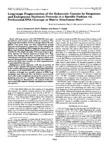
RESULTS AND DISCUSSION and presumably the enzyme contained 1 selenocysteine resiCharacterization of the Per@lusrnicHydrogenase-The en- due, the possibilities were considered that the extraselenium zymological and biochemical properties of the periplasmic was loosely bound to the hydrogenase or represented a nonhydrogenases purified from D. baculatus grown on low and specific incorporation of selenium as selenomethionine (1). In order to resolve the first question, the purified (NiFeSe) high selenium are shown in Table 111. The results show that hydrogenase was dialyzed anaerobically against a pH 9 soluthe hydrogenase prepared from bacteria grown in high seletion containing 0.1 M sodium sulfide for different periods of nium has twice the selenium content of those grown on low selenium andhasa 4-fold higher specific activity. These time (12-108 h). The Ni and Fe contents as well as activity observations raised important questions regarding the iden- were stable to this extended dialysis. However, during the tity of the purified hydrogenase and the significance of the first 12 h of dialysis, the selenium content decreased from 2.1 extra selenium. Because of the presence of different hydrog- to 1.1 mol/mol of hydrogenase and thereafter remained conenases in a single species of the sulfate reducing bacteria (35) stant. The result is clearly consistent with the presence of 1 selenocysteine/mol of hydrogenase; however, the and thepresence of multiple forms of hydrogenase (22), these molof amount of selenium was almost twice that of nickel. The results suggested the possibility that a new or different hyselenium content of the large and small subunits was invesdrogenase had been induced by growth in the presence of selenium. In order to resolve the question concerning the tigated and theresults arepresented in Table IV. Most of the Ni and Fe were lost during the isolation of the two subunits; identity of the periplasmic hydrogenase produced in thepreshowever, 1.2mol ofselenium were present in the large subunit ence of added selenium, Western blot analyses of the purified as predicted by the sequence of the gene. No evidence was hydrogenase were performed against polyclonal antibodies obtained from the presence of selenomethionine. It would raised against the (Fe) hydrogenase from D. vulgaris, the appear that the nonequivalence of the nickel and selenium (NiFe) hydrogenase from D. gigas, and the (NiFeSe) hydrogcontents may reflect the loss of nickel during the isolation of enase from D. baculatus. The results, shown in Fig. 1, demthe hydrogenase or a nickel deficiency during the growth of onstrate that thepurified hydrogenase cross-reacts only with antibodies to (NiFeSe) hydrogenase purified from bacteria the bacteria. EPR Spectroscopy-Previous EPR studies (22) have indigrown on a low selenium medium and does not cross-react cated that the “as-isolated” D. baculatus (NiFeSe) hydrogewith other characterized hydrogenases from the sulfate reducnase is almost EPR silent and exhibits a weak “isotropic” ing bacteria. These results demonstrate that thehydrogenase signal in the g = 2.02 region, similar to those observed for purified from selenium-supplemented media is the (NiFeSe) oxidized [3Fe-4S] clusters, and does not show prominent hydrogenase and that the presence of selenium causes an Ni(II1) EPR signals. In its partially reduced state, however, increased biosynthesis of the periplasmic (NiFeSe) hydrogethe (NiFeSe) hydrogenase does exhibit a signal at g, = 2.23, nase. g, = 2.17, and g, = 2.01, which is equivalent to the wellThe problem of the high selenium content was critical for characterized Ni-signal C in the D. gigas hydrogenase (39the interpretation of the EPRstudies since a form of selenium 41). Therefore, we have recorded EPR data from samples of other than selenocysteine bound to a metal center could lead (NiFeSe) hydrogenase poised at redox potentials between to broadening of the nickel EPR signal. As the gene encoding -340 and -380 mV, a region where the Ni-signal C is maxithe large and small subunits contained only one TGA codon mized.Fig. 2 shows the EPR spectra of the ?Se-enriched (trace A ) and theunenriched (trace B ) hydrogenase poised at TABLEI1 -360 mV. The data were recorded at 35 K with 2-milliwatt Purification of the (NiFeSe) hydrogenase fromD. baculatus microwave power and 1-millitesla modulation field. (Modumown on high selenium lation field of 0.1 millitesla has been applied to themeasureActivity ments but resolved hyperfine structure were not observed. Fraction Volume Protein Total Specific Applications of modulation field of either 1 milliteslas or 0.1 pmol pmol milliteslas yielded the same spectral shape.) Fig. 2 shows only mg ml HZ/min Hz/min/mg the g, = 2.23 and g, = 2.17 resonances, as the g = 2.0 region 122 5.75 X 10³ 7.02 X lo5 Crude extract 857 is masked by the radical signals originating from the reduced 3.84 X lo5 DE52 column 56.3 810 480 mediators. Comparison of the spectra shows that with the 18.4 269.3 1102 Sephacryl S-200 296,790 77Se-enrichedhydrogenase both resonances are appreciably column broadened by the spin of the 77Se nucleus (I = l/2). This (anion-exHPLC 6.65 93.8 186,800 1991 change column) observation demonstrated unambiguously that the unpaired (TSK DEAEelectron is shared by the Ni and Se atoms and that the Se is 3SW column) directly bound to the Ni. Identical results were obtained
TABLE 111 Enzymological properties of the (NiFeSe) hydrogenases fromD. baculatus grown o n low and high seleniumcontaining media Chemical analysis, 8.95 mol of Fe/mol of enzyme. Parameter
mass
Subunits molecular mass
Metal contents Fe
Ni
Se
Specific activity ( H evolution) ~
Am/Azm
Total hydrogenase in periplasm ”
kDa 100
Low Se hydrogenase (22) High Se hydrogenase 0.28 2,000 86.7 2.1
kDa
26,49 0.28 527 9.25 0.66 34,53 0.6 9.7
mollmol
%
0.69
47 90
Selenium as
a Ligand the to Active-site Nickel c
B
of a Hydrogenase
2681
2.228, g, = 2.174, and g, = 2.01, and with line widths of 1.95, 2.20, and 2.00 milliteslas, respectively. The same parameters were then used to simulate the spectrum of the 77Se-enriched hydrogenase. One 77Senucleus per each unpairedelectron and 70% enrichment were assumed for the simulations. Hyperfine coupling constants that best describe the observed broadening were found to be A, = 1.0 and A, = 1.8 milliteslas. (The value of A, was not determined.) The theoreticalsimulation is plotted above trace A in Fig. 2. These hyperfine coupling constants arevery similar in magnitudeas thoseobserved for the 77Se-substituted putidaredoxinfrom Pseudomonas putida FIG. 1. Western analysis of the purified periplasmic hydrogenase of D. baculatus. Lane 1, periplasmic (NiFeSe) hydrog- and adrenal ferredoxin (42). Both ferredoxins contain [2Feenase from D.baculatus grown on a selenium-rich medium. Lane 2, 2S] clusters, and, in the 77Se-substitutedproteins, the Se periplasmic (Fe) hydrogenase from D. uulgaris. Lane 3, periplasmic atoms are the bridging ligands between the Fe atoms. The (NiFe) hydrogenase from D. gigas. A , hydrogenasesreactedwith fact that similar magnitudes of hyperfine coupling constants antibodies raised against native enzyme of periplasmic hydrogenase were observed for the Ni signal C in the 77Se-enriched D. from D.baculatus (Norway 4). B, hydrogenases reacted withantibodies raised against native enzyme of periplasmic hydrogenase from D. baculatus hydrogenase strongly supports the conclusion that uulgaris. C, hydrogenases reacted with antibody raisedagainst native Se is a ligand to the Nicenter. The proximity of the nickel and selenium have also been enzyme of periplasmic hydrogenase fromD.gigas. demonstrated by complementary extended x-ray absorption TABLE IV fine structure studies of the unenriched (NiFeSe) hydrogen a ~ eT)e . ~ results shown that thedistance of the Ni-Se bond Metal contents of the large and small subunits of the (NiFeSe) hydrogenase is 2.46 A and that the selenium is bonded to a carbon atom, The recovery from SDS-PAGE plus electrophoretic elution was presumably selenocysteine. The nickel in the (NiFe)hydrogestimated tobe 50%. enase appears to be ligated to $wo or more sulfur atoms at an Fe Ni Se average bond distance of 2.20 A in a pseudooctahedral geometry with the otherligands being nitrogen and/or oxygen (23, mol/mol 43). The nickel at theactive site of the (NiFeSe) hydrogenase Large subunit 0.2 0.0 1.2 Small subunit 0.08 0.08 0.06 also appears tobe ligated in a pseudooctahedral geometry but to one or more sulfur atvms plus a selenium atcm: the former at A. As selenium at bond distancesof 2.2 A and the latter2.46 and sulfur share many chemical properties, it would therefore not be expected that substitution of a selenocysteine for a cysteine would dramatically alter either thecatalytic or physical properties of an enzyme; rather,the selenium/sulfur substitution probably represents a device for "fine tuning" a catalytic mechanism for a specific physiological requirement and only small differences, for example, in redox potential, EPR signals or catalyticactivity wouldbe expected. The nickel signal C (g = 2.19) is common to all nickel-containing hydrogenases and its redox potential has been estimated a t around -340 mV (40); however, this value is only approximate and small differences between the (NiFe) and (NiFeSe) hydrogenases cannot be ruled out. On the other hand, the g, values for the nickel signal C tend to be around 2.19 for (NiFe) hydrogenases (40) but around 2.23 for the (NiFeSe) hydrogenases (22, 40). This is also true for the 290 300 310 320 (NiFe) and (NiFeSe) hydrogenases characterized from the methanogenic bacteria4 (44) and may reflect the presence of Magnetic Field ( m T ) FIG. 2. EPR spectra of the 77Se-enriched (truce A ) and the selenium as a ligand to the nickel. With regard to catalytic un-enriched (truce B ) hydrogenases poised at -360 mV. The activity, the (NiFeSe) hydrogenase exhibits an HJHD ratio data were recorded at 35 K with microwave frequency of 9.43 GHz, of d . 0 in the proton deuterium exchange reaction while the 2-milliwatt microwave power and 1-millitesla modulation field. The (NiFe) hydrogenases show a ratio of >1.0 and this has been protein concentrations were 50 pM for the unenriched hydrogenase interpreted asa decreased stability of the hydride intermediand 105 p~ for the 77Se-enrichedhydrogenase. The spectrometer gain ate (45) due to the presence of selenium (22). Both (NiFe) was 2 X The smooth curues are theoretical spectra simulated and (NiFeSe) hydrogenases have been identified from methwith parameters cited in the text. anogenic bacteria4 (44,46) and,in some cases, the amount of selenium approaches2 mol/mol of hydrogenase (7).This whether the hydrogenase contained 1 or 2 mol of selenium/ observation suggests the possibility that more than one sulfur mol of hydrogenase. In order to estimate the strengthof the hyperfine interac- ligand to thenickel active site can be replaced by selenium. The sulfur/selenium substitution has also been shown to tion between the Senucleus and theunpaired electron of Ni, theoretical simulationswere performed. First, through a series M. K. Eidsness, R. A. Scott, B. Prickril, D. V. DerVartanian, J. of simulations, parameters thatyielded a theoretical spectrum LeGall, I. Moura, J. J. G. Moura, and H. D. Peck, Jr., unpublished that best resembles the experimental spectrum of the unen- results. riched hydrogenase were obtained. The smooth curve plotted S.-B. Woo, D. V. DerVartanian, J. LeGall, and H. D. Peck, Jr., below trace B in Fig. 2 was simulated with resonancesat g, = unpublished data. A
I
t
3
1
2
3
1
2
3
Selenium as a Ligand the to Active-site
2682
occur with at least two other selenium-containing enzymes, G S H peroxidase and the formate dehydrogenases.In addition to the Se-containing GSH peroxidase, a Se-independent GSH peroxidase has been reported (47) that exhibits greater activity with organic hydroperoxides than with HzOz(48);however, little is known concerning the structural homology between the two peroxidases. The formate dehydrogenases of prokaryotes are a diverse group of metalloenzymes, which contain non-heme iron, a molybdenum (49, 50) or a tungsten atom (51) liganded by a G-substituted pterin (52), and some contain selenium as selenocysteine (6, 53,54). The gene encodingthe selenopeptide of the formate dehydrogenase involved the in formate hydrogenlyase of Escherichia coli has been cloned and sequenced (9). The nucleotidesequence contains a TGA codon for selenocysteine and has a high level of homology with one of the genes encoding a non-selenium containing formate dehydrogenase from Methanobacterium formicicum(55). In the latter enzyme, the TGA codon is replaced by TGC as occurs in the (NiFe) and (NiFeSe) hydrogenases. In this case, the cysteine or selenocysteine probably servesas a ligand to molybdenum or iron rather t h a n t o nickel and modulates the catalytic activity of the metal center. Acknowledgments-We thank Liesje DerVartanian for her excellent technical expertise in preparation and characterization of the hydrogenase, and the staff of the University of Georgia Fermentation Plant for growing the bacterial cells.
REFERENCES 1. Stadtman, T.C. (1987) FASEB J . 1, 375-379 2. Forstrom, J. W., Zakowski, J. J., and Tappel, A. L. (1978) Biochemistry 17,2639-2644 3. Gunzler, W. A,, Steffens, G. L., Grossman, A., Kim, A.-M.A., Otting, F., Wendel, A., and Flohe, L. (1984) Hoppe Seyler’s 2. Physiol. Chem. 3 6 5 , 195-212 4. Stadtman, T. C. (1980) Annu. Reu. Biochem. 49,93-110 5. Cone, J. E., Martin Del Rio, R., Davis, J. N., and Stadtman, T. C. (1976) Proc. Natl. Acad. Sci. U. S. A . 7 3 , 2659-2663 6. Jones, J. B., and Stadtman, T. C. (1981) J. Biol. Chem. 2 5 6 , 656-663 7. Yamazaki, S. (1982) J. Biol. Chem. 2 5 7 , 7926-7929 8. Zinoni, F., Birkmann, A., Leinfelder, W., and Bock, A. (1987) Proc. Natl. Acad. Sci. U. S. A . 8 4 , 3156-3160 9. Zinoni, F., Birkman, A., Stadtman, T. C., and Bock, A. (1986) Proc. Natl. Acad. Sci. U. S. A. 8 3 , 4650-4654 10. Chambers, I., Frampton, J., Goldfarb, P., Affara, N., McBain, N., and Harrison, P. R. (1986) EMBO J . 5 , 1221-1227 11. Sukenaga, Y., Ishida, K., Takeda, T., and Takagi, K. (1987) Nucleic Acids Res. 1 5 , 7178 12. Sunde, R. A., and Evenson, J. K. (1987) J. Biol. Chem. 262,933937 13. Leinfelder, W., Zehelein, E., Mandrand-Berlhelot, M.-A., and Bock, A. (1988) Nature 3 3 1 , 723-725 14. Huber, R. E., and Criddle, R. S. (1967) Arch. Biochem. Biophys. 122,164-173 15. Tsibris, J. C. M., Namtvedt, M. J., and Gonsalus, I. C. (1968) Biochem. Biophys. Res. Commun. 30,323-327 16. Auric, P., Gaillard, J., Meyer, J., andModis,J.-M. (1987) Biochem. J. 242,525-530 17. Prickril, B. C., He, S.-H., Li, C., Menon, N., Choi, E.-S., Przybyla,
A. E., DerVartanian, D. V., Peck, H. D., Jr., Fauque, G., LeGall, J., Teixeira, M., Moura, I., Moura, J. J. G., Patil, D., and Huynh, B. H. (1987) Biochem. Biophys.Res.Commun. 1 4 9 , 369-377 18. Kruger, H. J., Huynh, B. H., Ljungdahl, P. O., Xavier, A. V.,
DerVartanian, D. V., Moura, I., Peck, H. D., Jr., Teixeira, M., Moura, J. J. G., and LeGall, J. (1982) J . Biol. Chem. 2 5 7 , 14620-14623
Nickel of a Hydrogenase 19. Rieder, R., Cammack, R., and Hall, D. 0. (1984) Eur. J. Biochem. 145,637-643 20. Li, C., Peck, H. D., Jr., LeGall, J., and Przybyla, A. E. (1987) D N A ( NY. . )6,539-551 21. Menon, N. K., Peck, H. D., Jr., LeGall, J., and Przybyla, A. E. (1987) J. Bacteriol. 1 6 9 , 5401-5407 22. Teixeira, M., Fauque, G., Moura, I., Lespinat, P. A., Berlier, Y.,
Prickril, B., Peck, H. D., Jr., Xavier, A. V., LeGall, J., and Moura, J. J. G. (1987) Eur. J. Biochem. 167,47-58 23. Scott, R. A,, Mallin, S. A. Czechowski, M., DerVartanian, D. V., LeGall, J., Peck, H. D., Jr., and Moura, I. (1984) J. Am. Chem. SOC.106,6864-6865 24. Hausinger, R. (1987) Microbiol. Reu. 5 1 , 22-42 25. Walsh, C. T., andOrme-Johnson, W. H. (1987) Biochemistry 2 6 , 4901-4906 26. Lespinat, P. A., Berlier, Y., Fauque, G., Czechowski, M., Dimon, B., and LeGall, J. (1986) Biochimie 68,55-61 27. Starkey, R. L. (1938) Arch. Mikrobwl. 9 , 268-304 28. Brewer, J. M., and Ashworth, R. B. (1969) J. Chem. Ed. 4 6 , 4145 29. Weber, K., and Osborn, M. (1968) J. Biol. Chem. 244,4406-4412 30. Peck, H. D., Jr., and Gest, H. (1956) J. Bacteriol. 7 1 , 70-80 31. LeGall, J., Ljungdahl, P. O., Moura, I., Peck, H. D., Jr., Xavier, A. V., Moura, J. J. G., Teixeira, M., Huynh, B. H., and DerVartanian, D. V. (1980) Biochem. Biophys. Res. Commun. 1 0 6 , 610-616 32. Gornall, A. G., Bardawill, C. J., and David, M. M. (1949) J. Biol. Chem. 1 7 7 , 751-766 33. Bradford, M. M. (1976) Anal. Biochem. 72,248-254 34. Massey, V. (1957) J. Biol. Chem. 2 2 9 , 763-770 35. Lissolo, T., Choi, E. S., LeGall, J., and Peck, H. D., Jr. (1986) Biochem. Biophys. Res. Commun. 1 3 9 , 701-708 36. Dutton, D. C. (1971) Biochem. Biophys. Acta 2 2 6 , 63-80 37. Laemmli, U. K. (1970) Nature 227,680-685 38. Hunkapiller, M. W., Lujon, E., Ostrander, F., and Hood, L. E. (1983) Methods Enzymol. 9 1 , 227-236 39. Moura, J. J. G., Moura, I., Huynh, B. H., Kruger, H. J., Teixeira,
40. 41. 42. 43.
M., DuVarney, R. C., DerVartanian, D. V., Xavier, A. V., Peck, H. D., Jr., and LeGall, J. (1982) Biochem. Biophys. Res. Commun. 108,1388-1393 Teixeira, M., Moura, I., Xavier, A.V., Huynh, B. H., DerVartanian, D. V., Peck, H. D., Jr., LeGall, J., and Moura, J. J. G. (1985) J. Biol. Chem. 260,8942-8950 Cammack, R., Patil, D. S., Hatchikian, E. C., and Fernandez, V. M. (1987) Biochem. Biophys. Acta 912,98-109 Orme-Johnson, W. H., Hansen, R. E., Beinert, H., Tsibris, J. C. M., Bartholomaus, R. C., and Gunsalus, I. C. (1968) Proc. Natl. Acad. Sci. U. S. A . 6 0 , 368-372 Scott, R. A., Czechowski, M., DerVartanian, D. V., LeGall, J., Peck, H. D., Jr., and Moura, I. (1985) Reu. Port. Quim. 27,67-
70 44. Kojima, N., Fox, J. A., Hausinger, R. P., Daniels, L., OrmeJohnson, W. H., and Walsh, C. (1983) Proc. Natl. Acad. Sci. U. S. A. 80,378-382 45. Krasna, A. I., and Rittenberg, D. (1954) J. Am. Chem. SOC.7 6 , 3015-3020 46. Muth. E.. Morschel., E... and Klein. A. (1987) Eur. J. Biochem. 169,571-577 47. Lawrence, R. A,, and Burke, R. F. (1976) Biochem. Biophys. Res. Commun. 71,952-958 48. Prohaska. J. R. (1980) Biochim. Bioohys. Acta 611,87-98 49. Enoch, H: G. and Lester, R. L. (1957) J. Bwl. Chem. 2 5 0 , 66936705 50. Scherer, P. A., and Thauer, R. K. (1978) Eur. J. Biochem. 8 5 , 125-135 51. Yamamoto, I., Saiki, T., Liu, S. M., and Ljungdahl, L. G. (1983) J. Biol. Chem. 2 5 8 , 1826-1832 52. Johnson, J. L., and Rajogopalan, K.V. (1982) Proc. Natl. Acad. Sci. U. S. A . 79,6856-6860 53. Jones, J. B, Dilworth, G. L., and Stadtman, T. C. (1979) Arch. Biochem. Biophys. 1 9 5 , 255-260 54. Jones, J. B., and Stadtman, T . C. (1981) J. Bwl. Chem. 2 5 6 , 656-663 55. Shuber, A. P., Orr, E. C., Recny, M. A. Schendel, P. F., May, H. D., Schauer, N. L., and Ferry, J. G. (1986) J . Biol. Chem. 2 6 1 , 12942-12947