Indian Journal of Biotechnology Vol 9, January 2010, pp 69-73
Evaluation and optimization of DNA extraction method for Dalbergia sissoo leaf H S Ginwal* and Shalini Singh Maurya Division of Genetics and Tree Propagation, Forest Research Institute, Kaulagarh Road, Dehradun 248 195, India Received 23 December 2008; revised 15 June 2009; accepted 28 August 2009 A modified protocol for Dalbergia sissoo genomic DNA isolation has been optimized based on a cetyl trimethyl ammonium bromide (CTAB) method, described for other forest species. Leaves obtained from macro-propagated clones and mature trees of D. sissoo were tested. The method involves mortar grinding of tissue, a modified CTAB extraction buffer incorporating high salt concentrations, polyvinyl pyrrolidone and successive isoamyl alcohol-chloroform extractions with modified temperature conditions. The modification involved the use of doubled concentration of polyvinyl pyrrolidone (4% instead of 2%), increased incubation time with extraction buffer (40 min instead of 30 min), use of freshly prepared CTAB buffer and increased timing of washing of DNA pellet with wash buffer (45 min instead of 30 min). The yield was approximately 100 to 400 µg DNA per 100 mg of leaf tissue. The genomic DNA obtained by this method was suitable to be used in RAPD and ISSR analysis. This extraction method would allow the molecular analysis of DNA from different clones of D. sissoo. Keywords: CTAB, Dalbergia sissoo, DNA extraction, forest tree, RAPD, ISSR
Introduction Dalbergia sissoo Roxb., commonly known as Shisham, is native to foothills of Himalayas of India, Pakistan, Nepal and Indo-Gangetic basin. It grows in the entire sub-Himalayan tract and also in the Himalayan valley up to an elevation of about 1,500 m. It naturally grows along streams gregariously on alluvial soil and descends down in the plains to some distance along stream banks. However, it is grown throughout Indo-Gangetic plain and Rajasthan as plantations. The species is used extensively for timber, shelterbelts and fuel wood in the sub-humid and drier areas. It’s a very important popular raw material for wood based industries, especially building construction and furniture because of strength, pleasant brown colour and beautiful grains. Being rich in protein, its leaves are suitable alternative of cattle fodder during scarcity period. It also enriches soil through atmospheric nitrogen fixation and rich leaf fall. Being an important timber species of the country, lots of effort is being put for the genetic improvement of D. sissoo. Knowledge of genetic relationship and variation in the species is pre-requisite for the long ___________ *Author for correspondence: Tel: 91-135-2755473; Fax: 91-135-2756 865 Mobile: 09412413158 E-mail:
[email protected];
[email protected]
term breeding program, because it permits the organization of germplasm including elite lines and provides for more efficient parental selection1. A wide array of techniques, including morphological and biochemical, have been used in the study of relationship and variation of forest trees, but the assessment of phenotypic traits at times are time consuming due to long gestation period of trees and are not always reliable measure of genetic traits. The analysis of DNA allows the direct assessment of variation in the genotype and, hence, genetic analysis of forest trees using molecular genetic markers provides a powerful tool in selection of genetic material in long rotation tree crops like Shisham. DNA isolation and purification are important steps for molecular biology studies. The quality and integrity of the isolated DNA directly affects the results of all subsequent scientific research. Like reagents, good quality DNA is essential to achieve good results in experiments2. Excess cell debris and proteins may inhibit polymerase chain reaction (PCR)3. Cellular components, polysaccharides, polyphenols, etc., interfere with extraction and purification of genomic DNA, which subsequently influence DNA restriction, amplification and cloning. For example, the presence of polysaccharides makes genomic DNA highly viscous with glue like texture that renders it unmanageable in pipetting and
70
INDIAN J BIOTECHNOL, JANUARY 2010
unsuitable for PCR by inhibiting Taq polymerase activity4. On the other hand, polyphenols, in their oxidized forms, covalently bind to DNA making it useless for most research applications5,6. Avoidance of extraneous cellular components has led to development of several protocols for DNA extraction from plants7-12. Various protocols are employed because of chemotypic heterogeneity among species. Even those very closely related, a single DNA protocol may not permit isolation of optimal genomic DNA13. The situation is precarious for forest trees, which exhibit a lot of variation within and between populations of a single species14. For example, the quantity and purity of genomic DNA in teak (Tectona grandis L.f.) depends upon age and type of donor trees15. With maturity, leaves contain increased quantities of polyphenols and polysaccharides that prevent extraction of good quality genomic DNA. To overcome such problems, in the present study, authors have standardized a procedure for the extraction of good quality genomic DNA from young and mature trees of D. sissoo for which no standard protocol was present. The extracted DNA was further tested for RAPD and ISSR analysis. Materials and Methods Leaves of mature trees (15-yr-old trees) and macropropagated clones (1-yr-old) of D. sissoo were collected from the germplasm bank of Forest Research Institute (FRI), Dehradun and stored at –80°C before subjecting to for four CTAB based protocols for genomic DNA extraction, which comprised of Doyle and Doyle7 (Protocol 1), Stange et al16 (Protocol 2), GeNei Spin Plant Genomic DNA Prep Kit17 (Protocol 3) and Modified CTAB-polyvinyl pyrrolidone (PVP)/ascorbic acid method (Protocol 4) developed at our laboratory. Protocol 1 and 2 incorporated PVP-40 and β-mercaptoethanol but Protocol 2 additionally had ascorbic acid and DIECA in CTAB extraction buffer. Protocols 1 and 2 are executed as described earlier7,16, while Protocol 3 was executed following the instructions of the manufacturer17. The procedure for Protocol 4 has been described below. Protocol 4
The young leaves (500 mg) without mid rib, veins and margins were chopped and ground to fine powder using liquid nitrogen in a pre-chilled mortar and pestle. The fine powder was transferred to 2 mL
Eppendorf tubes containing 1 mL of preheated (at 60°C) freshly prepared CTAB extraction buffer (2% CTAB; 1.42 M NaCl; 20 mM EDTA; 100 mM Tris HCl, pH 8.0; 4% w/v PVP40; 5 mM ascorbic acid) and 3 µL of β-merceptoethanol. The mixture was gently mixed by inversion to prepare slurry and incubated for 40 min at 60°C in a water bath. After incubation, equal amount of chloroform:isoamylalcohol in ratio of 24:1 (v/v) was added with gentle inversion for 15 min and centrifuged for 5 min at 13,000 rpm at 25°C. The upper aqueous layer was carefully collected and transferred to another fresh Eppendorf tube. Again a phase is separated with chloroform:isoamylalcohol (24:1, v/v), as has been previously done, and the upper phase was separated out. To this tube, 500 µL of cold isopropanol was added. The mixture was allowed to stand for 1-2 h at 4°C for precipitation of fibrous nucleic acid, which was pelleted by centrifugation for 15 min at 13,000 rpm. The DNA pellet was washed with 998 µL of 76% ethanol and 2 µL of 5 M ammonium acetate, and incubated for 45 min at room temperature on gel rocker. The mixture was again centrifuged for 5 min at 13,000 rpm, discarding supernatant. Again, the pellet was washed with 70% ethanol, following centrifugation for 15 min at 13,000 rpm. The supernatant was discarded and the pellet was dried in thermo-block. The dry genomic DNA pellet was dissolved in 100 µL of Tris-EDTA buffer (10 mM Tris-HCl; 1 mM EDTA; pH 8.0) and stored at –20°C. The quantity (µg) of extracted genomic DNA was estimated at 260 nm on Biophotometer (Eppendorf). The purity of the DNA was assessed on agarose gel electrophoresis (Biorad) (Fig. 1) as well as through absorbance ratio 260/280 nm on Biophotometer (Eppendorf). The deviation above and below from value 1.80 at 260/280nm absorbance ratio determines RNA and protein contamination in the extracted genomic DNA, respectively18. RAPD and ISSR Analyses
The extracted genomic DNA was tested for PCR amplification using RAPD decamer arbitrary primer (OPA 20 - GTT GCG ATC C) from a company (QIAGEN Operon, 1000 Atlantic Avenue, Almeda, CA 94501, USA) and ISSR primer (UBC 851 - GTG TGT GTG TGT GTG TYG) from University of British Columbia, Canada. The reaction mixture (25 µL volume) for RAPD and ISSR assays consists
GINWAL & MAURYA: DNA EXTRACTION METHOD FOR D. SISSOO LEAF
of genomic DNA, 10× Taq buffer, 2.5 mM each of dNTPs, 25 mM MgCl2, and 5 U of Taq DNA polymerase. However, 4 ng genomic DNA was used for RAPD assay and 25 ng for ISSR assay. The ultra pure distilled water was used to make the reaction volume. For RAPD assay the first cycle was programmed as denaturation for 1 min at 94°C, annealing for 1 min at 37°C and extension for 1 min at 72°C. Subsequently, 41 cycles each of denaturation was given for 45 sec at 94°C, annealing for 1 min at 37°C and primer extension for 1 min at 72°C. An additional extension at 72°C for 10 min was performed to facilitate complete extension. For ISSR assay, PCR amplification was carried out at 94°C for 5 min for initial denaturation, followed by 35 cycles of denaturation at 94°C for 30 sec, primer annealing for 30 sec at 54°C, extension at 72°C for 60 sec, and termination by 5 min at 72°C. The amplification products were electrophoresed using 1.5% agarose gels with 1× TBE buffer at pH 8.0 for 3 h. Gel was visualized by 0.5 µg/mL ethidium
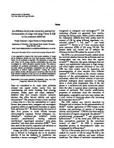
Fig. 1 (a-d)—Gel images of total genomic DNA isolated from leaves of adult (lane 1) and juvenile (lane 2) D. sissoo trees: Genomic DNA isolated following Protocol 17 (a), Protocol 216 (b), Protocol 317 (c), and modified Protocol 4 (d).
bromide staining and photographed under UV light using Gel Documentation System (GelDoc-It System, UVP Ltd., Unit 1, Trinity Hall Estate, Nuffield Rd. Cambridge, CB4 1 TG, UK). Reagents and chemicals used in the present study were of analytical grades and obtained from Sigma (Sigma-Aldrich, Inc., 3050 Spruce Street, St. Louis, MO, USA). Results and Discussion D. sissoo, being source of natural products mainly wood and bioactive substances like isoflavonoids, produces large amounts of secondary metabolites and substances of medicinal or industrial importance. Hence, DNA extraction from leaves of D. sissoo is complicated by the abundance of secondary metabolites. Also, the leaves exhibit oxidation when left under humid conditions after collection, because of which it is necessary to dry or freeze them as fast as possible to preserve the quality of DNA19. Age of tissue is important in determining DNA yield and quality20. Leaves from juvenile D. sissoo invariably extracted better yield and quality of genomic DNA than those from mature D. sissoo. The gel image of DNA extracted from mature tree leaves showed a smear from high to low mol wt position without having any intact band, indicating an impression of degraded nature of DNA (Fig. 1). On the other hand, Protocol 4 registered better values for both parameters of genomic DNA than other three protocols included in the study (Table 1). Genomic DNA of both types of samples obtained in Protocol 4 also exhibited single consolidated band without contamination of RNA (Fig. 1d) in comparison to the remaining protocols (Fig. 1a-c) on agarose gel. DNA yield and quality would likely to vary throughout the year21, however as per our observation, the best results were achieved when leaves were harvested in spring season, i.e., March and April. At this time the new flush of juvenile leaves appears which are more suitable for obtaining good quality and quantity
Table 1—DNA extraction protocols and yield of extracted DNA from D. sissoo leaves
No.
1 2 3 4
Protocol
Protocol 17 Protocol 216 Protocol 317 Protocol 4 (modified)
From mature tissue Yield OD 260/280 (µg per 100 mg ratio of leaf tissue) 40 to 50 20 to 30 60 to 90 100 to 400
71
1.29 to 2.03 1.42 to 2.09 1.10 to 1.34 1.69 to 1.99
From juvenile tissue Yield OD 260/280 (µg per 100 mg ratio of leaf tissue) 50 to 60 20 to 40 50 to 200 100 to 400
1.58 to 1.97 1.64 to 2.01 1.65 to 1.97 1.71 to 1.90
72
INDIAN J BIOTECHNOL, JANUARY 2010
Fig. 2—Gel photograph of the genomic DNA of studied D. sissoo clones isolated by optimized protocol
Fig. 3 (a & b)—a. RAPD pattern obtained from DNA extracted from modified Protocol 4: фX-174 DNA/BsuRI (HaeIII) molecular weight marker (lane 1); Eighteen DNA samples (lane 2-19) amplified using primer OPA-20. b. ISSR pattern obtained from DNA extracted from modified Protocol 4: фX-174 DNA/BsuRI (HaeIII) molecular weight marker (lane 1); Eighteen DNA samples (lane 2-19) were amplified using UBC primer 851.
of DNA. Sytsma et al22 has also emphasized the use of fresh and young leaf tissue to obtain good quality of DNA. Out of the various protocols tried, the CTAB method7,16 with modifications (Protocol 4) was found suitable for extraction of desired quantity and quality of genomic DNA from the leaf tissues. Approximate 100-400 µg of DNA/100 mg of foliage material was obtained with this protocol from both juvenile and mature tissues, which is enough to carry out 1000-1500 typical RAPD reactions. The principle
modification in this method included use of 4% PVP instead of 2% PVP used in Protocols 1 and 2, increased incubation time with extraction buffer (40 min instead of 30 min), use of freshly prepared CTAB buffer and an extended washing time (45 min instead of 30 min) with wash buffer and a repeated phase separation step with chloroform-isoamyl alcohol. High PVP concentration removes the polyphenols as it forms a complex with polyphenols through hydrogen bonding, allowing them to be separated from the DNA, and reducing levels of polyphenol in the product23. Extended incubation and washing durations coupled with the separation phase appears to have excluded DNA contaminants like polysaccharides, polyphenols and other secondary metabolite lysates, which otherwise hamper DNA isolation and purification. The isolated DNA using Protocol 4 of various samples of D. sissoo (Fig. 2) was tested in PCR amplification for RAPD and ISSR profiling with several primers. The PCR products produced by RAPD and ISSR analysis shows clear banding patterns (Fig. 3). Thus, this protocol proved to be advantageous because of its simplicity and affordable reagents, besides achieving intact high mol wt and purity of genomic DNA. The isolated DNA proved amenable to PCR amplifications including RAPD and ISSR analysis. The DNA extraction protocol described here consistently produced high yields of pure genomic DNA, while allowing for high sample throughput. Acknowledgement Authors are grateful to Punjab Forest Department, Government of Punjab, India for providing necessary financial support for conducting the present study. References 1
2
3
4
Karp A and Edwards K, DNA Markers: A global overview, in DNA markers: Protocols , applications and overviews, edited by G C Anolles & P M Gresshoff (Willy-Liss, Inc., New York) 1998, 1-14. Hoy M A, DNA amplification by the polymerase chain reaction: Molecular biology made accessible, in Insect molecular genetics: An introduction, principles and applications, (Academic Press, San Diego) 1994, 206-244. Saiki R K, Amplification of genomic DNA, in PCR protocols: A guide to methods and applications, edited by M A Iinnis, D H Gelfand, J J Sninski & White T J. (Academic Press, San Diego) 1990 13-20. Fang G, Hammar S & Grumet R, A quick and inexpensive method for removing. polysaccharides from plant genomic DNA, Biotechniques, 13 (1992) 52-54.
GINWAL & MAURYA: DNA EXTRACTION METHOD FOR D. SISSOO LEAF
5
6
7 8 9
10
11
12
13
14
Katterman F R H & Shattuck V I, An effective method of DNA isolation from the mature leaves of Gossypium species that contain large amounts of phenolic terpenoids and tannins, Prep Biochem, 13 (1983) 347-359. Peterson D G, Boehm K S & Stack S M, Isolation of milligram quantities of nuclear DNA from tomato (Lycopersicon esculentum), a plant containing high levels of polyphenolic compounds, Plant Mol Biol Rep, 15 (1997) 148-153. Doyle J J & Doyle J L, Isolation of plant DNA from fresh tissue, Focus, 12 (1990) 13-15. Scott K D & Playford J, DNA extraction technique for PCR in rain forest plant species, Biotechniques, 20 (1996) 974-979. Csaikl U M, Bastian H, Brettscheneider R, Gauch S, Meir A et al, Comparative analysis of different DNA extraction protocols: A fast, universal maxi-preparation of high quality plant DNA for genetic evaluation and phylogenetic studies, Plant Mol Biol Rep, 16 (1998) 69-86. Sharma K K, Lavanya M & Anjaiah V, A method for isolation and purification of peanut genomic DNA suitable for analytical applications, Plant Mol Biol Rep, 18 (2000) 393a-393h. Shepherd M, Cross M, Stokoe R L, Scott L J & Jones M E, High-throughput DNA extraction from forest trees, Plant Mol Biol Rep, 20 (2002) 425a-425j. Haymes K M, Ibrahim I A, Mischke BS, Scott D & Saunders J A, Rapid isolation of DNA from chocolate and date palm tree crops, J Agric Food Chem, 52 (2004) 5456-5462. Weishing K, Nybom H, Wolff K & Meyer W, DNA isolation and purification, in DNA fingerprinting in plants and fungi, edited by K Weishing, H Nybom, K Wolff & W Meyer (CRC Press, Boca Raton, Florida) 1995, 44-59. Narayanan C, Wali S A, Shukla N, Kumar R, Mandal A K et al, RAPD and ISSR markers for molecular characterization of teak (Tectona grandis L.f.), J Trop For Sci, 19 (2007) 218-225.
73
15 Narayanan C, Dubey S, Wali S A, Shukla N, Kumar R et al, Comparative efficacy of different DNA extraction methods for PCR-based assay in Tectona grandis L.f., Indian J Biotechnol, 7 (2008) 133-136. 16 Stange C, Prehn D & Arce-Johnson P, Isolation of Pinus radiata genomic DNA suitable for RAPD analysis, Plant Mol Biol Rep, 16 (1998) 1-8. 17 GeNei Spin Plant Genomic DNA Prep Kit (Cat # KT88) Bangalore Genei No. 6, 6th Main, BDA Indl. Suburb, Near SRS Rd., Peenya, Bangalore 560 058. 18 Linacero R, Rueda J & Vazquez AM, Quantification of DNA, in Molecular tools for screening biodiversity–Plants and animals, edited by A Karp, P G Isaac & D S Ingram (Chapman & Hall, London) 1998, 18-21. 19 Ribeiro R A & Lovato M B, Comparative analysis of different DNA extraction protocols in fresh and herbarium specimens of the genus Dalbergia, Genet Mol Res, 6 (2007) 173-187. 20 Ostrowska E, Muralitharan M, Chandler S, Volker P, Hetherington S et al, Technical review: Optimizing conditions for DNA isolation from Pinus radiata, In Vitro Cell Dev Biol (Plant), 34 (1998) 108-111. 21 Tibbits J F G, McManus L J, Spokevicius A V & Bossinger G, A rapid method for tissue collection and high-throughput isolation of genomic DNA from mature trees, Plant Mol Biol Rep, 24 (2006) 81-91. 22 Sytsma K, Givnish T J, Simt J F & Hahn W J, Collection and storage of land plant samples for macromolecular comparisions, in Methods in enzymology–Molecular evolution: Producing the biochemical data, vol 224, edited by E A Zimmer, T J White, R L Cann & A C Wilson (Academic Press, San Diego) 1993, 23-38. 23 Maliyakal E J, An efficient method for isolation of RNA and DNA from plants containing polyphenolics, Nucleic Acids Res, 20 (1992) 2381.