JOURNAL OF CLINICAL MICROBIOLOGY, Jan. 2008, p. 274–280 0095-1137/08/$08.00⫹0 doi:10.1128/JCM.01299-07 Copyright © 2008, American Society for Microbiology. All Rights Reserved.
Vol. 46, No. 1
Evaluation of a Commercialized In Situ Hybridization Assay for Detecting Human Papillomavirus DNA in Tissue Specimens from Patients with Cervical Intraepithelial Neoplasia and Cervical Carcinoma䌤 Ming Guo,1* Yun Gong,1 Michael Deavers,1 Elvio G. Silva,1 Yee Jee Jan,3 David E. Cogdell,1 Rajyalashmi Luthra,1 E. Lin,2 Hung Cheng Lai,4 Wei Zhang,1 and Nour Sneige1 Departments of Pathology1 and of Biostatistics and Applied Mathematics,2 The University of Texas M. D. Anderson Cancer Center, Houston, Texas; Department of Pathology, Taichung Veterans General Hospital, Taichung, Taiwan3; and Department of Obstetric/Gynecology, Tri-Service General Hospital, National Defense Medical Center, Taipei, Taiwan4 Received 27 June 2007/Returned for modification 15 September 2007/Accepted 24 October 2007
To evaluate a commercialized in situ hybridization (ISH) assay for detecting human papillomavirus (HPV) DNA, we compared the ability of a new ISH probe, Inform HPV III (Ventana Medical Systems, Tucson, AZ), to that of PCR assays to detect HPV DNA in cervical tissue specimens with normal cervix (20 cases), cervical intraepithelial neoplasia (CIN; CIN 1, 27 cases; CIN 2, 28 cases; and CIN 3, 33 cases), and cervical carcinoma (29 cases). General HPV DNA was detected using consensus primer-mediated PCR assays. HPV genotyping was performed by using EasyChip HPV blot (King Car Yuan Shan Institute, I-Lan, Taiwan). HPV16 integration status (E2/E6 ratio) was determined by using quantitative real-time PCR. Our findings showed that the ISH and PCR had fair to good agreements in detecting HPV DNA across all CIN categories without significant differences (Kappa coefficient, 0.34 to 0.63; P ⴝ 0.13 to 1.0). However, ISH detected significantly fewer HPV-positive cases in carcinoma than PCR did (Kappa coefficient, 0.2; P ⴝ 0.03). Eleven cases with ISHⴚ PCRⴙ results had HPV types that can be detected by Inform HPV III. Five carcinoma cases with ISHⴚ PCRⴙ results showed a significantly higher level of integrated HPV16 (P ⴝ 0.008) than did the ISHⴙ cases. As a consequence, lower copy numbers of episomal HPV16 in carcinoma might be the cause for the false-negative ISH results. Although the punctate signal pattern of HPV significantly increased with the severity of disease (P trend ⴝ 0.01), no significant difference in the HPV16 integration status was observed between the cases with a punctate signal only and the cases with mixed punctate and diffuse signals (P ⴝ 0.4). In conclusion, ISH using the Inform HPV III probe seems comparable to PCR for detecting HPV DNA in cervical tissue with CINs. False-negative ISH results appear to be associated with the lower copy numbers of the episomal HPV16 but not with the ability of the Inform HPV III probe to detect specific HPV types. In addition, signal patterns, especially a mixed punctate and diffuse pattern of HPV, cannot be reliably used to predict viral integration status. carcinoma (1, 26, 27, 34), and (iii) to provide valuable information in cervical cancer research. Different HPV DNA testing assays in FFPE cervical tissue specimens have been used, such as PCR and in situ hybridization (ISH). Each has its strengths and drawbacks. Although the PCR assay is highly sensitive for detecting HPV DNA, there are disadvantages to using PCR for HPV DNA detection in FFPE tissue specimens (5, 34). In particular, PCR inhibitors in DNA extracts from FFPE cervical tissue can interfere with the ability of PCR to detect HPV (10). In addition, although PCR assays for HPV DNA testing are commercially available, highly trained laboratory personnel are required to perform the assays, and strict laboratory conditions are needed to avoid contamination. More importantly, morphological context, which is essential for histopathological specimen interpretation, is lost when a PCR assay is used. In contrast, ISH, a direct signal detection assay, has the advantage of preserving the morphological context with HPV DNA signals. The ISH assay can be automated along the same lines as immunohistochemistry staining to minimize intra- or interassay inconsistency. The interpretation of ISH is also similar to that of immunohistochemistry staining in tissue sections. Furthermore, the ISH signal patterns of HPV DNA have been
Oncogenic human papillomavirus (HPV) infection is a wellestablished major risk factor for the development of more than 99% of cervical carcinomas and precancerous lesions (highgrade cervical intraepithelial neoplasia [CIN]; i.e., CIN 2/3) (6, 35, 39) In recent years, HPV DNA testing has been utilized as a molecular maker in gynecological cytology either for the triage of women with mildly abnormal Papanicolaou (Pap) test results (36) or in conjunction with Pap tests for predicting CIN 2/3 in women who are 30 years or older (37). In histopathology, HPV DNA testing in formalin-fixed, paraffin-embedded (FFPE) cervical tissue can be used for the following purposes: (i) to help resolve diagnostic discrepancy in patients with CINs and distinguish endocervical immature squamous metaplasia from high-grade dysplasia or endocervical glandular reactive changes from glandular dysplasia, (ii) to provide valuable information for assessing the risk of CIN progression or of disease recurrence in women who were treated for CIN 2/3 or
* Corresponding author. Mailing address: Department of Pathology, Unit 58, The University of Texas M. D. Anderson Cancer Center, 1515 Holcombe Blvd., Houston, TX 77030-4009. Phone: (713) 563-1842. Fax: (713) 563-1848. E-mail:
[email protected]. 䌤 Published ahead of print on 31 October 2007. 274
VOL. 46, 2008
INFORM HPV TESTING
reported to be associated with the physical status of HPV in infected cells, i.e., episomal or integrated forms (9, 11). Because the integration of oncogenic HPV into the human genome is a critical step for cervical cancer carcinogenesis, the signal pattern of HPV, which may suggest viral integration status, could be a useful marker for predicting precancerous lesion progression. Despite these advantages, the ISH assay has a low sensitivity, which is a major concern for pathologists who use ISH to detect HPV in tissue (10). In recent years, ISH assays that use improved signal-detecting methods, such as the enzyme-categorized signal detecting system, have shown a higher sensitivity (8, 31–33). Inform HPV (Ventana Medical Systems), a commercially available ISH assay for HPV DNA testing, can be used in both cytological and histological specimens. Recently, Inform HPV III, a new generation of ISH probe, became available for HPV DNA testing in tissue specimens. Inform HPV III utilizes a stacked antibody approach (the iView Blue Plus kit) to enhance its sensitivity. Briefly, the primary antibody is directed against the DNP hapten. Signal amplifications are generated through antibody stacks consisting of a secondary antibody and a biotinylated tertiary antibody. Finally, a streptavidin-alkaline phosphatase conjugate is added to generate color precipitates. Published studies of systemic analyses of the sensitivity of ISH using Inform HPV III to detect HPV DNA in cervical tissue are limited (38). Knowing the efficacy of the ISH assay using the Inform HPV III probe in cervical tissue not only would allow us to better use ISH-based HPV DNA testing in tissue specimens but also would provide valuable information for the cytological application of ISH in Pap specimens. Therefore, in the present study, we compared the ISH assay using the Inform HPV III probe to PCR assays using consensus primer sets for detecting HPV DNA in FFPE cervical tissue specimens with CINs and carcinoma. Using Inform HPV III, the nuclear signal patterns of HPV were also compared to HPV16 integration status by using quantitative real-time PCR (qRT-PCR) assay to determine whether the nuclear signal pattern of HPV might be reliable for estimation of HPV integration status and therefore a potential useful marker to predict precancerous lesion progression. MATERIALS AND METHODS The Institutional Review Board of The University of Texas M. D. Anderson Cancer Center approved this study. Cervical tissue specimen selection. Archived, FFPE cervical tissue specimens obtained from 2004 to 2005 were retrieved from the Department of Pathology at M. D. Anderson Cancer Center. In consecutive order, 200 cervical tissue specimens from punch biopsies, loop electrosurgical excision procedures, cone biopsies, and hysterectomies were selected. Three pathologists independently reviewed the hematoxylin and eosin (H&E)-stained slides. Specimens were eliminated from the study for any of the following reasons: (i) no consensus in diagnosis; (ii) no lesion in the last section of the specimen; (iii) insufficient DNA for analysis by spectrophotometry; and (iv) negative -globin amplification by PCR. After exclusion of specimens for these various reasons, there were 137 specimens with the following pathological diagnoses: normal cervix (20 cases), CIN 1 (27 cases), CIN 2 (28 cases), CIN 3 (33 cases), and cervical squamous cell carcinoma (29 cases). Tissue sectioning for in situ hybridization and PCR. Serial sections were cut to a thickness of 4 m and mounted on positively charged glass slides for ISH. The paraffin rolls were cut from each block (10 sections, 5 m in thickness) for DNA extraction. The extra sections cut before and after each tissue sections were stained with H&E and used to determine specimen quality for testing. To avoid cross-contamination, the microtome was cleaned with a new blade for each case.
275
Inform HPV III ISH. Inform HPV III probe sets able to detect 13 types of oncogenic HPV (i.e., types 16, 18, 31, 33, 35, 45, 51, 52, 56, 58, 59, 68, and 70) were provided by Ventana Medical Systems. The ISH assay was performed according to the manufacturer’s guidelines using the BenchMark automated slide staining system (Ventana Medical System). HPV control slides consisted of formalin-fixed, paraffin-embedded sections containing three separate collections of cells on a single slide (Ventana Medical Systems). These cells consisted of the CaSki cervical cancer cell line (containing 200 to 400 copies of HPV16 per cell); the HeLa cervical cancer cell line (containing 10 to 50 copies of HPV18 per cell); and the C-33A cell line, which served as a negative control. Reagent negative control was set using negative control probes provided by Ventana Medical Systems. Three pathologists independently reviewed the Inform HPV III slides. The pathologists agreed on the results of ISH in 95% of the cases (130 of 137). Consensus was obtained in the remaining seven cases after review. Nuclear staining was considered a positive result for HPV DNA. The signal patterns of HPV in nucleus using Inform HPV III were classified as follows: diffuse, signals that are condensed and uniformly packed in the nucleus (Fig. 1A); and punctate, signals that are dot-like and sparsely distributed in the nucleus (Fig. 1A). DNA extraction. DNA was extracted from tissue specimens using the DNeasy kit (catalog no. 69506; Qiagen, Valencia, CA) according to the manufacturer’s instructions. The extracted DNA was quantified by using a Nanodrop spectrophotometer (Nanodrop Technologies, Wilmington, DE). HPV DNA testing using consensus primer-mediated PCR. HPV DNA positive cases was determined by using consensus primer-mediated PCR assays independently performed in the genomic labs at M. D. Anderson Cancer Center, Houston, TX, and Tri-Service Hospital, National Defense Medical Center, Taipei, Taiwan. All of the HPV DNA testing was carried out randomly without the knowledge of the pathological diagnoses. At M. D. Anderson Cancer Center, PCR assays for HPV DNA detection were performed as described previously (16). Briefly, the amplification of -globin (268 bp) was performed to determine the quality of the extracted DNA using primer pc04/gh20 according to the method of Bauer et al. (4) and using AmpliTaq Gold polymerase (Applied Biosystems, Foster City, CA). A 25-ng aliquot of genomic DNA was added to a PCR master mixture containing 1⫻ PCR buffer (100 mM Tris-HCl, 500 mM KCl [pH 8.3]), 200 M concentrations of each deoxynucleoside triphosphate, 200 nM primer, 1.5 mM MgCl2, and 2.5 U of AmpliTaq Gold DNA polymerase. PCR was performed under the following permissive cycling conditions: 10 min at 94°C, followed by 1 min at 94°C, 1 min at 40°C, 90 s at 72°C, and 5 min at 72°C for 39 cycles. Specimens positive for -globin were screened for HPV DNA using GP5⫹/ GP6⫹ consensus primer-mediated PCR, which generates 150-bp amplicons and can detect all of the oncogenic HPV genotypes that Inform HPV III detects. PCR was performed according to the method of Jacobs et al. (21). A 25-ng aliquot of genomic DNA was added to the PCR master mixture, which contained 1⫻ PCR buffer, 200 M concentrations of each deoxynucleoside triphosphate, 200 nM primer, 3.5 mM MgCl2, and 2.5 U of AmpliTaq Gold DNA polymerase. The cycling conditions were the same as those used for -globin. Specimens that were positive for -globin but negative for GP5⫹/GP6⫹ were rescreened for HPV DNA with the second consensus primer set (PGMY09/11), which generates 450-bp amplicons and also detects all of the oncogenic HPV genotypes that Inform HPV III detects. A 25-ng aliquot of genomic DNA was used for amplification in a PCR master mixture containing 1⫻ PCR buffer, 200 M concentrations of each deoxynucleoside triphosphate, 200 nM primer, 4.0 mM MgCl2, and 7.5 U of AmpliTaq Gold DNA polymerase. The PCR was performed under the following cycling conditions: 9 min at 94°C, followed by 1 min at 94°C, 1 min at 55°C, and 1 min at 72°C for 39 cycles. Specimens with known HPV were used as positive controls. Sterile water was used as a negative control for each set of amplifications. The PCR products were visualized with ethidium bromide staining on a 4% low-melting-point agarose gel. At the Tri-Service Hospital, National Defense Medical Center, Taiwan, specimens were tested for HPV DNA using MY11/GP6⫹ primer sets to amplify a fragment of 192 bp as described previously (19, 24). GAPDHF/GAPDHR primer sets were used to amplify a 136-bp amplicon of GAPDH (glyceraldehyde-3phosphate dehydrogenase) for validation. Briefly, PCR for HPV DNA was performed in a final reaction volume of 26 l with 20-ng aliquot of genomic DNA in a PCR master mixture containing 15 mM Tris-HCl (pH 8.0), 2.0 mM MgCl2, 50 mM KCl, 0.25 mM concentrations of each deoxynucleoside triphosphate, 0.6 M primer, and 0.5 U of DNA polymerase (HP High-Performance HotStart Taq DNA polymerase; DNA Technologies, Ltd.). A PCR assay was performed as follows: 10 min at 95°C, followed by 40 cycles of 30 s at 95°C, 30 s at 45°C, and 30 s at 72°C, with a final extension of 5 min at 72°C. PCR for GAPDH was performed in a final reaction volume of 25 l with 10-ng aliquot of genomic DNA
276
GUO ET AL.
J. CLIN. MICROBIOL.
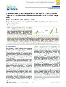
FIG. 1. (A) HPV signal patterns in CIN: punctate pattern (arrow) and diffuse pattern (arrow head) (Inform HPV III; magnification, ⫻400). (B) Focally distributed HPV in CIN1 (Inform HPV III; magnification, ⫻200). (C) Diffusely distributed HPV in CIN 3 (Inform HPV III; magnification, ⫻400). (D) Diffusely distributed HPV in cervical carcinoma (Inform HPV III; magnification, ⫻200).
in a PCR master mixture containing 15 mM Tris-HCl (pH 8.0), 2.5 mM MgCl2, 50 mM KCl, 0.2 mM concentrations of each deoxynucleoside triphosphate, 0.2 M primer, and 0.5 U of HotStart Taq DNA polymerase. A PCR assay was performed as follows: 10 min at 95°C, followed by 40 cycles of 15 s at 95°C, 1 min at 57°C, and 30 s at 72°C, with a final extension of 5 min at 72°C. An aliquot of 5 l of PCR product was analyzed by electrophoresis on a 2% agarose gel and stained with ethidium bromide. Specimens that tested positive for HPV DNA in one of the consensus HPV PCRs were subjected to genotyping. HPV genotyping by EasyChip. Specimens with positive results of consensus primer-medicated PCR were genotyped by using EasyChip HPV Blot (King Car, I-Lan, Taiwan) (24). EasyChip array can genotype 39 HPV types (HPV types 6, 11, 16, 18, 26, 31, 32, 33, 35, 37, 39, 42, 43, 44, 45, 51, 52, 53, 54, 55, 56, 58, 59, 61, 62, 66, 67, 68, 69, 70, 72, 74, 82, CP8061, CP8304, L1AE5, MM4, MM7, and MM8, as well as three intrinsic controls). The HPV type-specific probes are immobilized on a 14.4-mm-by-9.6-mm nylon membrane, which is used for reverse blot hybridization and detects HPV DNA in a single assay. The hybridization was performed according to the manufacturer’s guide. Briefly, the blot membrane was equilibrated with 2⫻ saline-sodium citrate (SSC; 1⫻ SSC is 0.15 M NaCl plus 0.015 M sodium citrate) at room temperature for 10 min. The blot was preincubated in hybridization buffer (2⫻ SSC, 0.5% blocking reagent, 5% dextran sulfate, 0.1% sodium dodecyl sulfate [SDS], 50 g of denatured salmon sperm DNA/ml) with shaking at 35°C for 30 min. The membrane was hybridized with 500 l of hybridization buffer containing 20 l of the denatured amplicons (15 l of HPV and 5 l of GAPDH PCR products) by shaking at 35°C for at least 3 h. The blot was washed twice in washing buffer 1 (2⫻ SSC, 0.1% SDS) for 5 min at 25°C and then washed twice in washing buffer 2 (0.2⫻ SSC, 0.1% SDS) for 5 min at 35°C. The blot was equilibrated with buffer 1 (1⫻ phosphate-buffered saline [pH 7.4], 0.05% Tween 20, 0.1% SDS) by shaking at 25°C for 5 min and then buffer 2 (1⫻ phosphate-buffered saline [pH 7.4], 0.05% Tween 20, 0.1% SDS, 0.5% blocking reagent) at 25°C for 1 h. The blot was incubated in 500 l of buffer 2 containing streptavidin-AP (Calbiochem; alkaline phosphatase conjugates and biotinylated antibodies, 1:1,000 dilution) at 25°C for 40 min. The blot was then washed in buffer 1 and rinsed with buffer 3 (0.1 M Tris-HCl [pH 9.5], 0.1 M NaCl) for 5 min. Then, 80 l of nitroblue tetrazolium-BCIP (5-bromo-4chloro-3-indolylphosphate) was added, followed by incubation for 30 min at 25°C. The reaction was stopped by adding distilled water. The HPV types were determined by a visual assessment protocol provided by King Car. qRT-PCR for HPV16 viral integration status. HPV16 DNA was amplified with primers and probe targeting the E6/E7 oncogenes of HPV16 using qRT-PCR
according to the method of Gravitt et al. (15). The TaqMan minor groove binder probes were used for qRT-PCR. Minor groove binder probes were labeled with a carboxyfluorescein reporter dye at the 5⬘ end and a nonfluorescent quencher at the 3⬘ end. Primers described by Flores-Munguia et al. (12) for the E6/E7 regions were used for HPV16. For the HPV16 integration assay, primers for HPV16 E2, as described by Peitsaro et al. (28), were used. Minor groove binder probe for the assay was designed by using Primer Express software; both probe and primers were purchased from Applied Biosystems. We used the plasmid HPV16 clone for positive controls (American Type Culture Collection, Manassas, VA). All qRT-PCR assays for HPV genotyping were performed by using an ABI Prism 7900 HT with a 96-well plate (Applied Biosystems). Briefly, 2.5 ng of genomic DNA of each specimen and control, including water as a no-template control, was added to a 25-l reaction mixture containing 1⫻ TaqMan Universal PCR Master Mix without AmpErase uracil-N-glycosylase, 1⫻ gene expression assay (Applied Biosystems), 250 nM fluorogenic probe, and 900 nM primer under the following cycling conditions: 10 min at 95°C, followed by 15 s at 95°C and 1 min at 60°C for 50 cycles. HPV16 was assayed on a single 96-well plate, with 40 specimens in duplicate. A standard curve to determine HPV quantity was established, also in duplicate, with a 10⫻ dilution series ranging from 10 to 107 copies per well using plasmid-cloned HPV, including water as a no-template control. A linear relationship was obtained between the log value of the viral copy numbers and the threshold cycle (data not shown). The integration status of HPV16 was determined by measuring the E2/E6 ratio (28), The protocol for the E2/E6 assay was modified to include dual standard curves of E2 and E6. The E2/E6 ratio was calculated from the same reaction. An E2/E6 ratio greater than or equal to 1 was classified as a complete episomal form. No amplification for E2 was classified as a complete integration. A low E2/E6 ratio represented predominantly integrated forms. Conversely, a high E2/E6 ratio indicated high levels of episomal forms. Statistical analysis. Descriptive statistics were calculated by using Kappa statistics to assess the agreement between Inform HPV III and PCR HPV DNA testing. McNemar’s tests were used to assess the homogeneity between paired categorical variables. The chi-square or Fisher exact test was used to assess the association between categorical variables. The Cochran-Armitage trend test or the Jonckheere-Terpstra trend test was used to assess the trend of the Inform HPV III results and the severity of disease. The Kruskal-Wallis test was used to assess the association between the Inform HPV III results and the HPV16 E2/E6 ratio. P values (two-sided test) of ⬍0.05 were considered significant. All statistical analyses were carried out by using SAS 8.0 (SAS Institute, Cary, NC).
VOL. 46, 2008
INFORM HPV TESTING
TABLE 1. Agreement between ISH using Inform HPV III and consensus primer-mediated PCR to detect oncogenic HPV DNA in CINs and cervical carcinoma Diagnosis and ISH result
No. of cases (%) examined by PCR
Kappa coefficient
P
Negative
Positive
CIN 1 Negative Positive
7 (77.8) 4 (22.2)
2 (22.2) 14 (77.8)
0.53
0.69
CIN 2 Negative Positive
5 (100) 4 (17.4)
0 19 (82.6)
0.63
0.13
CIN 3 Negative Positive
2 (40.0) 2 (7.1)
3 (60.0) 26 (92.6)
0.34
1.0
Carcinoma Negative Positive
1 (14.3) 0
6 (85.7) 22 (100.0)
0.2
0.03
RESULTS The age of women in the study ranged from 18 to 79 years, with a mean age of 40 years and a median age of 41 years. HPV DNA detection by ISH and PCR assays. Twenty cases of normal cervical tissue were negative for HPV DNA by ISH assay using Inform HPV III and consensus primer-mediated PCR assays using the GP5⫹/GP6⫹, PGMY09/11, and MY11/ GP6⫹ primer sets. In 117 cases of cervical specimens with diseases, we found that 91 cases (78%) were positive for HPV DNA by Inform HPV III, and 92 cases (79%) were determined to be positive by the PCR assays. Compared to the PCR assays, Inform HPV III showed similar HPV DNA positivity in CIN 1 (67% [18 of 27] versus 59% [16 of 27]) and CIN 2 (82% [23 of 28] versus 68% [19 of 28]) but lower HPV DNA positive rates in CIN 3 (85% [28 of 33] versus 88% [29 of 33]) and carcinoma (76% [22 of 29] versus 97% [28 of 29]). Combined HPV DNA positivities by Inform HPV III and PCR in CIN 1, 2, and 3 and carcinoma were 74% (20 of 27), 82% (23 of 28), 94% (31 of 33), and 97% (28 of 29), respectively. We found no association between HPV DNA positivity by Inform HPV III and the HPV genotypes (data not shown). The agreement between the Inform HPV III and PCR assays in detecting HPV DNA is shown in Table 1. The two tests were concordant in 85% (116 of 137) of all cases. The Inform HPV III and PCR assays had moderate to good agreements (Kappa coefficients of 0.53 and 0.63, respectively) in detecting HPV DNA in CIN 1 and CIN 2 and a fair agreement (Kappa coefficient, 0.34) in CIN 3 without significant differences (P ⫽ 0.13 to 1.0). However, in cervical carcinoma, the two tests showed a significant disagreement with regard to HPV DNA positivity, with the positive rate being lower for Inform HPV III than for the PCR assay (Kappa coefficient, 0.2; P ⫽ 0.03). There were 21 cases with discrepancy in results between PCR and ISH tests. Of these, 10 cases showed ISH⫹ PCR⫺ results, and 11 cases showed ISH⫺ PCR⫹ results. In the 10 ISH⫹ PCR⫺ cases, repeat DNA extractions with consensus primer-mediated PCR testing for HPV DNA were negative. Of the 11 cases with ISH⫺ PCR⫹ results, 6 were carcinoma
277
TABLE 2. Distribution of HPV genotypes in cases with ISH⫺ PCR⫹ results Case no.
Disease
HPV type(s)
1 2 3 4 5 6 7 8 9 10 11
CIN 1 CIN 1 CIN 3 CIN 3 CIN 3 Carcinoma Carcinoma Carcinoma Carcinoma Carcinoma Carcinoma
45 18 59 52 52 16 16 16 16 16/35 33/58
cases, with 4 cases positive for HPV16, 1 case positive for HPV16/35, and 1 case positive for HPV33/58. In the remaining five cases with ISH⫺ PCR⫹ results, all showed HPV types that can be detected by Inform HPV III probe, including HPV52 (2 cases), HPV45 (1 case), HPV18 (1 case), and HPV59 (1 case) (Table 2). Distribution of HPV-positive cells and HPV signal patterns. Figure 1 presents the ISH signal of HPV shown by Inform HPV III. Nonspecific background bindings were not observed, as frequently seen using Inform HPV II probes (data not shown). In the majority of cases (87%), HPV-positive cells were observed in ⬍50% of the lesions, with more HPV-positive cells focally located in the upper epidermis in CIN 1 (Fig. 1B) or CIN 2 and more HPV-positive cells diffusely distributed in CIN 3 (Fig. 1C) or carcinoma (Fig. 1D). A comparison of HPV signal patterns in the nucleus shown by Inform HPV III with cervical disease categories is illustrated in Table 3. Three cases with diffuse patterns were observed in CIN 1 and CIN 2 but not in CIN 3 or carcinoma. Mixed diffuse and punctate patterns were predominantly found in CIN 1 and 2. Punctate patterns were predominantly observed in CIN 3 and carcinoma cases, either alone or mixed with diffuse patterns. The punctate patterns significantly increased with the severity of disease (P ⫽ 0.01) (Table 3). HPV16 viral integration status and ISH signal patterns. The signal patterns of the HPV shown by Inform HPV III were compared to the HPV16 E2/E6 ratio. The mean of the E2/E6 ratio was lower in the specimens with a punctate pattern only (E2/E6, 0.293) than in cases with mixed punctate and diffuse patterns (E2/E6, 0.532). However, no significant difference in the E2/E6 ratio was observed between these two groups (P ⫽ 0.4) (Table 4). Five carcinoma cases with ISH⫺ PCR⫹ results were positive for HPV16, showing a significantly lower E2/E6 ratio (E2/E6, 0.05; P ⫽ 0.008) compared to the cases shown to
TABLE 3. Association between HPV signal patterns and CINs or cervical carcinoma as determined by Inform HPV III No. of cases (%) P-trend
HPV signal pattern
Diffuse Punctate ⫹ diffuse Punctate
CIN 1
CIN 2
CIN 3
Carcinoma
2 (11) 9 (50) 7 (39)
1 (4) 13 (57) 9 (39)
0 11 (41) 16 (59)
0 6 (27) 16 (73)
0.01
278
GUO ET AL.
J. CLIN. MICROBIOL.
TABLE 4. Association between HPV signal patterns as determined by Inform HPV III and HPV16 E2/E6 ratio HPV signal pattern
No. of cases
Mean E2/E6 ratio
SD
P
Negative Punctate Punctate ⫹ diffuse
5 20 17
0.050 0.293 0.532
0.101 0.291 0.407
0.008 0.4
be positive by Inform HPV III with either punctate or mixed signal patterns (Table 4) (Fig. 2). DISCUSSION In this study, we observed that the ISH assay using the Inform HPV III probe is comparable to the PCR assays in detecting oncogenic HPV DNA in FFPE cervical tissue from patients with CINs and can be used in cervical tissue specimens. Further, the punctate pattern of HPV is closely associated with CIN 2/3 and carcinoma. However, the use of signal patterns, especially the mixed HPV signal pattern to estimate the viral integration status, is limited because of the wide range of viral integration status in cases with mixed HPV signal patterns. Finally, we conclude that low copy numbers of episomal HPV16, as a result of high level of viral integration, may contribute in part to the false-negative results of ISH in carcinoma specimens, and it may represent a limitation of ISH in detecting HPV DNA in carcinoma specimens. Comparison of Inform HPV III with PCR. The ISH assay using the Inform HPV III probe to detect HPV DNA showed fair to good agreement with PCR assays in FFPE cervical tissue specimens with CINs. This represents a significant improvement in sensitivity for the Inform HPV III probe. We compared the Inform HPV III probe to the previous generation of Inform HPV probe, Inform HPV II, and observed significantly higher HPV-positive rates shown by Inform HPV III
compared to Inform HPV II, especially in CIN 3 and carcinoma cases (data not shown). Recently, Kong et al. reported similar observations on Inform HPV II and Inform HPV III. (23). With regard to the cause of false-negative ISH results in carcinoma specimens, it seems unlikely that the majority of such cases were due to undetected HPV types, which were not covered by the ISH probes. In our study group, all 11 cases with ISH⫺ PCR⫹ results tested positive for one or more specific HPV genotypes that were actually covered by the Inform HPV III probe. Of these, five cases of cervical carcinoma were positive for HPV16. These five cases showed a significantly lower HPV16 E2/E6 ratio compared to those in ISH⫹ cases, either with a punctate signal pattern only or with mixed signal patterns. This indicates a significantly higher proportion of integrated HPV16 in ISH⫺ PCR⫹ carcinoma cases than in ISH⫹ cases (16). We therefore speculate that the low level of episomal HPV16, as a consequence of high viral integration status, might be associated with the false-negative results in these carcinoma cases. Based on our observation, since the punctate signal pattern is more frequent in CIN 3 and cervical carcinoma, it becomes more difficult to recognize or interpret the ISH signals, especially when these signals are weak and sporadic. In a few cases of CIN 3 and carcinoma, high-power objective lenses were required to identify the punctate signals. Other factors that may contribute to the false-negative ISH results include HPV types that were not covered by Inform HPV III probes. In our study, such cases were not observed. In addition, the heterogeneous distribution of HPV in low-grade CIN might cause signal absence in tissue sections, resulting in false-negative results. Our study demonstrated the limitation of PCR assays for HPV DNA detection in FFPE tissue specimens, and optimizing the PCR assay to detect HPV DNA in FFPE tissue specimens is required. Limitations of PCR assays in detecting HPV DNA in FFPE tissue specimens have been observed. Even with the combination of type-specific and consensus primer sets, the
FIG. 2. Comparison of HPV16 integration status (E2/E6 ratio) with HPV signal patterns determined by Inform HPV III. P, punctate pattern; D, diffuse pattern.
VOL. 46, 2008
sensitivity is limited (3). In our study, ISH⫹ PCR⫺ results were observed mainly in cases with CINs. Unger et al. reported similar findings in FFPE cervical carcinoma using ISH and PCR (34). In both studies, optimizing the PCR assay for detecting HPV DNA was attempted. In our study, all of the specimens were selected within 2 years of the study; strict measures were followed for tissue sectioning, DNA extraction, and the PCR assays to avoid cross-contaminations among specimens; and general PCR and HPV genotyping tests were randomized without the knowledge of the pathological diagnoses and independently performed using three primer sets at two centers. Repeat DNA extraction with consensus primer-mediated PCR was conducted in the specimens with ISH⫹ PCR⫺ results. Even though the PCR assays were optimized, factors that affect the efficiency of the PCR assay in detecting HPV DNA in FFPE tissue specimens cannot be completely eliminated. The ISH⫹ PCR⫺ results in our study could be partially due to the presence of PCR inhibitors for amplification (5, 34). The heterogeneous distribution of HPV in CINs, especially when the viral load is low, can result in the target molecule missing, leading to a negative result. DNA extraction can also affect PCR efficiency for HPV DNA (6, 10). In our study, the amplification was suboptimal in 8% of all initially selected cases because of no -globin amplification. The results are similar to those in carcinoma cases in which the PCR assay was required to be optimized in tissue specimens (34). The primer sets selected for PCR assays can also affect the PCR testing results. Recently, Chan et al. compared GP5⫹/GP6⫹ and PGMY09/11, the two most commonly used primer sets for HPV DNA testing. These researchers found that although both the GP5⫹/GP6⫹ and the PGMY09/11 primer sets broadly cover most of the oncogenic HPV types, each primer set has a certain complementary coverage, with PGMY09/11 detecting more cases with multiple viral infections (7). Using GP5⫹/GP6⫹ consensus primers, we demonstrated a high HPV positivity, especially in cervical carcinoma, a finding which is consistent with the published pooled data (25). Using the second primer set, PGMY09/11, 6% more positive results were obtained, predominantly in CIN 3 and carcinoma cases (16). Four additional positive cases were identified by using the GP6⫹/MY11 primer sets. Using three consensus primer sets, Baay et al. reported only 72.8% positive rate of HPV DNA in carcinoma cases and an 87.6% positive rate with adding typespecific primer sets (3). Compared to the limitation of using the PCR assay in FFPE to detect HPV DNA, the application of ISH with Inform HPV III and the interpretation of the ISH results are relatively simple. With improved sensitivity for detecting HPV DNA, the ISH assay using Inform HPV III has potential use in cervical tissue specimens with CINs. HPV signal pattern by ISH and HPV16 integration status. The punctate pattern of HPV observed by ISH has been linked to the integrated HPV forms in the host genome using a Southern blot assay (9) and has been reported to be associated with CIN 2/3 or cervical carcinoma (18, 20, 22). Since HPV integration is an important step in the carcinogenesis of cervical carcinoma (29, 30), it has the potential to be used as a marker for CIN progression. In evaluating HPV signal patterns in CINs and cervical carcinoma, we found that a punctate pattern was more frequently observed in CIN 3 and cervical carcinoma, while diffuse or mixed patterns were predominantly
INFORM HPV TESTING
279
found in CIN 1 and 2, a finding consistent with those of previous studies (18, 20, 22). To evaluate the association between HPV signal patterns and HPV integration status, we compared HPV signal patterns with the HPV16 E2/E6 ratio using qRT-PCR. The E2/E6 ratio has been used to estimate HPV viral integration into the host genome. HPV16 integration occurs by disruption or deletion of the viral E1 and/or E2 open reading frame. Therefore, lower E2 amplification indicates a higher integrated form of HPV16. The E2/E6 ratio has been reported to be correlated well with the severity of CINs or cervical carcinoma (2, 13, 17, 28). Using qRT-PCR to measure the HPV16 E2/E6 ratio, we demonstrated that HPV16 was integrated more significantly into cervical carcinoma cells than into CIN 2/3 cells (16). In comparing the HPV16 E2/E6 ratio to the HPV signal patterns, however, we did not observe a significant difference in the E2/E6 ratio between cases with a punctate signal only and cases with mixed punctate and diffuse signals. We speculate that this could be caused by a variation in the percentage of the two patterns of HPV signals in cases with mixed signal patterns; this makes quantitative analysis difficult and therefore affects an accurate estimation. The presence of episomal forms of HPV DNA or RNA also has been reported to mask the integrated HPV DNA when the amount of the integrated viral forms is small (18), causing an inaccurate estimate of the level of integrated HPV. Furthermore, the heterogeneous distribution of HPVpositive cells shown by ISH assay in tissue sections may cause inaccurate classification. Although our observation supports the notion that the pure punctate pattern of HPV indicates a high level of viral integration and may be a marker for CIN progression, the level of HPV integration cannot be accurately determined in the cases with mixed signal patterns that encompass a wide range in viral integration status. Recently, Fijii et al. reported similar findings in their comparison of HPV signal patterns shown by ISH with the HPV16 E2/E6 ratio in CINs and cervical carcinoma (14). In our study, since the majority of CINs showed mixed signal pattern of HPV, the use of HPV signal patterns as a marker for viral integration may be limited. ACKNOWLEDGMENTS We thank Ventana Medical Systems for providing the Inform HPV III probe for the ISH assay. We thank Alen Silverman, Applied Biosystems, for designing the probes and part of the primers for HPV genotyping using real-time PCR and Valerie Dunmire and Erik E. Guerra at M. D. Anderson Cancer Center for technical contributions. We also thank Martha Morrison, Scientific Publications, M. D. Anderson Cancer Center, for editing the manuscript. This study was supported by the University of Texas M. D. Anderson Cancer Center. REFERENCES 1. Acladious, N. N., C. Sutton, D. Mandal, R. Hopkins, M. Zaklama, and H. Kitchener. 2002. Persistent human papillomavirus infection and smoking increase risk of failure of treatment of cervical intraepithelial neoplasia (CIN). Int. J. Cancer 98:435–439. 2. Andersson, S., H. Safari, M. Mints, I. Lewensohn-Fuchs, U. Gyllensten, and B. Johansson. 2005. Type distribution, viral load, and integration status of high-risk human papillomaviruses in pre-stages of cervical cancer (CIN). Br. J. Cancer 92:2195–2200. 3. Baay, M. F., W. G. Quint, J. Koudstaal, H. Hollema, J. M. Duk, M. P. Burger, E. Stolz, and P. Herbrink. 1996. Comprehensive study of several general and type-specific primer pairs for detection of human papillomavirus DNA by PCR in paraffin-embedded cervical carcinomas. J. Clin. Microbiol. 34:745–747. 4. Bauer, H. M., Y. Ting, C. E. Greer, J. C. Chambers, C. J. Tashiro, J. Chi-
280
5.
6.
7.
8.
9. 10.
11.
12. 13.
14.
15.
16.
17.
18. 19. 20.
21.
GUO ET AL. mera, A. Reingold, and M. M. Manos. 1991. Genital human papillomavirus infection in female university students as determined by a PCR-based method. JAMA 265:472–477. Biedermann, K., N. Dandachi, M. Trattner, G. Vogl, H. Doppelmayr, E. More, A. Staudach, O. Dietze, and C. Hauser-Kronberger. 2004. Comparison of real-time PCR signal-amplified in situ hybridization and conventional PCR for detection and quantification of human papillomavirus in archival cervical cancer tissue. J. Clin. Microbiol. 42:3758–3765. Bosch, F. X., M. M. Manos, N. Munoz, M. Sherman, A. M. Jansen, J. Peto, M. H. Schiffman, V. Moreno, R. Kurman, K. V. Shah, et al. 1995. Prevalence of human papillomavirus in cervical cancer: a worldwide perspective. J. Natl. Cancer Inst. 87:796–802. Chan, P. K., T. H. Cheung, A. O. Tam, K. W. Lo, S. F. Yim, M. M. Yu, K. F. To, Y. F. Wong, J. L. Cheung, D. P. Chan, M. Hui, and M. Ip. 2006. Biases in human papillomavirus genotype prevalence assessment associated with commonly used consensus primers. Int. J. Cancer 118:243–245. Cheung, A. L., A. H. Graf, C. Hauser-Kronberger, O. Dietze, R. R. Tubbs, and G. W. Hacker. 1999. Detection of human papillomavirus in cervical carcinoma: comparison of peroxidase, Nanogold, and catalyzed reporter deposition (CARD)-Nanogold in situ hybridization. Mod. Pathol. 12:689– 696. Cooper, K., C. S. Herrington, J. E. Stickland, M. F. Evans, and J. O. McGee. 1991. Episomal and integrated human papillomavirus in cervical neoplasia shown by non-isotopic in situ hybridization. J. Clin. Pathol. 44:990–996. Dabic, M. M., L. Hlupic, D. Babic, S. Jukic, and S. Seiwerth. 2004. Comparison of polymerase chain reaction and catalyzed signal amplification in situ hybridization methods for human papillomavirus detection in paraffinembedded cervical preneoplastic and neoplastic lesions. Arch. Med. Res. 35:511–516. Evans, M. F., S. L. Mount, B. G. Beatty, and K. Cooper. 2002. Biotinyltyramide-based in situ hybridization signal patterns distinguish human papillomavirus type and grade of cervical intraepithelial neoplasia. Mod. Pathol. 15:1339–1347. Flores-Munguia, R., E. Siegel, W. T. Klimecki, and A. R. Giuliano. 2004. Performance assessment of eight high-throughput PCR assays for viral load quantitation of oncogenic HPV types. J. Mol. Diagn. 6:115–124. Fontaine, J., C. Hankins, M. H. Mayrand, J. Lefevre, D. Money, S. Gagnon, A. Rachlis, K. Pourreaux, A. Ferenczy, and F. Coutlee. 2005. High levels of HPV-16 DNA are associated with high-grade cervical lesions in women at risk or infected with HIV. AIDS 19:785–794. Fujii, T., N. Masumoto, M. Saito, N. Hirao, S. Niimi, M. Mukai, A. Ono, S. Hayashi, K. Kubushiro, E. Sakai, K. Tsukazaki, and S. Nozawa. 2005. Comparison between in situ hybridization and real-time PCR technique as a means of detecting the integrated form of human papillomavirus 16 in cervical neoplasia. Diagn. Mol. Pathol. 14:103–108. Gravitt, P. E., C. L. Peyton, T. Q. Alessi, C. M. Wheeler, F. Coutlee, A. Hildesheim, M. H. Schiffman, D. R. Scott, and R. J. Apple. 2000. Improved amplification of genital human papillomaviruses. J. Clin. Microbiol. 38:357– 361. Guo, M., N. Sneige, E. G. Silva, et al. 2007. Distribution and viral load of eight oncogenic types of human papillomavirus (HPV) and HPV 16 integration status in cervical intraepithelial neoplasia and carcinoma. Mod. Pathol. 20:256–266. Ho, C. M., T. Y. Chien, S. H. Huang, B. H. Lee, and S. F. Chang. 2006. Integrated human papillomavirus types 52 and 58 are infrequently found in cervical cancer, and high viral loads predict risk of cervical cancer. Gynecol. Oncol. 102:54–60. Hopman, A. H., M. A. Kamps, F. Smedts, E. J. Speel, C. S. Herrington, and F. C. Ramaekers. 2005. HPV in situ hybridization: impact of different protocols on the detection of integrated HPV. Int. J. Cancer 115:419–428. Huang, L. W., S. L. Chao, P. H. Chen, and H. P. Chou. 2004. Multiple HPV genotypes in cervical carcinomas: improved DNA detection and typing in archival tissues. J. Clin. Virol. 29:271–276. Hudelist, G., M. Manavi, K. I. Pischinger, T. Watkins-Riedel, C. F. Singer, E. Kubista, and K. F. Czerwenka. 2004. Physical state and expression of HPV DNA in benign and dysplastic cervical tissue: different levels of viral integration are correlated with lesion grade. Gynecol. Oncol. 92:873–880. Jacobs, M. V., P. J. Snijders, A. J. van den Brule, T. J. Helmerhorst, C. J. Meijer, and J. M. Walboomers. 1997. A general primer GP5⫹/GP6(⫹)mediated PCR-enzyme immunoassay method for rapid detection of 14 high-
J. CLIN. MICROBIOL.
22.
23.
24.
25.
26.
27.
28.
29.
30.
31.
32.
33.
34.
35.
36.
37.
38.
39.
risk and 6 low-risk human papillomavirus genotypes in cervical scrapings. J. Clin. Microbiol. 35:791–795. Klaes, R., S. M. Woerner, R. Ridder, N. Wentzensen, M. Duerst, A. Schneider, B. Lotz, P. Melsheimer, and M. von Knebel Doeberitz. 1999. Detection of high-risk cervical intraepithelial neoplasia and cervical cancer by amplification of transcripts derived from integrated papillomavirus oncogenes. Cancer Res. 59:6132–6136. Kong, C. S., B. L. Balzer, M. L. Troxell, B. K. Patterson, and T. A. Longacre. 2007. p16INK4A immunohistochemistry is superior to HPV in situ hybridization for the detection of high-risk HPV in atypical squamous metaplasia. Am. J. Surg. Pathol. 31:33–43. Lin, C. Y., H. C. Chen, R. W. Lin, S. L. You, C. M. You, L. C. Chuang, M. H. Pan, M. H. Lee, Y. C. Chou, and C. J. Chen. 2007. Quality assurance of genotyping array for detection and typing of human papillomavirus. J. Virol. Methods 140:1–9. Munoz, N., F. X. Bosch, S. de Sanjose, R. Herrero, X. Castellsague, K. V. Shah, P. J. Snijders, and C. J. Meijer. 2003. Epidemiologic classification of human papillomavirus types associated with cervical cancer. N. Engl. J. Med. 348:518–527. Nagai, Y., T. Maehama, T. Asato, and K. Kanazawa. 2000. Persistence of human papillomavirus infection after therapeutic conization for CIN 3: is it an alarm for disease recurrence? Gynecol. Oncol. 79:294–299. Nobbenhuis, M. A., C. J. Meijer, A. J. van den Brule, L. Rozendaal, F. J. Voorhorst, E. K. Risse, R. H. Verheijen, and T. J. Helmerhorst. 2001. Addition of high-risk HPV testing improves the current guidelines on follow-up after treatment for cervical intraepithelial neoplasia. Br. J. Cancer 84:796–801. Peitsaro, P., B. Johansson, and S. Syrjanen. 2002. Integrated human papillomavirus type 16 is frequently found in cervical cancer precursors as demonstrated by a novel quantitative real-time PCR technique. J. Clin. Microbiol. 40:886–891. Pett, M. R., W. O. Alazawi, I. Roberts, S. Dowen, D. I. Smith, M. A. Stanley, and N. Coleman. 2004. Acquisition of high-level chromosomal instability is associated with integration of human papillomavirus type 16 in cervical keratinocytes. Cancer Res. 64:1359–1368. Pett, M. R., M. T. Herdman, R. D. Palmer, G. S. Yeo, M. K. Shivji, M. A. Stanley, and N. Coleman. 2006. Selection of cervical keratinocytes containing integrated HPV16 associates with episome loss and an endogenous antiviral response. Proc. Natl. Acad. Sci. USA 103:3822–3827. Player, A. N., L. P. Shen, D. Kenny, V. P. Antao, and J. A. Kolberg. 2001. Single-copy gene detection using branched DNA (bDNA) in situ hybridization. J. Histochem. Cytochem. 49:603–612. Plummer, T. B., A. C. Sperry, H. S. Xu, and R. V. Lloyd. 1998. In situ hybridization detection of low copy nucleic acid sequences using catalyzed reporter deposition and its usefulness in clinical human papillomavirus typing. Diagn. Mol. Pathol. 7:76–84. Sano, T., T. Hikino, Y. Niwa, K. Kashiwabara, T. Oyama, T. Fukuda, and T. Nakajima. 1998. In situ hybridization with biotinylated tyramide amplification: detection of human papillomavirus DNA in cervical neoplastic lesions. Mod Pathol. 11:19–23. Unger, E. R., S. D. Vernon, D. R. Lee, D. L. Miller, and W. C. Reeves. 1998. Detection of human papillomavirus in archival tissues: comparison of in situ hybridization and polymerase chain reaction. J. Histochem. Cytochem. 46: 535–540. Walboomers, J. M., M. V. Jacobs, M. M. Manos, F. X. Bosch, J. A. Kummer, K. V. Shah, P. J. Snijders, J. Peto, C. J. Meijer, and N. Munoz. 1999. Human papillomavirus is a necessary cause of invasive cervical cancer worldwide. J. Pathol. 189:12–19. Wright, T. C., Jr., J. T. Cox, L. S. Massad, J. Carlson, L. B. Twiggs, and E. J. Wilkinson. 2003. 2001 consensus guidelines for the management of women with cervical intraepithelial neoplasia. Am. J. Obstet. Gynecol. 189:295–304. Wright, T. C., Jr., M. Schiffman, D. Solomon, J. T. Cox, F. Garcia, S. Goldie, K. Hatch, K. L. Noller, N. Roach, C. Runowicz, and D. Saslow. 2004. Interim guidance for the use of human papillomavirus DNA testing as an adjunct to cervical cytology for screening. Obstet. Gynecol. 103:304–309. Yang, B., N. Prescott, J. D. Pettay, R. Arciaga, and R. R. Tubbs. 2006. Evaluation of an automated enhanced sensitivity HPV in situ hybridization assay in cervical and vulvar squamous dysplasia (CIN/VIN), abstr. Modern Pathol. 19:202A. zur Hausen, H. 2002. Papillomaviruses and cancer: from basic studies to clinical application. Nat. Rev. Cancer 2:342–350.