and 26 African elephants {Loxodonta africana) from 16 zoos and circuses throughout the United States. Infection status of each animal was determined by ...
Journal of Zoo and Wildlife Medicine 31(3): 291-302, 2000 Copyright 2000 by American Association of Zoo Veterinarians
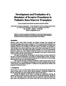
EVALUATION OF A MULTIPLE-ANTIGEN ENZYME-LINKED IMMUNOSORBENT ASSAY FOR DETECTION OF MYCOBACTERIUM TUBERCULOSIS INFECTION IN CAPTIVE ELEPHANTS R. Scott Larsen, D.V.M., M.S., M. D. Salman, B.V.M.S., Ph.D., Susan K. Mikota, D.V.M., Ramiro Isaza, D.V.M., M.S., Richard J. MontaU, D.V.M., and Joni Triantis, M.S. Abstract: Mycobucterium tuberculosis has become an important agent of disease in the captive elephant population of the United States, although current detection methods appear to be inadequate for effective disease management. This investigation sought to validate a multiple-antigen enzyme-linked immunosorbent assay (ELISA) for screening of M. tuberculosis infection in captive elephants and to document the elephant's sérologie response over time using a cross-sectional observational study design. Serum samples were collected from 51 Asian elephants {Elephas muximus) and 26 African elephants {Loxodonta africana) from 16 zoos and circuses throughout the United States. Infection status of each animal was determined by mycobacterial culture of trunk washes. Reactivity of each serum sample against six antigens was determined, and the linear combination of antigens that accurately predicted the infection status of the greatest number of animals was determined by discriminant analysis. The resulting classification functions were used to calculate the percentage of animals that were correctly classified (i.e., specificity and sensitivity). Of the 77 elephants sampled, 47 fit the criteria for inclusion in discriminant analysis. Of these, seven Asian elephants were considered infected; 25 Asian elephants and 15 African elephants were considered noninfected. The remaining elephants had been exposed to one or more infected animals. The specificity and sensitivity of the multiple-antigen ELISA were both 100% (91.9•100% and 54.4•100%, respectively) with 95% confidence intervals. Mycobacterium bovis culture filtrate showed the highest individual antigen specificity (95%; 83.0•100%) and sensitivity (100%; 54.4•100%). Serum samples from 34 elephants were analyzed over time by the response to the culture filtrate antigen; four of these elephants were culture positive and had been used to calculate the discriminant function. Limitations such as sample size, compromised ability to ascertain each animal's true infection status, and absence of known-infected African elephants suggest that much additional research needs to be conducted regarding the use of this ELISA. However, the results indicate that this multiple-antigen ELISA would be a valuable screening test for detecting M. tuberculosis infection in elephant herds. Key words: Elephant, Mycobacterium tuberculosis, ELISA, Elephas muximus, epidemiology, Loxodonta africana.
INTRODUCTION
African elephant (Loxodonta africana) in France
Mycobacterium tuberculosis, the etiologic agent of human tuberculosis, has caused morbidity and mortality in elephants and is a potential zoonosis.'^'
^as been documented.'' Elephant mycobacteriosis has become a disease of great concern to the public, ^oo and circus communities, and regulatory authorities.'
Several recent infections in captive elephants in the United States have been reported-^'»*>"^'5 29 Most reports document infection in Asian elephants {Elephas maximus), although one infection in a captive
From the Center of Veterinary Epidemiology and Ani, ^. ^ .,, ^ V, ,, r^T • mal Disease Surveillance Systems, College of Veterinary ..... , •• j, c • /-^ Í J c, » • Medicine and Biomédical Sciences, Colorado State TT Urnversity. Fort Collins, Colorado 80523, USA (Larsen, Salman, Triantis); the Audubon Center for Research of Endangered Species, 14001 River Road, New Orleans, Louisiana 70131, USA (Mikota); the Department of Clinical Sciences, College of Veterinary Medicine, Kansas State University, Manhattan, Kansas 66506, USA (Isaza); and the Department of Pathology, Smithsonian Institution Na, f" , . , , , . • tional Zoological Park, N. W. Washington, D.C. 20008, TTC A /n/i , T\ r. » jj /r \ T3 • »Í USA (Montali). Present address (Larsen): Environmental Medicine Consortium, Department of Clinical Sciences, College of Veterinary Medicine, North Carolina State University, Raleigh, North Carolina 25606, USA.
I" humans and domestic animals, screening for tuberculosis has been widely performed using the intradermal (i.d.) tuberculin test, the official test for live animals. Although this procedure has been described for nondomestic animals, systematic studies , , , , ,,.,.. have not been conducted to evaluate biologic activ. • , , ...... . . ity, optimal dose,' or suitable iniection sites in most J-> f j species.^" Other indirect laboratory tests that have been used for diagnosis of tuberculosis include lymphocyte transformation, gamma Interferon, enzyme-linked immunosorbent assay (ELISA), and the blood tuberculosis test (BTB)."""" These tests j^^^^ ^^j (^^^j, validated, uniformly administered, • » »i • .^ » j • ^ j nor consistently interpreted in most nondomestic . , "' ... species.^''In elephants, ^ ±' I.d. and sérologie 0 test results have not correlated well with culture status.^' Postslaughter testing remains the primary means of surveillance for cattle; however, this method is
291
292
JOURNAL OF ZOO AND WILDLIFE MEDICINE
impractical and undesirable in endangered and threatened species such as the Asian elephant and African elephant. Recently, guidelines have been developed for tuberculosis control and diagnostic investigation in captive elephants^^ that include mycobacterial culturing of respiratory secretions that are obtained from trunk wash material. Investigation of alternate testing modalities is being encouraged, however, in order to collect further information and to develop better tests.^^ Mycobacterial culture, although highly specific, has limited sensitivity. Culture results may be falsely negative because of insufficient mycobacterial numbers in trunk secretions ( Q To
* B1 - 33 yr old > B2 - 39 yr old - B 'B3 - 35 yr old U 84 - 31 yr old -
Dec-96 May-97 Oct-97 Mar-98 Aug-98 Jan-99 MI f Treatment Begun (B4, B6) f I Treatment Begun (82) | Transport of 82 to different facility Deatti of B1
Treatment Ended (82)
Date of Sampling
Figure 3. Optical density (OD) values of ELISA to Mycobucterium hovis AN5 culture filtrate (CF) for six elephants in herd B. Two female Asian elephants had cultures positive for Mycobucterium tuberculosis: elephant Bl was positive at postmortem exam in March 1997; elephant B6 had positive trunk wash cultures in March 1997, April 1997, and October 1997. Elephant B6 was transported to a different facility in April 1997 and had a positive trunk wash culture for Mycobucterium uvium in March 1999. No other elephant in the herd had a positive trunk wash culture. - - represents a "cut-off" value (OD = 0.58) for infected vs. noninfected as determined by discriminant analysis of CF.
herd of five elephants, one with a positive culture for M. tuberculosis,^^ and antigens that included heat-killed cells of M. bovis, purified protein derivative of M. bovis (PPD), and heat-killed cells of M. avium. Two elephants that were i.d. tuberculin test positive showed substantial seroreactivity to heatkilled M. bovis and PPD, whereas two of three i.d. tuberculin test-negative animals showed seroreactivity to heat-killed M. bovis only. A subsequent investigation in 1998, with a herd of four presumed-negative elephants,^' suggested that tuberculin exposure influences sérologie analysis with ELISA and may affect the BTB test. The antigens used in this sérologie investigation were M. tuberculosis CF protein, M. avium CF protein, M. avium sonicate antigen, PPD, avian purified protein derivative, and a lipoarabinomannan antigen. Elephants showed minimal to no reactivity to these antigens prior to i.d. injection but seroreactivity increased dramatically afterward. The BTB results were also affected because all elephants tested negative prior to antigenic stimulation, whereas two of three stimulated elephants tested positive for M. bovis antigen afterward. Elephants are constantly exposed to saprophytic Mycobacteria spp. and can be colonized
by atypical mycobacteria because of their behavior of dusting and bathing with their trunks. This exposure may account for nonspecific sérologie reaction to mycobacterial antigens.^' In this report, the significant differences in seroreactivity between infected and noninfected groups indicate a substantial humoral response in elephants with active M. tuberculosis infection. Between infected and noninfected groups, there were significant differences in the ODs for all antigens except MPB, and these differences do not appear to be associated with age-related exposure. It is not surprising that low reactivity to MPB was observed in both groups because MPB has been used to increase specificity for M. bovis screening in cattle and cervids.' '''-^'' It is also not surprising that good specificity and sensitivity were achieved by a combination of antigens derived from M. bovis and M. tuberculosis because these mycobacteria are closely related, sharing 85•100% of homology at the DNA level."" Shared antigenicity of these organisms has been documented.'''*^'' This multiple-antigen ELISA was able to accurately discriminate between infected and noninfected elephants. The classification function that
299
LARSEN ET AL.•DIAGNOSIS OF M. TUBERCULOSIS IN ELEPHANTS
Elephant Herd E 2.500
2.000
1 -
1.500
y
0.500
» El - » £2 - •>* 'ES ---1-- E4 - C3 'ES
- 10 - 40 - 24 - 26 - 15 - 18 ^ E7 - 19 • E8 - 15 • E9 - 19 -