endodermis (Bell and McCully, 1970; Baker, 1973). The stele ..... 511-517, Anderson, W.P., ed. Lon- ... Bell, J.K., McCully, M.E.: A histological study of lateral root.
Planta 9 by Springer-Verlag 1980
Planta 148, 116-123(1980)
Evidence for Proteins Specific for Vascular Elements in Intact and Cultured Tissues and Ceils of Maize E.E. Khavkin, E.Yu. Markov, and S.I. Misharin Siberian Institute of Plant Physiologyand Biochemistry,SiberianBranch, Academyof Sciencesof USSR, P.O. Box 1243, Irkutsk 33, USSR 664033
Abstract. A group of antigenically distinct proteins
characteristic for the tissue complex of the vascular cylinders was found in maize (Zea mays L.) seedlings using an immunofiltration technique. Specific stelar antigens present in the fully developed stele (vascular cylinder) of the primary root were also found in steles extracted from adventitious roots and from the mesocotyl but were absent, within the limits of sensitivity of the immunodiffusion tests employed, in root cortex and epidermis. Some of the stelar antigens were also evident in the meristem of the primary root and were present in traces in the scutellum, the mesocotyl node, and the primary leaves plus coleoptile. The specific stelar antigens could be traced in 13- and 15-day-old developing embryos and were definitely expressed by the 21st day after pollination. Several stelar-specific antigens were found in embryo-derived callus tissues and in stem-derived cells maintained in serial suspension culture. Higher resolution of the stelar antigens by a modified technique of crossed immunoelectrophoresis was used to demonstrate several minor stelar antigens that were presumably characteristic exclusively of the completely differentiated stele. This technique along with sequential immunoprecipitation of labelled proteins provided a semiquantitative estimate of the specific stelar antigens in the meristem and the stele of the primary root, and in suspension-cultured cells which were devoid of noticeable signs of vascular differentiation. Key words: Antigen - Callus Cell suspension cultures Proteins (in differentiation) - Seedling - Vascular cylinder (stele) - Zea.
Introduction
Several immunochemical studies of cytodifferentiation in higher plants have proven the existence
of characteristic stage (phase)-specific proteins (Boutenko and Volodarsky, 1968; Khavkin et al., 1979). Much less is known of the rearrangement of protein patterns at higher levels of organization, i.e., in the course of tissue and organ development. Several organ-specific antigens have been demonstrated in intact and isolated tissues of tobacco, maize and cherry (Boutenko and Volodarsky, 1968; Khavkin etal., 1979; Misharin et al., 1974; Raft et al., 1979), yet no data were presented to associate these antigens with definite cell types or to relate the accumulation of these specific proteins to the temporal pattern and crucial events of tissue differentiation and to the development of specific physiological functions. The pattern and the development of vascular elements in maize have been studied extensively (see Esau, 1943, 1965; Euxovfi, 1975; Walch and Evert, 1975; Feldman, 1977, and literature cited therein). The fact that the stele and the cortex plus the epidermis can be easily separated at least in maize roots (Laties and Budd, 1964) and mesocotyls considerably facilitates the biochemical investigation of stele specificity. Preliminary studies of maize seedling roots (Misharin et al., 1974) indicated, though by no means proved, the existence of specific stelar antigens. It was therefore obvious to raise antisera against the proteins of two tissue complexes, stele and cortex + epidermis, and to search for specific stelar antigens absent in the latter tissue complex. In this paper we report evidence for the existence of characteristic root stelar antigens and describe the stelar antigen patterns in the various parts of maize seedlings and in maize tissue and cell cultures of different origin. Some of these data have been briefly reported elsewhere (Markov et al., 1979). In addition, we describe procedures, including a novel technique of crossed immunoelectrophoresis with an immunoabsorbing barrier, which have been used to obtain semi-quantitative evidence of the expression of the
0032-0935/80/0148/0116/$01.60
E.E. Khavkin et al. : Evidence for Proteins Specific for Vascular
117
2. From 5-Day-Old Seedlings. Scutellum, and coleoptile plus leaves. 3. From 30- to 40-Day-Old Plants. Segments of internodes including the intercalary meristems, and the leaf bases. Callus and cell-suspension cultures were established and maintained as described in Khavkin et al. (1979) except that the suspension-culture cycle was shortened to 7 days. Two types of embryoderived callus tissues, one from the scutellum and one from the root tip, and one stem-derived cell-suspension culture sampled on the 3rd (proliferation phase) and the 7th day (stationary phase) were investigated. The tissue samples were frozen and stored in liquid nitrogen.
Preparation of Tissue Extracts
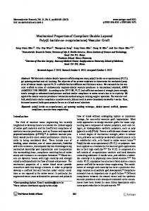
Fig. 1 A and B. Extraction of vascular cylinder (stele) from different parts of maize seedlings. A Succesive stages (1, 2, 3) of extraction of the stele from the primary root. B Isolated steles from different parts of a 3-day-old seedling: 1 primary root ; 2 adventitious roots ; 3 mesocotyl
The frozen samples were homogenized in 100 mM Tris (trishydroxymethylaminomethane)-HC1buffer, pH 8.3, containing 10 mM 2-mercaptoethanol. The tissue:buffer ratio (w/v) depended on the protein content of the samples and was 1 :I for stelar samples from roots and mesocotyl, for the root cortex+epidermis, the leaves, the intercalary meristem of stems, and the samples of callus tissues and 7-day-old cell suspensions; i:2 for the primary root meristem, the mesocotyl node and the 3-day-old cell suspensions; 1:3 for the scutellum. Extraction was carried out for 20 min at 4~ and followed by centrifugation at 18000xg for 20 min at 4~ (Janetzki K-24; VEB MLW Zentrifugenbau, Engelsdorf, GDR). The protein content of extracts was measured according to Lowry et al. (195 l) after 3-fold precipitation with trichloroacetic acid.
lmmunochemical Methods s t e l a r a n t i g e n s in d i f f e r e n t t i s s u e s a n d cells a n d t o correlate the stelar-antigen patterns with the development of vascular elements.
Material and Methods Plant Material Kernels of maize (Zea mays L.), hybrid Bukovinsky 3, obtained commercially from the Krasnodar region, were germinated and grown at 27 ~ C in the dark or in a greenhouse as described earlier (Khavkin etaI., 1977). Developing ears of the inbred W155, 12-21 day after pollination, were collected in the field in the Krasnodar region. Parts and tissues used for analysis were as follows:
i. From 3-Day-OM Seedlings. Root apical meristem (0-2 mm from the tip, the root cap excluded); vascular cylinders (stele) isolated from the primary and adventitious roots and from the mesocotyl (Fig. 1 B); a 2-ram-long segment of the mesocotyl (coleoptile) node incIuding the plumule. To extract the mature part of the stete of the root, a 15-mm-long tip portion of the latter was cut off with a razor blade, the root was slightly fractured by bending at its base, and the epidermis+cortex "tube" was carefully pulled off, unsheathing the stele (Fig. 1), Fifteen-mm segments were cut from either tissue complex, and the completeness of their separation was checked either visually or by blowing lightly through the cortex+epidermis "tube". The two tissue complexes of the root are separated because of rupture of the outer periclinal walls of the endodermis (Bell and McCully, 1970; Baker, 1973). The stele of the mesocotyl is easily extracted in the same manner, even without having to cut off the node.
(iocal
Antisera. Rabbits breed, obtained commercially) were injected with protein preparations from the mature part (15-25 mm from the tip) of cortex+epidermis, from the stele of the primary root of seedlings, and from the 2-mm-long segments of the mesocotyi node, to obtain three multivalent sera. The protocols of immunization and 7-globulin enrichment of the sera were as described in Khavkin et at. (1977). Absorption of Common Antigens. Batch absorption of common antigens was carried out by mixing crude tissue extracts with a moderate excess of cortex+epidermis antiserum and incubating first for 1 h at 37~ C and then for 18 h at 4~ C, followed by centrifugation in Janetzki K-24 at 3000• for 10min. The procedure was checked by the routine Ouchterlony test. Intra-gel absorption of the common antigens in the course of electrophoresis was provided by addition of cortex + epidermis antiserum to the separation media. Barrier absorption in the double immunodiffusion tests was as described previously (Khavkin et al., I977). Immunoprecipitation of Labelled Proteins. Two-day-old seedlings were labelled with [14C]leucine (7.5 gCi/ml, 28.7 mCi/mmol) for 24 h, and tissue extracts were prepared as described above. The common antigens were precipitated 3 times by batch procedure with cortex+epidermis antiserum, centrifuged off, and the stelar antigens were precipitated in the same way with stele antiserum. Proteins of the initial extracts, the common antigen precipitates, the stelar-antigen precipitates, and the residual non-precipitated proteins were reprecipitated with 5% trichloroacetic acid in the presence of an excess of non-labelled leucine, and their radioactivity was estimated by the method of Hall and Cocking (1965) in a liquid-scintillation /Lspectrometer (model 8100; LKB-Wallac, Stockholm, Sweden).
118
E.E. Khavkin et al. : Evidence for Proteins Specific for Vascular
Proteins from cell suspensions were labelled heavily in order to carry out subsequent radioautography of immunoprecipitated polypeptides (data not presented). One mCi [14C]leucine (21 mCi/ mmol) was added under sterile conditions to a flask with a 3-dayold cell suspension (final radioactivity 16 gCi/ml). Two days later the cells were harvested by filtration under suction, washed, frozen, and prepared as described above.
Immunoelectrophoresis. Two procedures were employed for onedimensional separation: routine separation in agarose B (Reachim, Olaina, USSR) as described by Crowle (1973), after batch absorption of the common antigens, and separation after batch plus intragel absorption of the common antigens. In the latter case ca. 500 mg protein of cortex +epidermis antiserum, dialyzed against barbitalacetate buffer, pH 8.6, ionic strength 0.05 (Crowle, 1973), was added to 20 ml of molten 1% agarose. The current was 25-30 mA at 80 V. The common antigens formed "rocket" precipitin flares, while the stelar antigens migrated freely for about 2.5 h and were precipitated with stele antiserum. To control the specificity of the procedure a parallel slot was filled with cortex+epidermis antiserum. The technique of crossed immunodisc electrophoresis combined the procedure of immunoabsorption in stacking polyacrylamide gels (Maurer, 1968) and that of" rocket" immunoelectrodiffusion (Crowle, 1973; Smyth et al., 1977). Vertical slab gels of 8 x 13.5 x 0.1 cm 3 were prepared between 0.1-cm-thick glass plates. The separation gel, 12cm high, was made of 6% acrylamide+0.16% bis-acrylamide in 376 mM Tris-HC1 buffer, pH 8.8. The stacking gel was made of 2.5% acrylamide+0.625% bis-acrylamide and contained ca. 8 mg/ml enriched preparation of cortex+ epidermis antiserum. The electrode buffer for :the first run was made up of 38 mM glycine and 4.9 mM Tris, p H 8:3. Tissue extracts partially freed from the common antigens by batch absorption were dialyzed against 56.4mM Tris-HC1 buffer, pH 6.9, concentrated ca. 2-fold with dry Sephadex G-25 (coarse; Pharmacia, Uppsala, Sweden), then 2-mercaptoethanol, glycerol and bromophenol blue were added to a final concentration of 10 mM, 10% and 0.001%, respectively, and 0.4 ml of this solution (ca. 2 3 mg plant protein) was loaded on the surface of the stacking gel containing cortex + epidermis antiserum. The current was 6 mA per slab, and after 7 h at 4 ~ C the marker dye had reached the bottom of the slab. Next, the slabs were sliced lengthwise. Some of the 2-cm-wide strips (without the stacking gel) were kept as controls, while other strips were embedded into agarose gels containing ca. 6 mg protein per 1 ml of stele antiserum or cortex+ epidermis antiserum (to control the absorption procedure). The second run was performed at 2 V/cm for 16 h at 4~ at right angle to the direction of the first run. The completeness of the electrodiffusion transfer of antigens was checked by subsequent staining of the used polyacrylamide strips for protein. The agarose gels were finally soaked in 150 mM NaC1 for 48 h to remove any non-specific precipitates and soluble proteins, and in distilled water to remove NaC1; the gel plates were then air-dried, stained with Coumassie Brilliant Blue R-250, and destained according to the usual protocol.
Results
The Patterns of the Stelar Antigens in the Isolated Stele and in Non-Separated Samples of Intact and Cultured Tissues and Cells T h r e e s t e l a r - s p e c i f i c a n t i g e n s w e r e f o u n d by t h e d o u b l e - i m m u n o d i f f u s i o n test in p r e p a r a t i o n s f r o m m a t u r e steles i s o l a t e d f r o m t h e p r i m a r y a n d a d v e n t i t i o u s
Fig. 2A and B. Spectra of specific stelar antigens in different tissue complexes and suspension-cultured cells of maize. A Stelar antigens in crude cell-free extract (I) and after a single batch-absorption of the common antigens (II) in the stele of the primary root. B Comparison of stelar antigens from different samples. Antigens 1 3 stele of the primary root, adventitious roots, and mesocotyl, respectively, of 3-day-old seedlings; 4 3-day-old cell suspension culture; 5 6 leaf base and stem internode of 30-day-old plants, respectively. Antisera: ASt, against the stele of the primary root; ACo, against epidermis + cortex of the primary root; ANo, against the mesocotyl node of 3-day-old seedlings
r o o t s o f m a i z e s e e d l i n g s (Fig. 2). A p p a r e n t l y i d e n t i c a l stelar a n t i g e n s w e r e f o u n d in t h e m e s o c o t y l stele. N o f u r t h e r stelar a n t i g e n s c o u l d be d e m o n s t r a t e d in t h e m e s o c o t y l stele w i t h m e s o c o t y l - n o d e antiserum (Fig. 2B). F u r t h e r m o r e , o n e o r t w o p r e c i p i t a t e lines w e r e s o m e t i m e s also e v i d e n t in p r e p a r a t i o n s f r o m m e s o c o t y l n o d e , t h e leaves, a n d callus a n d cell s u s p e n s i o n c u l t u r e s w h i c h h a d b e e n m a i n t a i n e d for 19 a n d 52 t r a n s f e r s , r e s p e c t i v e l y ( T a b l e 1). I n d e v e l o p i n g e m b r y o s o n l y t r a c e s o f t h e stelar a n t i g e n s c o u l d be seen in d o u b l e - i m m u n o d i f f u s i o n tests by t h e 13th a n d 15th d a y a f t e r p o l l i n a t i o n t h o u g h v a s c u l a r d i f f e r e n t i a t i o n was e v i d e n t at this t i m e (Sass, 1977). L a t e r , in 2 1 - d a y - o l d e m b r y o s stelar a n t i g e n s
E.E. Khavkin et al. : Evidence for Proteins Specific for Vascular Fable l. Specific stelar antigens in intact organs and in cultured tissues and ceils of maize Investigated sampIe
Antigens"
3-day-old seedlings: Root apical meristem Primary root stele Adventitious root stele Mesocotyl stele Mesocotyl node
+ + + + +
5-day-old seedlings: Scutellum Leaves + coleoptile
tr + +
30-40-day-old plants : Stem internodes Leaf bases
0 +
Developing embryos: 13-15-day-old 21-day-old
tr +
Embryo-derived callus cultures: Scutellum-derived Root-derived
+ +
Stem-derived cell suspension culture: 3-day-old 7-day-old
+ +
a
+ + + +
+ , + + =definitely expressed; tr=trace; 0=absent
were expressed quite definitely (Table 1). The apparent absence of the stelar antigens in internodes seems to result from the low content of the antigens and an inadequate titer of the stele antiserum. The two most mobile anodic stelar antigens were apparent in one-dimensional electropherograms after batch absorption of the common antigens (Fig. 3A). The discrimination of the stelar antigens was considerably facilitated by an additional intra-gel absorption with cortex+epidermis antiserum (Fig. 3B), and in this case the anodic stelar antigens were likewise the most prominent in the samples from the root and the mesocotyl stele. Crossed immunodisc-electrophoresis after intragel absorption of the common antigens produced a much
A
119
better resolution, especially in regard to the slowlymigrating stelar antigens (Fig. 4). Faint precipitin iines congruent with four antigens were observed in some of the control agarose gels containing cortex + epidermis antiserum, indicating an inadequate absorption of the common antigens by the barrier in the stacking gel; the specificity of the stelar antigens 4, 5, 14 and 15 seemed therefore to be somewhat questionable. Several stelar antigens, including the major antigen 3 and several minor proteins, were common to all samples examined. Other stelar antigens (e.g. I, 2, 7, 9-12) were apparently absent from the suspension-cultured cells and were therefore presumably characteristic of the root. Both the root meristem and the suspension-cultured cells seemed to lack the stelar antigens 1, 2, 7, 9, and 10; these can be tentatively related to the final stages of vascular differentiation.
Quantitative Studies of the Stelar Antigens in Relation to Stelar Development Double-immunodiffusion tests were employed to estimate the lower threshold amount of protein sufficient to produce visible precipitin lines of stelar antigens. In the completely developed stele and in the meristem of the primary root these values were ca. 30 and 150 gg, respectively. However, when we examine Fig. 4A and B where the areas of the precipitin arcs can be related to the content of the respective individual antigens, the patterns do not seem to indicate a 5-fold increase in the concentration of the major antigens 3, 8a and 8b. Therefore either the former evidence is not very reliable, or most of the quantitative variation in the stelar proteins is caused by the minor stelar antigens. To resolve this dilemma we employed another experimental technique, namely, a sequential, quantitative immunoprecipitation of the common antigens and the stelar antigens. For doing this the proteins of seedlings and suspension-culture cells were heavily
B
Fig. 3A and B. One-dimensional immunoelectropherograms of stelar proteins from primary root (l) and mesocotyI (2) of 3-day-old seedlings of maize. A Single batch-absorption of the common antigens; B Batch-plus intragel absorption of the common antigens. Otherwise as in Fig. 2
120
E.E. Khavkin et al. : Evidence for Proteins Specific for Vascular
+ I
0
3 3 \/,,,~
8a
8b
5
8a
Fig. 4A-C. Photographic (left) and graphic (right) patterns of the stelar antigens of maize after crossed immunodisc electrophoresis with immunoabsorption of the common antigens in the stacking gel. I agarose gel containing stele antiserum, lI agarose gel containing cortex+epidermis antiserum (a control for completeness of immunoabsorption). A Mature part of the stele of the primary seedling root; B Apical meristem of the primary root; C 7-day-old cell suspension culture. 1-I5 antigens. Initial protein concentrations were 3.5, 3.75 and 4.2 mg/ml, respectively. The shaded band in A(I) at the right corresponds to the stelar antigen 3 fraction used to raise an antiserum of narrow specificity
labelled with [14C]leucine and the stelar antigens were precipitated from extracts already free of the common antigens. The completeness of the immunoprecipitation procedure was checked immunologically and could be additionally judged from the extent of nonprecipitated plant proteins. The latter was usually negligible (up to 5% of the total radioactivity of the protein), except in the case of the root meristem where it constituted 15%, and in the suspension-cultured cells where it amounted to 36% (Table 2), apparently in consequence of the high content of proteins related to proliferation (meristematic, embryonal; see Khavkin et al., 1979) but unrelated to antisera against the mature part of the root. In addition, the data of Table 2 provide two kinds of evidence. The first concerns the relative content of the stelar antigens. This varies unexpectedly little in all investigated samples, thus corroborating the evidence of Fig. 4, and the stele specific antigen-antibody complex seems to constitute about one third of the total immunoprecipi-
tate. The second evidence of Table 2 concerns the incorporation of label into the common and stelar antigens. The absolute values of total incorporation per mg plant protein differed about 50-fold (compare root stele and scutellum), depending mainly on the transport and redistribution of the label, the size of the amino acid-pools, etc. ; yet the relative incorporation into the stelar and common antigens was comparable within each of investigated samples. Though the results of immunoprecipitation experiments with these highly heterogeneous systems can not be interpreted quantitatively, the data seem to indicate greatest synthesis of stelar antigens in the stele of the primary root and in the growing leaves (mesocotyl node + plumule and leaves + coleoptile).
Further Studies of the Stetar Antigen 3 We obtained an antiserum of narrow specificity against the band corresponding to the stelar antigen 3 in the polyacrylamide gel strips after the first elec-
E.E. Khavkin et al. : Evidence for Proteins Specific for Vascular
121
Table 2. Label distribution in the common antigens (CA) and specific stelar antigens (StA) in various parts of 3-day-old seedlings and cultured cells of maize
~
Total protein radioactivity, 103 dpm, in the celI-free extracts
Distribution of total protein radioactivity, 103 dpm, after batch immunoprecipitation
per mg plant protein per ml extract
o~
o~
8
~
~.
-
=~
135. l
445.1
219.1
101.7
59.9
46.1
5.7
5,880
384.9
587.6
285.0
132.2
97.0
150.4
17.5
23,522
10,480 4,552 8,549
per ml extract
CA StA NpP"
178.2 148.9 57.7
206.8 355.5 25.3
137.6 133.1 14.2
68.5 61.0 2.6
30,5 64.1 2.4
60.8 85.0 4.5
12.3 4.4 0.8
% of total
CA StA NpP ~
46.3 38.7 i5.0
35.2 60.5 4.3
48.3 46.7 5.0
51.8 46.2 2.0
31.5 66.0 2.5
40,4 56.6 3.0
70.3 25.2 4.5
10.56 6.34
12.00 8.17
Immunoprecipitate (antigens + antibodies) protein, mg/ml initial extract
CA StA StA CA + StA %
Specific radioactivity of immunoprecipitates, 103 dpm per mg protein of antigen-antibody complex
CA StA StA/CA
6.24 3.56 36.3 24.0 41,8 1.74
6.30 3.72
4.08 1.28
5.30 3.05
37.2
23.9
36.6
37.5
40.5
32.8 95.6 2.9
33.6 I04.0 3.I
12.9 20.0 1.6
2.9 10.1 3.5
5.05 10.4 2.1
9.36 6.77 42.0 1.3 0.65 0.5
44.5 19.2 36.3 4.0 3.3 45.2 2,620 137 0.5
NPP=non-precipitable residual protein
trophoretic run (the position of this band is shaded in Fig. 4A). Frozen gel segments were stored, the stelar antigen 3 eluted by electrodiffusion and checked for specificity (Fig. 5A), and each rabbit was injected with a total of 7-8 mg of this preparation. The antiserum developed one distinct precipitin line with the total stele extract, yet contained traces of antibodies cross-reacting with the common antigen (Fig. 5B). Further purification of the antigens is evidently necessary to produce monovalent antisera.
Fig. 5A and B. Experiments with the stelar antigen 3 (St 3) of maize and the antiserum against this preparation (ASt3). A Specificity of the St3 preparation obtained by electrophoretic separation (see Fig. 4A); B Specificity of ASt3. Otherwise as in Fig, 2
Discussion In the primary root of maize seedlings several proteins were found that were absent from the epidermal and cortical cells and were characteristic of a well-defined tissue complex, the stele. These proteins seem to lack all organ specificity. Most of them appear at the very earliest stages of vascular development, in the developing embryos and the root meristem; some are also present in suspension-cultured cells which seem to be devoid of any microscopically discernable vascular structures. We therefore suggest that at least the major stelar antigens, e.g., stelar antigen 3, are related to such vascular elements that have undergone only early differentiation, or to the associated ground tissue of the vascular cylinder. In addition to the major stelar antigens, minor stelar antigens were found but only in the completely developed stele. These need further investigation in relation to the time course of vascular differentiation. At present, we are not able to detail the distribution of the stelar antigens within the tissue complex of the stele. However, some of the procedures described in this paper provide a promising approach to the preparation of sera of very narrow specificity appropriate for immunohistochemical localization of specific stelar proteins.
122
Perhaps it is appropriate to mention here two alternative approaches to the search for specific proteins in vascular elements of higher plants, namely, biochemical investigations of phloem exudate (for a review, see Sabnis and Hart, 1979) and electron-microscopic studies of P-protein which is unequivocally associated with the sieve tubes (for a recent review see Evert, 1977) although this protein is absent in maize (Welsh and Evert, 1975). It seems, however, premature to compare the results of such divergent studies of vascular proteins. Some additional facts are also relevant to the evidence in Table 2. Dudley and Northcote (1978) reported a selective increase in the production of messenger RNAs coding for two characteristic polypeptides when development of vascular elements was induced in bean cell-suspension cultures by increasing the kinetin/auxin ratio. Recently we estimated the relative rates of incorporation of labelled leucine into the stelar and the common antigens, separated as shown in Fig. 2 in this paper, in the roots of 3- and 9-day-old maize seedlings. The synthesis of the stelar specific antigens was considerably greater than that of the common antigens, especially in the younger roots (Markov et al., 1979). If the same criterion, the ratio of the specific radioactivities of immunoprecipitates, is applied to the data of Table 2, it is apparent that synthesis and turnover of the stelar antigens are ahead of those of the common antigens in all parts of the seedling we investigated, except the scutellum and the suspension-cultured cells, and especially so in the root stele and the plumule. Cell growth and differentiation in maize tissues clearly involve profound switches in metabolic patterns, including protein synthesis. Nonetheless, evidence available from immunochemical studies in most instances indicates relatively small changes in the patterns of genes functioning in diverse non-differentiated, differentiated and dedifferentiated cell types. Does then differentiation of plant cells at all produce any specialized protein products, as is claimed by the current concept of developmental biochemistry? This paper completes the series (Khavkin et al., 1977, 1978, 1979) describing our quest for specific proteins related to cell differentiation in maize, mostly by comparative immunofiltration methods. As compared to the more frequently used batch absorption of sera, immunofiltration proved to be more reliable and efficient in screening for minor specific proteins whose content differed widely in various tissue samples, and for semi-quantitative estimation of these antigens. Antisera of narrow specificity obtained against antigens purified either by protein fractionation, as in the case o f reserve globulins (Khavkin et al., 1978), or by immunofiltration, as in the case
E.E. Khavkin et al. : Evidence for Proteins Specific for Vascular
of the stelar antigen 3 described in this paper, are however necessary for further discrimination between qualitative and quantitative interpretations of the changes in the developmental patterns of these antigens. At present, the relatively abundant reserve globulins of developing embryos seem to be the only stagespecific antigens that have been chemically defined and unambigously related to characteristic events of cytodifferentiation and embryonal growth in maize. Similar patterns have been reported for developing bean, broad bean and pea cotyledons (Manteuffel etal., 1976; Millerd etal., 1971, 1978; Sun et al., 1978). In maize there exist apparently other stagespecific antigens (embryonal and meristematic proteins), the tissue-specific proteins of the vascular elements, and root- and shoot-specific antigens although these latter are scarcely traceable. A shootspecific and several meristematic antigens have been clearly demonstrated also in tobacco (Boutenko and Volodarsky, 1968; Moiseeva et al., 1979). However, in all these cases the minor specific antigens have not been identified as characteristic products of differentiation either by their association with a histologically distinct cell type, or by biochemical criteria. We feel that the next, obvious step of obtaining monovalent antisera to the specific proteins can now be taken, in order to determine conclusively the localization of these proteins and to provide quantification of the changeable facets of gene expression in relation to cell and tissue differentiation in plants.
References Baker, D.A.: The radial transport of ions in maize roots. In: Ion transport in plants, pp. 511-517, Anderson, W.P., ed. London, New York: Academic Press 1973 Bell, J.K., McCully, M.E.: A histological study of lateral root initiation and development in Zea rnays. Protoplasma 70, 179-205 (1970) Boutenko, R.G., Volodarsky, A.D. : Analyse immunochimique de la differentiation cellulaire dans les cultures de tissus de Tabac. Physiol. V+g. 6, 299-309 (1968). Crowle, A.J.: Immunodiffusion, 2nd edn. London, New York: Academic Press 1973 Dudley, K., Northcote, D.H. : Comparison of the in vitro translation products of mRNA isolated from suspension cultures of Phaseolus vulgaris grown on maintenance and induction medium. Planta 138, 41-48 (1978) Esau, K. : Ontogeny of the vascular bundle in Zea mays. Hilgardia 15, 327-368 (1943) Esau, K. : Vascular differentiation in plants. New York, Chicago, San Francisco, Toronto, London: Holt, Rhinehart & Winston 1965 Evert, R.F.: Phloem structure and histochemistry. Annu. Rev. Plant Physiol. 28, 199-222 (1977) Fetdman, L.J. : The generation and elaboration of primary vascular tissue patterns in roots of Zea. Bot. Gaz. 138, 393-401 (1977)
E.E. Khavkin et al. : Evidence for Proteins Specific for Vascular Hall, T.C., Cocking, E.C.: High-efficiency liquid-scintillation counting of 14C-labelled material in aqueous solution and determination of specific activity of Iabelled proteins. Biochem. J. 96, 626-633 (1965) Khavkin, E.E., Misharin, S.I., Ivanov, V.N., Danovich, K.N.: Embryonal antigens in maize caryopses: the temporal order of antigen accumulation during embryogenesis. Planta 135, 225 231 (1977) Khavkin, E.E., Misharin, S.I., Markov, Ye.Yu., Peshkova, A.A. : Identification of embryonal antigens of maize: globulins as primary reserve proteins of the embryo. Planta 143, 11-20 (1978) Khavkin, E.E., Misharin, S.I., Polikarpochkiua, R.T.: Identical embryonal proteins in intact and isolated tissues of maize (Zea mays L.). Planta 145, 245-251 (1979) Laties, G.G., Budd, K. : The development of differential permeability in isolated steles of corn roots. Proc. Natl. Acad. Sci. USA 52, 462-469 (1964) Lowry~ O.H., Rosebrough, N.J., Farr, A.L., Randall, R.J. : Protein measurement with the Folin phenol reagent. J. Biol. Chem. 193, 265 276 (1951) Luxovh., M.: Some aspects of the differentiation of primary root tissues. In: The development and function of roots, pp_ 73-90, Torrey, J.G., Clarkson, D.T., eds. London, New York: Academic Press 1975 Manteuffel, R., Miintz, K., P(ichel, M., Schoiz, G.: Phase dependent changes in DNA, RNA and protein accumulation during the ontogenesis of broad bean seed. Biochem, Physiol. Pflanz. 169, 595 605 (1976) Markov, Ye.Yu., Mazel', Yu.Ya., Khavkin, E.E. : Rate of protein synthesis in the root stele of maize seedlings. [In Russ.] Fiziol. Rast. 26, 953-960 (1979)
123 Maurer, H.R.: Disk-Elektrophorese. Berlin: de Gruyter 1968 MilIerd, A., Simon, M., Stern, H. : Legumin synthesis in developing cotyledons of I/'iciafaba L. Plant Physiol, 48, 419-425 (1971) Mi[lerd, A., Thompson, J.A., Schroeder, H.E. : Cotyledonary storage proteins in Pisum sativum. III. Patterns of accumulation durirlg development. Aust. J. Plant Physiol. 5, 519-534 (1978) Misharin, S.I., Antipina, A.I., Reimers, F.E., Khavkin, E.E.: Phase-, tissue- and organ specific proteins of maize seedlings. [In Russ.] DokI. Acad. Nauk SSSR 219, 473-476 (1974) Moiseeva, N.A., Volodarsky, A.D., Butenko, R.G.: Detection of antigens-markers in tobacco stem meristem. [In Russ.] Fiziol. Rust. 26, 479-484 (1979) Ruff, J.W., Hutchinson, J.F., Knox, R.B., Clarke, A.E.: Cell recognition : antigen determination of plant organs and their cultured.callus cells. Differentiation 12, 179-186 (1979) Sabnis, D.D., Hart, J.W. : Heterogeneity in phloem protein complements from different species. Consequences to hypotheses concerned with P-protein function. Planta 145, 459-466 (1979) Sass, J.E. : Development of the caryopsis. In: Corn and corn improvement, pp. 89-110, Sprague, G.F., ed. Madison, Wis., USA: Am. Soc. Agron. 1977 Smyth, C.J., S6derholm, J., Wadstr6m, T.: Laying-on technique for crossed immunoelectrofocusing, Appl. Note 269, Bromma, Sweden: LKB-Produkter 1977 Sun, S.M_, Mutschler, M.A., Bliss, F.A., Hail, T.C.: Protein synthesis and accumulation in bean cotyIedons during growth. Pla1~t Physiol. 61, 9I 8-923 (I978) Walsh, M.A., Evert, R.F.: Ultrastructure of metaphIoem sieve elements in Zea mays. Protoplasma 83, 365-388 (1975)
Received 9 July; accepted 8 November 1979