Current Pharmaceutical Design, 2006, 12, 261-271
261
Evolution of Resistance to Cancer Therapy Franziska Michor1,2*, Martin A. Nowak1 and Yoh Iwasa3 1
Program for Evolutionary Dynamics, Department of Organismic and Evolutionary Biology, Department of Mathematics, Harvard University, Cambridge, MA 02138, USA; 2Harvard Society of Fellows, Cambridge, MA 02138, USA and 3 Department of Biology, Kyushu University, Fukuoka 812-8581, Japan Abstract: Acquired drug resistance is a major limitation for successful treatment of cancer. Resistance emerges due to drug exclusion, drug metabolism and alteration of the drug target by mutation or overexpression. Depending on therapy, the type of cancer and its stage, one or several genetic or epigenetic alterations are necessary to confer resistance to treatment. The fundamental question is the following: if a genetically diverse population of replicating cancer cells is subjected to chemotherapy that has the potential to eradicate it, what is the probability of emergence of resistance? Here, we review a general mathematical framework based on multi-type branching processes designed to study the dynamics of escape of replicating organisms from selection pressures. We apply the general model to evolution of resistance of cancer cells and discuss examples for diverse mechanisms of resistance. Our theory shows how to estimate the probability of success for any treatment regimen.
Key Words: Cancer Therapy; Evolution of Resistance; Evolutionary Dynamics. 1. INTRODUCTION Drug resistance can result from two general causes [1]: (i) host factors such as poor absorption and rapid metabolism reduce the maximum achievable serum levels of the drug this is sometimes referred to as intrinsic resistance, and (ii) specific genetic or epigenetic alterations enable resistant cancer cell clones to outgrow and escape from otherwise effective treatment. The main mechanisms of cellular resistance are depicted in Fig. (1). Some of these mechanisms, such as loss of a cell surface receptor or transporter, specific metabolism and an increase or alteration in the drug target, result in resistance to only a small number of related chemotherapeutic agents. Other mechanisms, however, lead to simultaneous resistance to many structurally and functionally unrelated drugs. This phenomenon is known as multidrug resistance [1, 2] and can result from changes that limit accumulation of drugs within cells by decreasing uptake, enhancing efflux, or affecting membrane lipids [3], block apoptosis which is activated by most anticancer drugs [4], induce general response mechanisms that detoxify drugs and repair DNA damage [5], and modulate the cell cycle [6] and checkpoints [7]. Many genes have been identified that contribute to diverse mechanisms of resistance to chemotherapy [8]: amplification or overexpression of the P-glycoprotein family of membrane transporters (e.g., MDR1, MRP, LRP) which decrease intracellular drug accumulation; changes in cellular proteins involved in detoxification (e.g., glutathione Stransferase pi, metallothioneins, human MutT homologue, bleomycin hydrolase, dihydrofolate reductase) or activation of the chemotherapeutic drugs (DT-diaphorase, NADP: *Address correspondence to this author at the Program for Evolutionary Dynamics, Department of Organismic and Evolutionary Biology, Department of Mathematics, Harvard University, Cambridge, MA 02138, USA; E-mail:
[email protected] 1381-6128/06 $50.00+.00
cytochrome P-450 reductase); changes in molecules involved in DNA repair (e.g., O6-methylguanine-DNA methyltransferase, DNA topoisomerase II, hMLH1, p21WAF1/CIP1); and activation of oncogenes such as Her-2/neu, bcl-2, bclXL, c-myc, ras, c-jun, c-fos, and MDM2 as well as inactivation of tumor suppressor genes like p53 can all confer resistance to therapy. The number of genetic or epigenetic alterations necessary for escape from therapy depends on the patient’s genetic background, the therapy administered, and the type of cancer and its stage. Here, we will discuss mechanisms of resistance brought about by one or two genetic alterations. The following listing is not intended to be exhaustive, but serves to demonstrate the principle of escape from therapy via one or two alterations. One alteration. Several genes confer cancer drug resistance if affected by a single genetic alteration. For example, single point mutations in alpha- or beta-tubulin confer resistance to hemiasterlins, which are sponge-derived tripeptides that inhibit cell growth by depolymerizing existing microtubules and inhibiting microtubule assembly [9]. Overexpression of Bcl-2 abrogates the short-term apoptotic response to chemotherapy and correlates with a poor long-term outcome [10]. Activation or overexpression of c-myc can induce and modulate drug resistance [11]. Similarly, mutation or overexpression of PI3K or Ras leads to increased radioresistance [12]. Upregulation of the transcription factor Ets-1 confers resistance to the DNA damaging agent cisplatin via transcriptional regulation of metallothioneins and DNA repair enzymes [13]. Several N-terminal and core-domain mutations have been identified in human topoisomerase II alpha, each of which is sufficient to confer bisdioxopiperazine resistance [14]. Overexpression of glutathione-S-transferases, a family of detoxification enzymes, is also implicated in the development of resistance toward chemotherapy agents [15]. Increased expression of the transcription factor NF-κB induces drug resistance through MDR1 expression in cancer © 2006 Bentham Science Publishers Ltd.
262 Current Pharmaceutical Design, 2006, Vol. 12, No. 3
Michor et al.
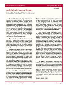
Fig. (1). Cellular factors that cause drug resistance. Cancer cells become resistant to anticancer drugs by several mechanisms. One way is to pump drugs out of cells by increasing the activity of efflux pumps, such as ATP-dependent transporters. Alternatively, resistance can occur as a result of reduced drug influx - a mechanism reported for drugs that enter on intracellular carriers or by means of endocytosis. Through compartmentalization, drug concentrations in the cytosol can be limited. In cases in which drug accumulation is unchanged, activation of detoxifying proteins can promote drug resistance. Cells can also activate mechanisms that repair drug-induced DNA damage, and disruptions in apoptotic pathways allow cells to become resistant to drug-induced cell death. Finally, alterations of cell cycle checkpoints or drug targets confer resistance to cancer therapy.
cells [16]. Overexpression of HER-2 represents a resistance mechanism to hormonal therapy in breast cancer [17]. Amplification or increased expression of p-glycoprotein confers multidrug resistance [18]. Two alterations. Other mechanisms of escape from therapy require two genetic alterations, either because of haplosufficiency of a gene such that one recessive mutation cannot confer resistance, or because of the use of combination therapy targeting two different positions in the cancer genome. For example, homozygous or compound heterozygous inactivation of p53 leads to acquired resistance to gamma irradiation and chemotherapy [4, 19]. Similarly, inactivation of both alleles of ATM confer resistance to therapy [20]; so does homozygous inactivation of Rb [21]. The cyclin dependent kinase inhibitors p16 and p18 can also be involved in resistance if inactivated in both alleles [22]. Loss of DNA mismatch repair due to hMLH1 hypermethylation or inactivation causes resistance to 5-fluorouracil in colorectal cancer [23]. The accumulation of specific genetic alterations leading to acquired drug resistance is greatly accelerated by genetic instability. Genetic instability is a defining characteristic of human cancers [24]. Two main types of genetic instabilities have been identified: in a small fraction of colorectal, endometrial, gastric and some other cancers, inactivation of the mismatch repair pathway leads to an elevated point mutation rate called microsatellite instability (MIN) [25, 26]; the majority of cancers, however, has chromosomal instability (CIN) [27]. CIN refers to an increased rate of losing or gaining whole chromosomes or large parts of chromosomes
during cell division. The consequence of CIN is an imbalance in chromosome number (aneuploidy) and an increased rate of loss of heterozygosity (LOH). An elevated rate of LOH is an important property of CIN, because it accelerates the inactivation of tumor suppressors and other recessive genes [28-30]. 2. CALCULATING RESISTANCE
THE
PROBABILITY
OF
Consider a population of cancer cells that grows according to a continuous time branching process [31, 32]. At each time step, a cell either produces an offspring or dies. If each cell produces on average more than one new cell, then the basic reproductive ratio [33] is larger than one, r > 1, and the cancer grows over time. If a cell dies with probability a and divides with probability b per time step, then the basic reproductive ratio is given by r = b/a. Therapy reduces the basic reproductive ratio either by increasing the death rate, decreasing the growth rate or both. Denote the basic reproductive ratio during therapy by R. If R still exceeds one, then therapy can reduce the rate of cancer growth, but is not capable of eradicating it. If R is less than one, however, then each cell produces on average less than one new cell, and therapy can eradicate the cancer. At the time of initiating therapy, there are N cancer cells. In this first model, we assume that all these cells are genetically identical. Thus we ignore genetic heterogeneity and we do not consider the possibility of resistance mutations. Under these limiting assumptions, the probability that the cancer population is eradicated by therapy is given by
Evolution of Resistance to Cancer Therapy
p = 1 for R < 1
Current Pharmaceutical Design, 2006, Vol. 12, No. 3 263
(1)
N
p = 1/R for R > 1 Therefore, successful therapy requires the basic reproductive ratio during therapy to be less than one, R < 1. This means that the cancer cells are sensitive to therapy and the cancer cell population decreases over time because each cell produces on average less than one new cell. However, if R is larger than one and there is a reasonably large number of cancer cells, then treatment will certainly fail. This means that the cancer cells are resistant to therapy and the cancer cell population increases over time because each cell produces on average more than one new cell. The probability of success depends on the total number of cancer cells: the larger the population size, the less likely is extinction due to random (chance) events; in a small population, however, random extinction is possible. 2.1 One Step to Resistance Let us now consider genetic heterogeneity. In the simplest case, there are two types of cancer cells (Fig. 2). Type 0 cells are sensitive to therapy. Their basic reproductive ratio during therapy is less than one, R0 < 1. Type 1 cells are resistant to therapy. Their basic reproductive ratio during therapy is larger than one, R1 > 1. Suppose that resistant cells are not present in the cancer before the beginning of treatment. They could have a strong selective disadvantage in the absence of therapy and/or the mutation rate at which they are being produced could be very low. Hence the cancer consists of N sensitive cells at the beginning of therapy. During therapy, however, resistant cells are being produced from sensitive cells at rate u per cell division. The probability of successful therapy [34, 35] is given by P = exp - Nu
R0 1 - R0
R1 - 1 R1
(2)
This probability holds in the limit of a small mutation rate, 0 < u > N *0, then success is nearly impossible. However, if the cancer size is well below this critical size, N0 > N*, then success is nearly impossible. However, if the cancer size is well below this critical size, N 1. The rates at which the two positions are mutated per cell division are denoted by u1 and u2 respectively. (b) The individual contributions to the evolution of resistance depend on the basic reproductive ratio during therapy and the fitness values of the different cell types in the absence of therapy. Here ai = Ri /(1 - Ri) for i = 00, 01, 10 and bi = 1/(1 - wi) for i = 01, 10, 11.
The escape probability of a lineage starting with one cell of type 00, 01, 10, or 11 is denoted by p00, p01, p10 and p11,
2.3 Tumor Suppressor Gene Inactivation Some cancers can acquire resistance to therapy by means of the inactivation of a tumor suppressor gene (TSG). First, suppose that both alleles of the TSG are wild type, TSG+/+, in all cells at the beginning of therapy. During therapy, TSG+/+cells have basic reproductive ratio R00 < 1 and are sensitive to therapy. However, they can accumulate genetic alterations inactivating both TSG alleles. The first allele is usually inactivated by a point mutation, whereas the second allele can be inactivated either by a second point mutation or a loss of heterozygosity (LOH) event (Fig. 5). Denote the mutation rate per gene per cell division by u, and the rate of LOH by p. The first allele is inactivated at rate 2u per cell division, because either of the two alleles can be inactivated first. Once the first TSG allele has been inactivated, TSG+/-,
Evolution of Resistance to Cancer Therapy
Table 1.
Current Pharmaceutical Design, 2006, Vol. 12, No. 3 267
Probability of Success. (a) The table shows the probability of successful therapy if one genetic alteration is needed for resistance (Equation 4). The basic reproductive ratio of resident cancer cells is denoted by R0 and the relative fitness of resistant cancer cells prior to therapy by w1. Parameter values are u = 10-9, N0 = 108, w0 = l, and R1 = 2. (b) The table shows the probability of successful therapy and the critical population size if two genetic alterations are needed for resistance (Equation 7). The basic reproductive ratio during therapy is denoted by R and can differ for types 00, 01, 10, and 11. The fitness values prior to therapy are denoted by w. Parameter values are u1 = u2 = 10-7 and N = 1012.
Table 1a. Probability of Success (One Alteration) W1 0.10
0.50
0.90
0.99
0.10
94%
90%
60%
0.7%
0.50
90%
86%
58%
0.6%
0.90
60%
58%
39%
0.4%
0.99
0.7%
0.6%
0.4%
0%
R0
Table 1b. Probability of Success and Critical Population Size (Two Alterations)
type
00
01
10
11
R
0.00
0.00
0.00
1.10
w
1.00
0.00
0.00
0.00
R
0.10
0.50
0.50
1.10
w
1.00
0.50
0.50
0.10
R
0.90
0.90
0.90
1.10
w
1.00
0.10
0.10
0.10
R
0.10
0.10
0.10
1.10
w
1.00
0.90
0.90
0.90
R
0.90
0.90
0.90
1.10
w
1.00
0.90
0.90
0.90
R
0.99
0.99
0.99
1.10
w
1.00
0.99
0.99
0.99
cells have basic reproductive ratio R 01 < 1 and are sensitive to therapy. If their basic reproductive ratio is the same as the basic reproductive ratio of wild type cells, R00 = R01, then the TSG is strictly recessive and inactivation of one allele does not alter the reproductive capabilities or death rates of cells during therapy. If R00 < R01 < 1, then the TSG is haploinsufficient and inactivation of one allele can increase the growth rate or decrease the death rate of cells during therapy. The remaining allele is inactivated at rate u + p per cell division. Once both TSG alleles have been inactivated, TSG-/-, the cells are resistant to therapy and have basic reproductive ratio R11 > 1. At the beginning of therapy, the cancer consists of N wild type cells, TSG+/+. The probability of successful therapy [34, 35] is given by
P = exp - N2u (u + p)
R 00 1 - R 00
P
N*
100%
4·1014
99%
1·1014
84%
6·1012
83%
5·1012
60%
2·1012
0%
2·1010
1 +
R 01
R 11 - 1
1 - R 01
R 11 -7
(8)
Assume that the mutation rate is about u = 10 per allele per cell division; the point mutation rate has been measured to be around 10-10 per base per cell division [50], and a typical TSG allele might be inactivated by any one of 1000 point mutations. The rate of LOH might be about p = 10-6 in genetically stable cells; in genetically unstable cells, however, the rate of LOH has been determined to be p = 10-2 per cell division [51]. Suppose the basic reproductive ratios of both wild type cells and cells with one inactivated TSG allele are R00 = R01 = 0.1, and the basic reproductive ratio of cells with two inactivated TSG alleles is R11 = 1.1. Then the probability
268 Current Pharmaceutical Design, 2006, Vol. 12, No. 3
Michor et al.
Fig. (5). Inactivation of a tumor suppressor gene (TSG). First, both alleles of the TSG are wildtype, +/+. The first allele is inactivated at rate 2u per cell division; the mutation rate per allele per cell division is denoted by u, and either of the two alleles can be inactivated first. Once the TSG is heterozygously inactivated, +/–, the second allele of the TSG is inactivated at rate u+p, where p denotes the rate of loss of heterozygosity (LOH). Then the TSG is homozygously or compound heterozygously inactivated, –/–.
of successful therapy is very close to 100% if the cancer initially consists of N = 109 or N = 1012 genetically stable cells wild type with respect to the TSG. If the cancer initially consists of N = 10 9 genetically unstable cells, however, then the probability of successful therapy is 98%. This chance drops to 0% if N = 1012. Now assume basic reproductive ratios of R00 = R01 = 0.9 and R 11 = 1.1. In that case and with genetically stable cells, the probability of successful therapy is close to 100% for N = 109 and 17% for N = 1012. With genetically unstable cells, the probability is 0% both for N = 109 and N = 1012. Finally, consider R00 = 0.1, R01 = 0.5 and R11 = 1.1. Now the probability of success is close to 100% for N = 109 and 98% for N = 10 12 genetically stable cells, and 82% for N = 109 and 0% for N = 1012 genetically unstable cells. Cells with one or two inactivated TSG alleles, however, can preexist in the cancer before the onset of therapy. Suppose that at the start of therapy, the cancer consists of N00 cells wild type with respect to the TSG, TSG+/+, N01 cells with one inactivated TSG allele, TSG+/-, and N11 cells with two inactivated TSG alleles, TSG-/-. In that case, the probability of successful therapy [34, 35] is given by P = exp
- 2N 00u (u + p )
N01 (u + p)
R 00 1 - R 00 R 01
1 - R 01
1 +
- N 11
R 01 1 - R 01
What is the critical population size that correlates with a substantial probability of success if two alterations are needed for escape from therapy? Assume that at the beginning of therapy, the cancer consists of N wild type cells, TSG+/+. Then the maximum population size that can be contained by therapy is given by N * = 2u(u + p)
R00 1 - R00
1+
R01
R11 - 1
1 - R01
R 11
-1
For example, if R00 = R01 = 0.9, R11 = 1.1, u = 10 -7, and p = 10 -6, then the critical population size is N * = 6·1011. If the number of cancer cells in a patient is below this threshold, then the therapy will be successful; if it is above, failure is likely. If the cancer cells have chromosomal instability, however, then p = 10 -2 and the critical population size reduces to N* = 6·107. Thus the response to therapy crucially depends on whether or not a particular cancer has already evolved some form of genetic instability. 2.4 n Steps to Resistance
-
R 11 - 1 R 11
genetically unstable. Then the probability of success is 0% for all combinations of parameter values used above.
(9)
This probability holds in the limit of small mutation rates, (u + p) > N*, success is nearly impossible. If N