Open Access
et al. Romero 2005 Volume 6, Issue 8, Article R66
Research
Héctor Romero*†, Yan Zhang‡, Vadim N Gladyshev‡ and Gustavo Salinas§ Addresses: *Laboratorio de Organización y Evolución del Genoma, Dpto. de Biología Celular y Molecular, Instituto de Biología, Facultad de Ciencias, Iguá 4225, Montevideo, CP 11400, Uruguay. †Escuela Universitaria de Tecnología Médica, Facultad de Medicina, Piso 3 Hospital de Clínicas, Avda. Italia s/n, Montevideo, CP 11600, Uruguay. ‡Department of Biochemistry, University of Nebraska, Lincoln, NE 68588-0664, USA. §Cátedra de Inmunología, Facultad de Química/Ciencias, Instituto de Higiene, Avda. A. Navarro 3051, Montevideo, CP 11600, Uruguay.
comment
Evolution of selenium utilization traits
Correspondence: Gustavo Salinas. E-mail:
[email protected]
Received: 20 April 2005 Revised: 7 June 2005 Accepted: 27 June 2005
Genome Biology 2005, 6:R66 (doi:10.1186/gb-2005-6-8-r66)
reviews
Published: 27 July 2005
The electronic version of this article is the complete one and can be found online at http://genomebiology.com/2005/6/8/R66
reports
© 2005 Romero et al.; licensee BioMed Central Ltd. This is an Open Access article distributed under the terms of the Creative Commons Attribution License (http://creativecommons.org/licenses/by/2.0), which permits unrestricted use, distribution, and reproduction in any medium, provided the original work is properly cited. Evolution
Completely it>genes. selenium utilization.
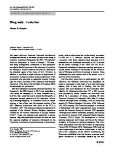
SelB of selenium sequenced and utilization genomes SelC traits were were analyzed found for occurrence to be signatures of SelA, for the Sec decoding B, trait,C, while SelD D defines and ybbB the overall 70) of maximum likelihood analysis and posterior probabilities (>0.90) of Bayesian analysis respectively. In all trees γ-proteobacteria are highlighted in red, β-proteobacteria in blue and Sinorhizobium meliloti (α-proteobacteria) in green. Red circles denote putative horizontal gene transfer events.
Genome Biology 2005, 6:R66
http://genomebiology.com/2005/6/8/R66
98 1.00
100
P. luminicum
75 100 100 1.00
88 0.99
ybbB
1.00
94 1.00 88
Figure 2 (see legend on previous page)
Genome Biology 2005, 6:R66
information
P. syringae P. aeruginosa P. putida B. bronchiseptica 99/1.00 E. coli 100 S. flexneri 1.00 S. thyphi 0.99 1.00 P. profundum S. oneidensis 1.00 N. europeae 99 B. bacteriovorus 1.00 D. psycrophila C. perfringes B. pseudomallei G. sulfurreducens W. succinogenes C. jejuni P. gingivalis D. vulgaris 76 1.00
interactions
79
96
refereed research
M. succiniciproducens H. influenzae P. multocida H. ducreyi 100 Y. pseudotuberculosis Y. pestis P. luminicum E. coli 72 97 S. flexneri 1.00 S. typhi P. profundum1 P. aeruginosa 0.93 P. putida 96 92 P. syringae 1.00 1.00 B. bronchiseptica S. oneidensis 1.00 92 B. mallei 85 100 B. pseudomallei 1.00 1.00 S. meliloti M. avium W. succinogenes 100 H. hepaticus 1.00 C. jejuni A. aeolicus D. psycrophila 100 T. denticola 1.00 E. faecalis T. tengcongensis C. perfringens P. profundum G. sulfurreducens N. europeae 100 B. bacteriovorus 1.00 D. vulgaris P. gingivalis S. termophilum M. maripaludis 100 M. jannaschii 1.00 M. kandleri H. sapiens (SPS1) 100 78 H. sapiens (SPS2) 100 1.00 1.00 D. melanogaster (SPS1) 1001.00 C. elegans 1.00 D. melanogaster (SPS2) 73
selD
deposited research
S. flexneri S. typhi E. coli Y. pestis Y. pseudotuberculosis P. luminicum S. oneidensis M. succiniciproducens H. ducreyi H. influenzae P. multocida P. aeruginosa P. putida B. pseudomallei B. mallei S. meliloti D. vulgaris G. sulfurreducens C. perfringens T. tengcongensis S. termophilus D. psycrophila M. avium T. denticola P. profundum C. jejuni W. succinogens H. hepaticus A. aeolicus M. jannaschii M. kandleri M. maripaludis C. elegans D. melanogaster H. sapiens
selC
reports
P. luminicum S. oneidensis P. multocida 93 H. influenzae 100 M. succiniciproducens 88 H. ducreyi 1.00 100 B. pseudomallei 71 1.00 B. mallei 1.00 S. meliloti 1.00 P. putida 100 P. aeruginosa 1.00 M. avium G. sulfurreducens 0.95 89 D. psycrophila 1.00 D. vulgaris T. tengcongensis 81 1.00 1.00 C. perfringens 100 0.99 S. thermophilum 1.00 W. succinogenes 100 C. jejuni 1.00 H. hepaticus P. profundum 100 T. denticola 1.00 A. aelicus H. sapiens 97 100 D. melanogaster 1.00 1.00 C. elegans M. maripaludis 100 M. jannaschii 1.00 M. kandleri 81
S. typhi
reviews
1.00
100 1.00
Romero et al. R66.5
100/1.00 E. coli 99 S.flexeneri 1.00 S. typhi 98 1.00 100 Y. pseudotuberculosis 73 Y. pestis 1.00 1.00
selB
100 E. coli 100 1.00S. flexeneri 1.00
M. succiniciproducens P. multocida H. influenzae 1.00 H. ducreyi 100 S. oneidensis 1.00 P. putida 100 1.00 P. aeruginosa 60 1.00 100 B. mallei 83 1.00 B. pseudomallei 0.60 S. melioti M. avium S. thermophilum T. tengcongensis C. perfringens D. vulgaris D. psycrophila G. sulfurreducens P. profundum 100 1.00 T. denticola W. succinogenes H. hepaticus C. jejuni A. aeolicus 1.00
Volume 6, Issue 8, Article R66
comment
100 Y. pestis 100 1.00 Y. pseudotuberculosis 1.00
selA
95
Genome Biology 2005,
R66.6 Genome Biology 2005,
Volume 6, Issue 8, Article R66
Romero et al.
The analysis of selA, selB, selC, selD and ybbB genes also revealed that, within the set of species that incorporate Sec, many, but not all, organisms, possess ybbB and vice versa. In other words, the set of species that incorporate Sec into protein overlaps with, but is different from, the set of species that possess ybbB (Figure 1). It is important to note that a lowidentity homolog to bacterial ybbB is present in Methanococcus jannaschii and Methanopyrus kandleri, and absent in other archaea, suggesting that this base modification might not be unique to bacteria. Finally, we investigated the presence of additional genes linked to the selenouridine synthesis trait by searching genomes for genes that occur in organisms possessing ybbB and are absent in organisms lacking ybbB. This search did not identify any additional gene associated with this trait. Thus, the overall analysis allows us to corroborate that the two products of these genes form a pathway with 2-selenouridine in the tRNA as the final product. However, only ybbB is the gene signature of this trait. On the other hand, the dual use of selenophosphate (for Sec decoding and 2-selenouridine biosynthesis) makes selD a signature of a broader trait of selenium utilization, and our data suggest that both Sec decoding and selenouridine traits are independently maintained, but both require selD.
Phylogeny of selA, selB, selC, selD and ybbB: evidence of horizontal gene transfer (HGT) of Sec-decoding and selenouridine synthesis traits The phylogenies of selA, selB, selC, selD and ybbB shown in Figure 2 are neither mutually coherent nor match the 'species tree' (Figure 1). This does not necessarily imply an error in the phylogenetic reconstruction since the evolutionary history of each gene could be different. Many nodes are mutually consistent across different methods and have high statistical support. Certain anomalous situations occur with distantly related organisms (deep nodes), which could be due to the limitations of these analyses. However, some of the inconsistencies may be considered as 'genuine' and raise HGT as the most likely alternative explanation. A striking observation is the clustering of P. profundum (a γproteobacterium) with T. denticola (a spirochete) at a basal position of the selA, selB and selC trees. This topology is consistent in various phylogenetic reconstruction methods and has high statistical support in all cases. The congruence of the trees sustains the idea that these genes were horizontally transferred to P. profundum. Several facts provide further support for this proposition. The P. profundum genome encodes four selenoproteins: two glycine reductases A, one glycine reductase B and selenophosphate synthetase. This selenoproteome is entirely distinct from that of γ-proteobacteria and very similar to that of T. denticola, which consists of glycine reductase A, two glycine reductases B, selenophosphate synthetase, glutathione peroxidase and thioredoxin. Furthermore, glycine reductase is absent in every other pro-
http://genomebiology.com/2005/6/8/R66
teobacterial genome. In addition, P. profundum is the single prokaryotic genome that has two selDs: one encodes a Seccontaining isoform that is located next to the selAB operon, on chromosome II; the second encodes a Cys-containing enzyme that is adjacent to ybbB on chromosome I. The phylogeny of selD places the Cys isoform within the γ-proteobacterial clade as expected according to the organismal phylogeny, whereas the Sec isoform does not cluster with T. denticola or with γ-proteobacteria. Altogether, these results indicate that it is highly unlikely that P. profundum has acquired the Sec-decoding trait by vertical descent, raising HGT as the obvious alternative. In addition, we analyzed the codon usage of selA and selB, looking for an anomalous pattern, using the method described by García-Vallvé [22]. These genes do not display biased values of codon usage with respect to the rest of the genes. This result could indicate that P. profundum has already adapted the codon usage of these genes to its internal values. A recent paper suggested that the compatible codon usage between foreign genes and recipient genomes increases the probability of HGT [23]. Since T. denticola and P. profundum do not share the same environment, it is likely that the HGT took place from a species of spirochete, a bacterial phylum exhibiting great variability in habitat and physiology [24]. An additional incongruence is observed when the selA, selB, selC and the species trees are compared among Pseudomomas spp. (γ-proteobacteria), Sinorhizobium meliloti (α-proteobacterium) and Burkholderia spp. (β-proteobacteria) (Figure 2). The evolutionary history of these genes is, however, difficult to solve. Conflicts relating to the selenouridine synthesis trait were also observed. The consistent and statistically supported cluster between Bordetella bronchiseptica (a β-proteobacterium) and Pseudomonas spp., within the γ-proteobacteria clade in both selD and ybbB gene trees, strongly suggests an event of HGT from Pseudomonas spp. to B. bronchiseptica. A situation that cannot be explained by vertical descent is also the cluster of Nitrosomonas europae (a β-proteobacterium) and Bdellovibrio bacteriovorus (δ-proteobacteria) in selD and ybbB phylogenies (Figure 2). The location of ybbB and selD genes also supports this possibility: while arranged in an operon in N. europeae and B. bacteriovorus; they are distant in the genomes of the other δ-proteobacteria (Figure 3). Furthermore, B. bacteriovorus is a predatory bacterium with a multiplication phase within many Gram-negative bacteria [25]. Thus, the ready access to the prey's genetic information and vice versa might be a possible explanation for this HGT event.
Discussion
The distribution of the Sec-decoding trait within the 'species tree' prompts the question of how it evolved. A supported conclusion from our data is the common origin of selA, selB,
Genome Biology 2005, 6:R66
Genome Biology 2005,
Volume 6, Issue 8, Article R66
M. kandleri M. jannaschii M. maripaludis
selA
W. succinogenes H. hepaticus C. jejuni G. sulfurreducens P-δ D. vulgaris D. psycrophila B. bacteriovorus P-γ P. syringae P. aeruginosa P. putida P. luminescens S. oneidensis S. typhi E. coli Y. pseudotuberculosis Y. pestis KIM S. flexneri P. profundum chr. I chr. II M. succiniciproducens P. multocida H. influenzae H. ducreyi P-β B. pseudomallei chr. II B. mallei chr. II B. bronchiseptica N. europaea S. meliloti P-α Firm C. perfringens T. tengcongensis
selC
Eury
selB
0
1
2
3
4 Size (Mbp)
5
6
7
28]. The emergence of the Sec-decoding trait before the division of the three domains has been previously postulated [18,29]. The evolution of the Sec insertion system only once is certainly the most parsimonious evolutionary scenario. However, this does not necessarily imply that every gene involved in Sec-decoding has a common origin. This is exemplified by selA: no clear ortholog has been found in Archaea and Eukarya. This suggests that the mechanism of Sec biosynthesis and insertion could have been adjusted during evolution.
Genome Biology 2005, 6:R66
information
selC and selD in the domain Bacteria. This is based on the absence of close paralogs for Sec-decoding genes in bacteria, the high bootstrap value for the bacterial node in all phylogenies, and the presence of bacterial sequence signatures in selA, selB, selC and selD sequences (see Additional data files). The phylogenies of selB, selC and selD also indicate that the archaeal and eukaryal Sec-decoding genes cluster together. This is further supported by the similar overall organization of the Sec-decoding machinery in Archaea and Eukarya [26-
interactions
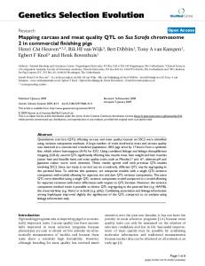
Figure Genome3location of selA, selB, selC, selD and ybbB Genome location of selA, selB, selC, selD and ybbB. Each bar represents one replicon of a species. On the vertical axis the species name, phylum, and domain are specified. The horizontal axis corresponds to the replicon size. Location of selA (yellow), selB (blue), selC (green), selD (red) and ybbB (black) is indicated; arrows denote direction of transcription.
refereed research
T. denticola P. gingivalis A. aeolicus Synechococcus sp. P. marinus
deposited research
Spiro Bact Aquif Cyano
ybbB
reports
M. avium S. thermophilum
selD
reviews
Bacteria
P-ε
Actin
Romero et al. R66.7
comment
Archaea
http://genomebiology.com/2005/6/8/R66
R66.8 Genome Biology 2005,
Volume 6, Issue 8, Article R66
Romero et al.
http://genomebiology.com/2005/6/8/R66
Assuming the common origin of the Sec-decoding trait, it is possible to sketch a scenario compatible with our results in order to explain the pattern of presence/absence of the Secdecoding trait. We propose that this pattern is the result of two mechanisms, primarily speciation and differential gene loss, with some contribution from HGT. Regarding the selenouridine synthesis trait, the results also suggest a common origin in the bacterial domain, as well as the possibility that 2selenouridine pathway can be acquired by HGT.
matching twofold codons might be a refinement in the base discrimination at the wobble position. The interaction of the first base of the anticodon with the third base of the codon plays an important role in the efficiency and accuracy of the translation process, suggesting that this base modification could be linked to certain aspects of codon usage. In any case, it should be stressed that ybbB null E. coli has no apparent phenotypic differences to wild type-E. coli and does not alter nonsense suppression phenotype [13].
An important issue in the evolution of Se utilization traits relates to the selective forces operating to maintain, loose or acquire the traits. Although it is not possible to draw conclusions, the search for a common biochemical, physiological or ecological trait in organisms possessing/lacking either or both traits provides interesting clues. The analysis of the prokaryotic selenoproteome revealed that formate dehydrogenase is present in most organisms capable of Sec decoding, exceptions being T. denticola, P. profundum, Clostridium perfringens and Thermoanerobacter tengcongensis [6]. Formate dehydrogenase plays a key role in anaerobic respiration. Indeed, most of these species are obligatory anaerobes or facultative aerobes; the sole exception was S. meliloti, a symbiotic nitrogen-fixing obligatory aerobe that lives in the oxygenlimited environment of the nodule [30]. Formate dehydrogenase is the single Sec-containing polypeptide encoded in the Sinorhizobium meliloti genome [6,30], suggesting that the presence of the trait may be important for respiration under conditions of restricted oxygen supply. On the other hand, glycine reductase is present in T. denticola, P. profundum and T. tengcongensis and several species of the genera Clostridium except C. perfringens. Glycine reductase is an enzymatic complex that allows certain anaerobic bacteria to conserve energy via a soluble substrate level phosphorylation system [31]. Sec is more reactive than Cys by virtue of the lower pKa and higher nucleophilicity of selenol group compared to that of the thiol group [12], and can increase the pH range at which certain enzymes are active [32]. This might have conferred a selective advantage improving catalytic efficiency of proteins.
One of the driving forces for the loss of the traits probably relates to the variability of selenium abundance in the environment. The absolute dependence of organisms on Se can compromise their existence if dietary Se becomes limiting. In these situations, enzymes containing Sec as catalytic residues could have evolved into Cys-containing proteins or, alternatively, both Sec-containing and Cys-containing forms could be maintained. This latter case is exemplified by the genome of M. maripaludis, which encodes several Sec-containing proteins and also homologs that contain cysteine in place of Sec. In a medium that contains adequate amounts of selenium, this organism represses the synthesis of the cysteine homologs, but this repression is not observed in a mutant with disrupted selB [35], suggesting that the cysteine homologs are a backup system in case of selenium scarcity. Nevertheless, the existence of organisms carrying only one of the selenium-utilization traits suggests that selenium availability might not be the sole factor involved in the loss of either trait. It is also possible that the higher reactivity of selenium over sulfur in biological molecules might have had a role in counterselecting the pervasive use of Sec and/or selenouridine in living systems.
Regarding selective forces operating on the evolution of the selenouridine synthesis trait, we begin from the fact that synthesis of 2-selenouridine is carried out exclusively at the wobble position (first of the anticodon) of the tRNAs for lysine, glutamate and glutamine (the only amino acids encoded by twofold purine-ending codons). Several modifications of this base have been reported to be essential for correct decoding; thiouridine, in particular, would convert the base into an ionized form that would favor pairing with A and G, and avoid pairing with U or C, contributing to the discrimination of twofold codons ending in purine from those ending in pyrimidine [33]. The low pKa value of 2-selenouridine of these tRNAs would be consistent with this argument and it has been suggested that this would also favor base-pairing with G [34]. Thus, we postulate that selenium modification of tRNAs
Conclusion
This paper provides an organismal map for Sec-decoding and 2-selenouridine synthesis traits within the tree of life, and defines selB and selC as the gene signature of the Sec-decoding trait, ybbB as the gene signature of selenouridine synthesis, with selD defining overall selenium utilization. We show that the set of species that incorporate Sec overlaps with, yet is distinct from, the set of species that synthesize 2selenouridine, and our data suggest that Sec decoding and 2selenouridine traits can be independently maintained, and both require selD. Analysis of the phylogenies of the Sec-decoding and 2-selenouridine synthesis genes provides evidence for the ancient origin of these traits and demonstrates that their evolution is a highly dynamic process that occurs at different evolutionary levels, namely phylum, class, order, family, genera, and even species. We show that this process can be explained as the result of speciation and differential gene loss, and provide conclusive evidence that the loss of these traits is not irreversible as previously thought, and that entire sets of genes can be acquired by HGT. It is striking that the genetic code of an
Genome Biology 2005, 6:R66
Genome Biology 2005,
organism and the amino-acid repertoire can be 'laterally' expanded.
The final alignment sets were the following: i) raw alignments using each software with two different sets of parameters ii) 'sub-alignments' obtained removing the unstable blocks from the raw alignments using g-blocks software, and iii) 'subalignment' obtained using the '-score' option of T-coffee for evaluation of the alignment, then the low scoring regions were removed manually.
The study of selenium-utilization traits, which directly associate protein synthesis with a discrete set of genes, can contribute to the understanding of basic questions regarding the evolution of the genetic code and the translation machinery.
Materials and methods Complete genome sequences of 194 prokaryotes were retrieved from GenBank [36] as of 20 October 2004, representing 151 species.
EF-Tu and EF-1α were recovered using a similar approach to that described for selA, selB, selD and ybbB, and aligned using T-coffee. An all-against-all BLAST search was performed sequentially and best reciprocal hits were identified as putative orthologs. A set of nine genes was obtained. These sequences were aligned with ClustalW and concatenated. In all cases we used the neighbor-joining (NJ) algorithm to build the trees from different distance matrices using MEGA software [43]. In the case of SSU-rRNA, the Tamura-Nei (TN93) distance with pairwise-deletion was calculated. For the amino-acid alignments we use the JTT transition matrix.
Genome Biology 2005, 6:R66
information
Several phylogenetic gene trees were built using different inference methods performed on different sequence alignments. Sequences were aligned with T-coffee version 1.37 [40], ClustalW 1.8 and Dialign-2 [41] using different parameters. The 'score' option of the T-coffee software was enabled to assess alignment quality. The alignments were then visually inspected, compared and uncertain sites were removed. In another approach, we applied the g-blocks software [42] to remove unstable blocks with 2 different sets of parameters.
Different species trees were initially constructed, based on small-subunit (SSU) rRNA, EF-Tu/EF-1α (a highly conserved translation elongation factor present in all organisms), and a concatenated set of 9 ortholog sequences present in all prokaryotes. A set of aligned SSU-rRNA sequences was retrieved from the Ribosomal Database Project (RDP) [46], release 2.1, missing sequences were retrieved from Genbank and aligned against the set from the RDP using the profile option of ClustalW.
interactions
Alignments and phylogenetic reconstruction of gene trees
Species tree refereed research
All sequences are provided aligned in the Additional data files.
Almost all trees yielded similar topologies and, more important, all of them supported the conclusions. In particular, the HGT results were reproduced with any of the alignments and phylogenetic trees.
deposited research
Most sequences of selC were retrieved from GenBank or identified using tRNAscan software with default parameters [38,39]. The sequences of Wolinella succinogenes, Helicobacter hepaticus, Burkholderia mallei and Thermoanaerobacter tencongensis, were found changing the parameters to 'Cove-only search mode' and lowering the tRNA Cove cutoff score to 6.
For each of these alignments, we applied several phylogenetic reconstruction methods including Neighbor Joining using MEGA software [43], Maximum Likelihood (ML) using phyML 2.4 software [44] and Bayesian approaches using MrBayes 3.0b4 [45]. For each of these methods, different transition matrices (WAG and JTT) and evolutionary models were tested. In total, more than 80 trees were analyzed for each gene. The gene trees presented in Figure 2 were built using the T-coffee alignment evaluated with the '-score' option and manually refined. The ML and Bayesian trees were built using WAG matrix and gamma+invar model of evolutionary change. In the ML method, the assessment of node reliability was done using 100 bootstrap replicates. In the case of Bayesian analyses, four heated Markov chains were started from random trees and run for 1,000,000 generations each. Chains were sampled every 500 generations to assure independence. Sample points prior to reach stationary (200) were discarded as 'burn-in'.
reports
Annotated sequences corresponding to selA, selB, selD and ybbB prokaryotic genes were retrieved from GenBank, and used as queries to perform local BLAST searches across a database generated with the 194 genomes. For selA, selD and ybbB, hits with an e-value below e-15 were recovered; for selB, the cutoff e-value was e-30 to decrease the number of hits corresponding to other translation factors. A total of 242 selB, 48 selA, 47 selD and 25 ybbB sequences were recovered. The sequences were aligned using ClustalW [37], a raw phylogenetic analysis was conducted and clear nonorthologous sequences were discarded. This dataset was manually curated, and the number of sequences was reduced to 29 selA, 32 selB, 41 selD, and 21 ybbB sequences. These datasets were used as queries for BLAST searches but no new sequences were identified. Finally, only one sequence by strain was included and the set was supplemented with sequences of three representative eukaryotes (Caenorhabditis elegans, Drosophila melanogaster and Homo sapiens).
Romero et al. R66.9
reviews
Sequences of selA, selB, selC, selD and ybbB
Volume 6, Issue 8, Article R66
comment
http://genomebiology.com/2005/6/8/R66
R66.10 Genome Biology 2005,
Volume 6, Issue 8, Article R66
Romero et al.
The different 'species trees' display some discrepancies. In any case, the conclusions drawn are maintained with any of the above mentioned 'species tree'. The SSU-rRNA was finally adopted because it is by large the commonest used, and trees inferred for this gene are sound descriptors of the general evolutionary history of prokaryotes. This tree also recovers the major groups described in Bergey's Manual [47].
http://genomebiology.com/2005/6/8/R66
7. 8.
9. 10.
Codon bias analyses Codon bias was evaluated according to the method described in [22]. This method uses the Mahalanobis distance measure for detecting outliers in a multivariate distribution.
Search for additional genes linked to Se-U trait To study the possible association of a certain gene with ybbB we run an all-against-all BLAST search with an e-value threshold of e-10 among organisms carrying ybbB, to pick up homologs present in all these genomes. Then we used this set of genes to run a new BLAST search against a control set of closely related species lacking a ybbB homolog. This search detected no gene. When we excluded Sec-decoding species from the control set, we were able to recover a single gene: selD.
11.
12. 13.
14.
15. 16.
17.
Additional data files
Additional data are available with the online version of this paper. Additional data file 1 is a table containing the gene locations of selA, selB, selC selD and ybbB in the genomes analyzed in this work. Additional data file 2 contains the sequence alignments of selA, selB, selC selD and ybbB of the genomes analyzed in this work.
18. 19. 20.
genomes Click Sequence ybbB Additional A table here incontaining the analyzed alignments for data genomes filefilein the 2 1 this analyzed of gene selA, work. location selB, in this selC ofwork selA, selD selB, and ybbB selC selD of the and 21.
Acknowledgements This work was supported by Fogarty International Research Collaboration Award TW006959 and NIH GM061603 grant to V.N.G. We thank Dr Alexey Lobanov for help in identifying tRNASec sequences and Héctor Musto (Universidad de la República, Uruguay) for critical reading of the manuscript. We also thank the faculty, teaching assistants and students of the 'Workshop on Molecular Evolution' 2004 at the Marine Biological Laboratory, Woodshole, attended by HR, for valuable general discussion.
22. 23.
24.
References 1. 2. 3. 4.
5. 6.
Low SC, Berry MJ: Knowing when not to stop: selenocysteine incorporation in eukaryotes. Trends Biochem Sci 1996, 21:203-208. Hatfield DL, Gladyshev VN: How selenium has altered our understanding of the genetic code. Mol Cell Biol 2002, 22:3565-3576. Driscoll DM, Copeland PR: Mechanism and regulation of selenoprotein synthesis. Annu Rev Nutr 2003, 23:17-40. Lee SR, Bar-Noy S, Kwon J, Levine RL, Stadtman TC, Rhee SG: Mammalian thioredoxin reductase: oxidation of the C-terminal cysteine/selenocysteine active site forms a thioselenide, and replacement of selenium with sulfur markedly reduces catalytic activity. Proc Natl Acad Sci USA 2000, 97:2521-2526. Kryukov GV, Castellano S, Novoselov SV, Lobanov AV, Zehtab O, Guigo R, Gladyshev VN: Characterization of mammalian selenoproteomes. Science 2003, 300:1439-1443. Kryukov GV, Gladyshev VN: The prokaryotic selenoproteome. EMBO Rep 2004, 5:538-543.
25.
26.
27. 28. 29. 30.
Leinfelder W, Zehelein E, Mandrand-Berthelot MA, Bock A: Gene for a novel tRNA species that accepts L-serine and cotranslationally inserts selenocysteine. Nature 1988, 331:723-725. Forchhammer K, Rucknagel KP, Bock A: Purification and biochemical characterization of SELB, a translation factor involved in selenoprotein synthesis. J Biol Chem 1990, 265:9346-9350. Bock A: Biosynthesis of selenoproteins - an overview. Biofactors 2000, 11:77-78. Copeland PR, Driscoll DM: Purification, redox sensitivity, and RNA binding properties of SECIS-binding protein 2, a protein involved in selenoprotein biosynthesis. J Biol Chem 1999, 274:25447-25454. Carlson BA, Xu XM, Kryukov GV, Rao M, Berry MJ, Gladyshev VN, Hatfield DL: Identification and characterization of phosphoseryl-tRNA[Ser]Sec kinase. Proc Natl Acad Sci U S A 2004, 101:12848-12853. Stadtman TC: Selenocysteine. Annu Rev Biochem 1996, 65:83-100. Wolfe MD, Ahmed F, Lacourciere GM, Lauhon CT, Stadtman TC, Larson TJ: Functional diversity of the rhodanese homology domain: the Escherichia coli ybbB gene encodes a selenophosphate-dependent tRNA 2-selenouridine synthase. J Biol Chem 2004, 279:1801-1809. Castellano S, Morozova N, Morey M, Berry MJ, Serras F, Corominas M, Guigo R: In silico identification of novel selenoproteins in the Drosophila melanogaster genome. EMBO Rep 2001, 2:697-702. Lescure A, Gautheret D, Carbon P, Krol A: Novel selenoproteins identified in silico and in vivo by using a conserved RNA structural motif. J Biol Chem 1999, 274:38147-38154. Martin-Romero FJ, Kryukov GV, Lobanov AV, Carlson BA, Lee BJ, Gladyshev VN, Hatfield DL: Selenium metabolism in Drosophila: selenoproteins, selenoprotein mRNA expression, fertility, and mortality. J Biol Chem 2001, 276:29798-29804. Jukes TH: Genetic code 1990. Outlook. Experientia 1990, 46:1149-1157. Bock A, Forchhammer K, Heider J, Baron C: Selenoprotein synthesis: an expansion of the genetic code. Trends Biochem Sci 1991, 16:463-467. Gladyshev VN, Kryukov GV: Evolution of selenocysteine-containing proteins: significance of identification and functional characterization of selenoproteins. Biofactors 2001, 14:87-92. Venter JC, Remington K, Heidelberg JF, Halpern AL, Rusch D, Eisen JA, Wu D, Paulsen I, Nelson KE, Nelson W, et al.: Environmental genome shotgun sequencing of the Sargasso Sea. Science 2004, 304:66-74. Deng W, Burland V, Plunkett G 3rd, Boutin A, Mayhew GF, Liss P, Perna NT, Rose DJ, Mau B, Zhou S, et al.: Genome sequence of Yersinia pestis KIM. J Bacteriol 2002, 184:4601-4611. Garcia-Vallve S, Romeu A, Palau J: Horizontal gene transfer in bacterial and archaeal complete genomes. Genome Res 2000, 10:1719-1725. Medrano-Soto A, Moreno-Hagelsieb G, Vinuesa P, Christen JA, Collado-Vides J: Successful lateral transfer requires codon usage compatibility between foreign genes and recipient genomes. Mol Biol Evol 2004, 21:1884-1894. Seshadri R, Myers GS, Tettelin H, Eisen JA, Heidelberg JF, Dodson RJ, Davidsen TM, DeBoy RT, Fouts DE, Haft DH, et al.: Comparison of the genome of the oral pathogen Treponema denticola with other spirochete genomes. Proc Natl Acad Sci USA 2004, 101:5646-5651. Rendulic S, Jagtap P, Rosinus A, Eppinger M, Baar C, Lanz C, Keller H, Lambert C, Evans KJ, Goesmann A, et al.: A predator unmasked: life cycle of Bdellovibrio bacteriovorus from a genomic perspective. Science 2004, 303:689-692. Fagegaltier D, Hubert N, Yamada K, Mizutani T, Carbon P, Krol A: Characterization of mSelB, a novel mammalian elongation factor for selenoprotein translation. EMBO J 2000, 19:4796-4805. Hubert N, Sturchler C, Westhof E, Carbon P, Krol A: The 9/4 secondary structure of eukaryotic selenocysteine tRNA: more pieces of evidence. RNA 1998, 4:1029-1033. Foster CB: Selenoproteins and the metabolic features of the archaeal ancestor of eukaryotes. Mol Biol Evol 2005, 22:383-386. Rao M, Carlson BA, Novoselov SV, Weeks DP, Gladyshev VN, Hatfield DL: Chlamydomonas reinhardtii selenocysteine tRNA[Ser]Sec. RNA 2003, 9:923-930. Barnett MJ, Fisher RF, Jones T, Komp C, Abola AP, Barloy-Hubler F,
Genome Biology 2005, 6:R66
http://genomebiology.com/2005/6/8/R66
32.
33. 34.
36. 37.
38.
40. 41.
43. 44. 45.
47.
refereed research
46.
deposited research
42.
reports
39.
Romero et al. R66.11
reviews
35.
Bowser L, Capela D, Galibert F, Gouzy J, et al.: Nucleotide sequence and predicted functions of the entire Sinorhizobium meliloti pSymA megaplasmid. Proc Natl Acad Sci USA 2001, 98:9883-9888. Andreesen JR: Glycine reductase mechanism. Curr Opin Chem Biol 2004, 8:454-461. Gromer S, Johansson L, Bauer H, Arscott LD, Rauch S, Ballou DP, Williams CH Jr, Schirmer RH, Arner ES: Active sites of thioredoxin reductases: why selenoproteins? Proc Natl Acad Sci USA 2003, 100:12618-12623. Takai K, Yokoyama S: Roles of 5-substituents of tRNA wobble uridines in the recognition of purine-ending codons. Nucleic Acids Res 2003, 31:6383-6391. Ching WM: Characterization of selenium-containing tRNAGlu from Clostridium sticklandii. Arch Biochem Biophys 1986, 244:137-146. Rother M, Mathes I, Lottspeich F, Bock A: Inactivation of the selB gene in Methanococcus maripaludis: effect on synthesis of selenoproteins and their sulfur-containing homologs. J Bacteriol 2003, 185:107-114. GenBank [http://ftp.ncbi.nlm.nih.gov/genomes/Bacteria] Thompson JD, Higgins DG, Gibson TJ: CLUSTAL W: improving the sensitivity of progressive multiple sequence alignment through sequence weighting, position-specific gap penalties and weight matrix choice. Nucleic Acids Res 1994, 22:4673-4680. Lowe TM, Eddy SR: tRNAscan-SE: A program for improved detection of transfer RNA genes in genomic sequence. Nucl Acids Res 1997, 25:955-964. tRNAscan [ftp://ftp.genetics.wustl.edu/pub/eddy/software/tRNAs can-SE.tar.Z] Notredame C, Higgins DG, Heringa J: T-Coffee: A novel method for fast and accurate multiple sequence alignment. J Mol Biol 2000, 302:205-217. Morgenstern B: DIALIGN 2: improvement of the segment-tosegment approach to multiple sequence alignment. Bioinformatics 1999, 15:211-218. Castresana J: Selection of conserved blocks from multiple alignments for their use in phylogenetic analysis. Mol Biol Evol 2000, 17:540-552. Kumar S, Tamura K, Jakobsen IB, Nei M: MEGA2: molecular evolutionary genetics analysis software. Bioinformatics 2001, 17:1244-1245. Guindon S, Gascuel O: A simple, fast, and accurate algorithm to estimate large phylogenies by maximum likelihood. Syst Biol 2003, 52:696-704. Ronquist F, Huelsenbeck JP: MrBayes 3: Bayesian phylogenetic inference under mixed models. Bioinformatics 2003, 19:1572-1574. Cole JR, Chai B, Farris RJ, Wang Q, Kulam SA, McGarrell DM, Garrity GM, Tiedje JM: The Ribosomal Database Project (RDP-II): sequences and tools for high-throughput rRNA analysis. Nucleic Acids Res 2005, 33:D294-D296. Taxonomic Outline of the Prokaryotes [http://www.bergeys outline.com/]
Volume 6, Issue 8, Article R66
comment
31.
Genome Biology 2005,
interactions information
Genome Biology 2005, 6:R66