1*Senior Research Engineer and Faculty, S. Abstract. Heat pipes are a closed ... São José dos Campos/SP - 18,19 e 20 de agosto de 201 ental Investigation of ...
The heat transfer degradation as a function of time at various gas approach ..... D.P (2002), Fundamentals of Heat and Mass Transfer, 5th edition, John ... Schlichting, H. (1979), Boundary Layer Theory, 7th edition, McGraw-Hill, New York.
(structural parts, pipe line elements, sanitary appliance etc.). In Fig. 1 some ... made by tube hydroforming are shown: rear axle component of the car, sanitary.
and MRU Delta 1600 V gas analyzer was used to measure exhaust gas emissions. Dynamometer and ... cryogenic tank which has capacity of 12.5 m3 and fixed on 200 bar pressure. A check ... second stage decreased the 12-14 to 1-0 bar.
carriers reported that about 40% of all damage comes from platform or ground handling and maintenance [2]. Up to now failure caused by delamination is ...
separation in the diffuser. More details are given in the caption of figure 1. Similar modelling of decelerating flow is still employed e.g. Marxen et al. [5] and Uruba.
ZHANG Jian-min, YANG Qing, WANG Yu-rong, XU Wei-lin, CHEN Jian-gang. State Key Laboratory of Hydraulics and Mountain River Engineering, Sichuan ...
The HVAC system includes air handling unit (AHU), supply and exhaust air ... Therefore the most significant noise sources are the air handler equipment and the.
Pasteurellosis among rabbit colonies causes a considerable economical loss especially in labora- tory health departments, and, besides, interferes with the col-.
accidents from the in-vessel components, to 150 kPa (abs). This is key ... densation at atmospheric pressure, it emerged that the attention of researchers ..... and steam flow rate excursions) when starting a condensation test run within the condensa
Brazil, the nuclear renaissance can be seen in the completion of construction of .... The TRIGA reactor is the only nuclear reactor in this category that offers true.
speed (1000Y2000 rpm), feed rate (250Y350 mm/min) and depth of cut (0.3Y0.9 mm) were ... mm/min and spindle speed of 2000 rpm in the direction of the feed.
At some level of input torque, the pressure on the suction side of the stator ... through suitable cavitation numbers and net positive suction head (NPSH) values.
Revolution rpm. -. Revolution per minute. RPD. -. Robust parameter design sec ..... Dimla (1999) classified the application of perceptron-type neural networks to .... simplicity, robustness, and good convergence properties (Kennedy and.
Aug 2, 2011 - in magnetic fluids: Influence of grainâgrain interaction ... investigations of field-induced anisotropy are useful to characterize the physical state ...
Experimental investigation of helium migration in an fcc aluminum matrix. Benny Glam a,b,*, Daniel ... b Ben-Gurion University of the Negev, Beer-Sheva, Israel. a r t i c l e i n f o ... In order to see the migration process of helium and bubble ...
in evaporators, but it was not possible to consider all of them, so parameters
including,. Reynolds ... types of evaporators are, forced circulation evaporators,.
Terminal S.A. (PCT) â a subsidiary of Cosco Pacific Limited) is presented and evaluated. ... performed in different quay cranes, in order to compare if there are ...
Tribology in Industry www.tribology.fink.rs ... 2016 Published by Faculty of Engineering. Corresponding ... S. Senhadji et al., Tribology in Industry Vol. 38, No.
The original message bandwidth. b(t). The binary ..... Then despreads it into the original data bandwidth at the receiver by using ...... fx15=sum(abs(b15-Q15)/2);.
ject detection for the visually impaired, as well as to study autism and the ... namely visual attention modelling, salient object detection and salient ... Several studies argue that such a mechanism is likely to be present in the human system .....
2008) and Prony's series (McDaniel J.G., 2000), and most concentrated on ... X Cong res so N acion al de Eng enha ria M e câni ca, 20 a 24 d e maio de 201 8, ...
the disadvantages of biodiesel usage in diesel engines, many experimental and ..... engine modifications (at the injection pump timing , prescribed for D100) [41]. ..... to biodiesel therefore has to be done with great care and only if a stable sourc
Experimental Investigation of Saliency Processing in ...
Visual Saliency and Attention Experimental Investigation of Saliency Processing in ADHD - Initial Findings
Benjamin Cowley, Cognitive Science Unit, Department of Behavioural Sciences
C O G N I T I V E S C I E NC E U N I V E R S I T Y OF H E L S I N K I
UNIVERSITY OF HELSINKI, FINLAND
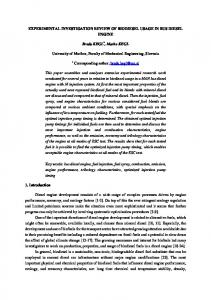
We investigate the role of visual salience processing in attention deficit disorders (ADHD) using a novel Event-Related Potential (ERP) protocol. Saliency refers to the order of importance attached to visual features by the attention system, making certain features ‘stand out’, with processing time 25-50ms. Task-related or top-down attention can modulate saliency but is much slower, 200ms or more [1].
a strong effect on processing, as expected. In general, amplitudes are higher for Incongruent trials, reflecting that an additional neuronal recruitment is required.
Findings of brain structural alteration in ADHD include significantly smaller volumes in the dorsolateral prefrontal cortex (dPFC) and associated regions: caudate, pallidum, anterior cingulate, and cerebellum [2]. This motivates the study of attentional neural processing in a task requiring top-down redirection of attention. To simulate this, our protocol manipulates the saliency of targets in a choice-response task, testing interference inhibition at the task-response level, and saliency-processing at the pre-attentive level. Here we present initial findings.
RESULTS On behavioural measures, Patients generally performed worse than Controls. In all hit trials, Patients had higher response time means and variability; in all conditions they had lower hit rates and higher false alarms; their d’prime index of sensitivity was lower overall: though it was higher for incongruent trials. This exception might be explained by the EEG results. Of all these comparisons, statistically significant ones are shown in Figure 2. ERPs from four conditions show that Incongruent stimuli had
Figure 1: ERPs from healthy controls and ADHD adults, across four conditions and eight bilateral electrodes (left hemisphere above - odd numbered electrodes; right hemisphere below - even numbered electrodes). Green highlighting shows where groups were different in all four conditions for 50ms timeslices, statistically significant at the p