© 2003 Nature Publishing Group http://www.nature.com/natureneuroscience
ARTICLES
Expert face processing requires visual input to the right hemisphere during infancy Richard Le Grand1, Catherine J Mondloch1, Daphne Maurer1,2 & Henry P Brent2 Adult expertise in face processing is mediated largely by neural networks in the right hemisphere. Here we evaluate the contribution of early visual input in establishing this neural substrate. We compared visually normal individuals to patients for whom visual input had been restricted mainly to one hemisphere during infancy. We show that early deprivation of visual input to the right hemisphere severely impairs the development of expert face processing, whereas deprivation restricted mainly to the left hemisphere does not. Our results indicate that the neural circuitry responsible for adults’ face expertise is not pre-specified, but requires early visual experience. However, the two hemispheres are not equipotent: only the right hemisphere is capable of using the early input to develop expertise at face processing.
Adults can recognize thousands of faces at a glance and are thus considered ‘experts’ at processing faces1. This ability requires encoding information about subtle differences among individual faces such as the shape of facial features (featural processing), the shape of the face (contour processing) and differences in the spacing among facial features, such as the distance between the eyes (a type of configural processing commonly referred to as second-order relational processing)2. Although adults use all three types of information to recognize faces, expertise in processing second-order relations is especially important3 and continues to improve past 14 years of age4. The right hemisphere is critically involved in processing secondorder relations. In adults, areas of the occipito-temporal cortex (including the ‘fusiform face area’) are more active when viewing faces versus a variety of non-face objects with which adults do not have the same expertise5,6. This face-sensitive activation is typically larger in the right than in the left hemisphere5,6, especially when the task encourages attending to the entire face rather than individual features7,8. Lesions involving these areas in both hemispheres, or just the right hemisphere, can lead to impairment in face recognition (prosopagnosia)9, which becomes more severe when faces must be recognized on the basis of second-order relational cues10. By 9 months of age, babies show evidence of similar hemispheric specialization. For example, only when face or non-face stimuli are presented to the right hemisphere do they show sensitivity to spacing information11,12. Perhaps as a result, infants learn to discriminate faces more rapidly if the stimuli are presented to the right hemisphere than to the left13. Together, these results support the existence of a neural substrate within the right hemisphere that is involved in processing spacing among features from infancy, and that underlies adults’ expertise in face processing. Some have argued for the existence of an innate face module that is pre-specified in the genome14. In a previous study,
however, we showed that early visual experience is necessary for the development of expert face processing. Deprivation of early visual input to both hemispheres, by dense bilateral congenital cataract, severely impairs the later development of sensitivity to secondorder relations in faces15. In the present study, we evaluated the degree of plasticity in the lateralization of networks underlying expert face processing by measuring the effects of early visual deprivation to the right versus left hemisphere. To do so, we took advantage of the fact that during infancy, visual input to each eye is predominantly transmitted to the contralateral hemisphere. During the first six months of life, the monocular visual field expands from the center outward, and sensitivity to stimuli in the temporal visual field develops much faster than sensitivity to stimuli in the nasal visual field16. Images from the temporal visual field are cast on the nasal hemi-retina, and its fibers project to the contralateral cerebral hemisphere. Unlike the adult nervous system, there appears to be no functional integration of visual information across the corpus callosum during early infancy. Cortically mediated transfer of visual information between the hemispheres is not evident before 24 months of age13,17–19. Therefore, during early infancy, input to the right hemisphere comes primarily from the left eye and input to the left hemisphere comes primarily from the right eye. A similar contralateral bias has been documented in anatomical and electrophysiological studies of infant kittens and ferrets20,21. As a consequence of this bias, a unilateral congenital cataract that blocks all patterned input to the left eye causes deprivation of input mainly to the right hemisphere, whereas a unilateral congenital cataract in an infant’s right eye causes deprivation of input mainly to the left hemisphere. After treatment by surgical removal of the cataract and fitting with a compensatory contact lens, input to both hemispheres is restored. Here we tested the developmental consequences of an initial period of deprivation affecting mainly the right or left hemisphere.
1Department
of Psychology, McMaster University,1280 Main Street West, Hamilton, Ontario L8S 4K1, Canada. 2The Hospital for Sick Children, 555 University Avenue, Toronto, Ontario M5G 1X8, Canada. Correspondence should be addressed to D.M. (
[email protected]). Published online 7 September 2003; doi:10.1038/nn1121
1108
VOLUME 6 | NUMBER 10 | OCTOBER 2003 NATURE NEUROSCIENCE
ARTICLES
© 2003 Nature Publishing Group http://www.nature.com/natureneuroscience
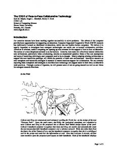
Figure 1 The face stimuli. (a) Featural stimulus set: variation in individual features (eyes and mouth). (b) Contour stimulus set: variation in the external contour of the face. (c) Spacing stimulus set: variation in spacing of the eyes and between the eyes and mouth.
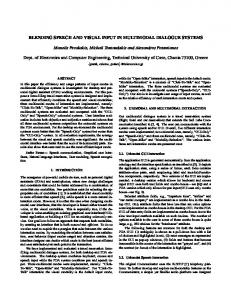
below the age norm). Thus, normal performance by a patient is indicated by a Z-score near zero. We found that early visual deprivation affecting the right hemisphere impairs the later development of sensitivity to second-order relational information in faces, but deprivation affecting the left hemisphere does not. Visual deprivation to either hemisphere had no apparent effect on the development of sensitivity to the shape of the internal features or the external contour.
We tested 20 patients treated for unilateral congenital cataract that blocked all patterned input to either the left eye (LE, n = 10) or the right eye (RE, n = 10; see Table 1 for patient details). Testing occurred later in life and after many (at least 8) years of visual experience to allow for recovery from the initial monocular deprivation. Testing was binocular; thus patients were able to use their unaffected eye when performing the task. This design allowed us to evaluate whether initial deprivation affecting the right hemisphere prevents the later development of expert face processing, despite years of visual input being available to both hemispheres beginning later in infancy, via both the expanded visual field of the unaffected eye and the recovery of the treated eye. Patients made same/different judgments about pairs of faces that were presented sequentially and that differed only in the shape of the eyes and mouth (featural set), only in the shape of the external contour (contour set) or only in the spacing among the internal features (spacing set; Fig. 1). The three stimulus sets were presented in separate blocks, with all sets first presented in an upright orientation, followed by an inverted orientation (see Methods section). In previous research, we showed that the three sets tap different aspects of face processing, which develop at different rates4,22. Moreover, inverting the stimuli greatly disrupts visually normal adults’ ability to discriminate faces in the spacing set, but not in the featural or contour sets15,22. These results are consistent with previous findings that inversion disrupts second-order relational processing but has little or no effect on other types of face processing23. Because face processing skills continue to improve through adolescence4, the mean accuracy and reaction time for each patient for each condition were compared to the mean of a group of 36 subjects of the same age with a normal visual history and then converted to Z-scores (units of standard deviation (s.d.) above or
RESULTS Accuracy Mean accuracy Z-scores for each condition are shown in Fig. 2 (see Supplementary Table 1 online for mean accuracy scores). To determine whether either patient group showed a deficit in any condition (performed worse than an expected mean Z-score of zero), we conducted one-sample t-tests. A Bonferroni correction was applied for the number of comparisons (adjusted alpha = 0.008). For the featural and contour sets, both patient groups performed normally whether the faces were upright or inverted (all, P > 0.1). For the spacing set, patients in the RE (left-hemisphere deprivation) group also performed normally on both orientations (both, P > 0.1). In contrast, patients in the LE (right-hemisphere deprivation) group were severely impaired on distinguishing faces from the upright spacing set (t9 = –4.94, P < 0.001; Fig. 2a). The LE group had a mean Z-score of –1.78, a value equivalent to the performance of the lowest 4% of the normal population. The second-order relational deficit in the LE group was manifested despite binocular testing (with the non-deprived right eye available) and did not correlate with acuity in either eye (both, P > 0.1). The deficit was evident even in cases when the initial deprivation lasted as little as 2–3 months (Fig. 3). The LE group also showed a small but significant impairment of about 0.5 s.d. on the inverted spacing set (t9 = –3.45, P < 0.007; Fig. 2b). We obtained similar results for the one-sample t-tests when accuracy scores were converted to the sensitivity statistic d′ (see Supplementary Notes and Supplementary Fig. 1 online). An analysis of variance (ANOVA) with group, stimulus set and orientation as factors confirmed that the LE and RE groups differed only on the spacing set. The ANOVA showed a significant interaction between group and stimulus set (F2,36 = 3.83, P < 0.01) that did not interact with orientation. Analysis of simple effects indicated that the two patient groups did not differ on the featural and contour sets (both, P > 0.1). They differed significantly only on the spacing set (F1,18 = 10.58, P < 0.01), with the LE (right-hemisphere deprivation) group performing worse than the RE (left-hemisphere deprivation) group. The same results were obtained when we used standardized d′ statistics as the dependent measure (see Supplementary Notes online).
Table 1 Details of the two patient groups Group
n
Age at test (yrs.) mean (range)
Duration of deprivation (d) Acuity of non-deprived eyea mean (range) median (range)
Acuity of deprived eyea median (range)
Patchingb (h/d) mean (range)
LE
10
17 (8–29)
199 (43–863)
20/20 (20/20–20/25)
20/200 (20/50–20/200)
4 (0.5–7)
RE
10
15 (9–23)
201 (56–483)
20/25 (20/16–20/32)
20/200 (20/32–20/200)
3 (0–5)
LE, left eye deprived group (right hemisphere deprivation); RE, right eye deprived group (left hemisphere deprivation). aAcuity
measured at time of test. bMean number of hours per day of patching the non-deprived eye until the age of 5 years.
NATURE NEUROSCIENCE VOLUME 6 | NUMBER 10 | OCTOBER 2003
1109
ARTICLES
© 2003 Nature Publishing Group http://www.nature.com/natureneuroscience
Figure 2 The effect of early left versus right hemisphere deprivation on face processing. (a) Upright orientation and (b) inverted orientation. Plotted are the mean accuracy Z-scores, which represent the difference between patients and age norms, for detecting featural, contour and spacing changes. Data are shown for the patient groups with deprivation affecting mainly the right hemisphere (LE group) or mainly the left hemisphere (RE group). A value of zero represents the normal mean, and negative values indicate deficits in units of standard deviation (s.d.) from the normal mean. The area outside the shaded portion (± 1.3 s.d.) represents the highest and lowest 10% of the normal population.
Previously, we documented a deficit in sensitivity to second-order relations of faces in patients treated for bilateral congenital cataract15. To compare the effect of early visual deprivation to both hemispheres versus that to the right hemisphere alone, we carried out a planned comparison between a group of ten patients treated for bilateral cataract (BE, both eyes group) and the LE group on Z-scores from the spacing set for both orientations. The LE and BE groups were equally impaired on the spacing set (Fig. 3), and both showed larger deficits for the upright than the inverted orientation. This was shown by a significant effect of orientation on the size of the deficit for the spacing set (F1,18 = 22.76, P < 0.001), but no significant effect of group or interaction (both, P > 0.1). The analyses showed similar results for standardized d′ scores (see Supplementary Notes online). Reaction times For each patient in each condition, median reaction times on correct trials were calculated and converted to standardized Z-scores based on age norms. One-sample t-tests revealed that both the LE and RE groups had normal response times on the three stimulus sets for both orientations (all, P > 0.2). These data demonstrate that the LE (righthemisphere deprivation) patients’ impairment on second-order relational processing (as shown by accuracy scores) cannot be attributed to a speed–accuracy trade-off (Table 2).
visual acuity because the size of the face processing deficit was not correlated with visual acuity and because patients could use their non-deprived eye, which had normal or nearly normal acuity, to do the task (Table 1). It also cannot be attributed to patients in the LE group performing poorly on any difficult perceptual task. There were no baseline differences between the upright spacing and contour sets for visually normal adults15,22 or in the normative groups to which the patients were compared (see Supplementary Table 1 online). Both patient groups were normal on the contour set, whereas only the RE group performed normally on the spacing set (Fig. 2a). Other results from the same cohort indicate that the deficit is not in object perception per se nor in all higher level visual capabilities that are mediated by the right hemisphere. Children treated for bilateral congenital cataract perform normally when asked to make judgments about the spacing among features in a simple pattern of five shapes26, discriminations for which there is evidence of lateralization favoring the right hemisphere in infants as young as 4 months old12. When tested monocularly with either eye, patients treated for unilateral congenital cataract are normal at matching
DISCUSSION Our findings indicate that neural networks in the right hemisphere are not pre-specified for second-order relational processing: this expert ability will not develop if patterned visual input is missing during early infancy. Deprivation of early visual input to the right hemisphere (LE group) led to impaired second-order relational processing that was as severe as the deficit following early deprivation to both hemispheres (BE group). In contrast, deprivation of early visual input to the left hemisphere (RE group) had no apparent effect on expert face processing. These results show that early visual input restricted mainly to the right hemisphere is adequate to set up or maintain the neural substrate responsible for expert face processing, whereas early input restricted mainly to the left hemisphere is insufficient. Thus, the two hemispheres are not created equal. Networks in the right hemisphere might be affected by early visual input because (i) they are biased to respond to stimuli that are more face-like (ii) relevant regions of the temporal cortex become functional earlier in the right hemisphere than do homologous regions in Table 2 Deprived patients’ reaction time Z-scores for each stimulus set the left hemisphere24 and/or (iii) they are better tuned to the low spatial frequencies that Upright Inverted the infant can perceive and that specify the Group Featural Spacing Contour Featural Spacing Contour spacing among features but provide little LE –0.12 (0.38) 0.43 (0.39) –0.47 (0.41) 0.09 (0.40) 0.27 (0.42) –0.42 (0.34) information about feature details25. 0.02 (0.52) –0.12 (0.41) 0.27 (0.54) –0.09 (0.48) 0.95 (0.74) –0.15 (0.49) Deficits in second-order relational pro- RE cessing following early right-hemisphere Z-scores given as mean (s.e.m.). LE, left-eye deprived group (right-hemisphere deprivation); RE, right-eye deprived deprivation cannot be attributed to poor group (left-hemisphere deprivation).
1110
VOLUME 6 | NUMBER 10 | OCTOBER 2003 NATURE NEUROSCIENCE
ARTICLES
© 2003 Nature Publishing Group http://www.nature.com/natureneuroscience
Figure 3 The effect of duration of visual deprivation on second-order relational processing. Individual Z-scores for accuracy on the upright spacing set for patients with deprivation affecting mainly the right hemisphere (LE group) are plotted as a function of the duration of visual deprivation from birth. For comparison, Z-scores are shown for patients (n = 10) with deprivation affecting both hemispheres (BE group). Negative scores represent deficits in units of standard deviation from the norm for the patient’s age.
METHODS
geometric shapes, even when the target and matching shape differ in luminance, size, contour or the presence of masking lines27. They do have small deficits in linking local elements into a global form, but unlike the results reported here, the deficit is only in the previously deprived eye and does not depend on whether the left or right eye was deprived28. Regardless of which eye is tested, children treated for unilateral congenital cataract perform normally on measures of visual spatial attention29 (for methodological details, see refs. 30,31), another function mediated largely by networks in the right hemisphere32,33. Nevertheless, it is possible that early visual input to the right hemisphere is necessary not only for the later development of normal processing of second-order relations in faces, but also for other types of expertise that depend on the same or similar networks in the right hemisphere. For example, it may be a prerequisite for the development of later expertise in differentiating among individual members of other homogeneous classes such as car experts’ ability to differentiate among similarly shaped cars34. Visually normal adults show a large inversion effect for the spacing set. This pattern is consistent with previous reports that inversion disrupts second-order relational processing22,23. However, inversion does not completely eliminate sensitivity to the spacing among features, as adults’ accuracy for the inverted spacing set is low but above chance. This finding may account for the LE groups’ severe deficit on the upright spacing set (almost 2 s.d. below normal), and slight impairment (0.5 s.d. below normal) on the inverted spacing set. For the upright spacing set, the impairment was pronounced compared to normal individuals with intact second-order relational processing (Fig. 2a). For the inverted spacing set, the LE patients’ deficits were minimal when compared to visually normal individuals who also have difficulty processing spacing information when these faces are inverted (Fig. 2b). Clearly not all face-processing abilities require early visual experience. Featural processing of faces emerges early in life and becomes lateralized to the left hemisphere by 9 months of age11, yet our results indicate that early deprivation to either or both hemispheres does not prevent it from developing to a normal level. Contour face processing is also spared following early visual deprivation4, although it too emerges during infancy35. Individuals treated for bilateral congenital cataract can lip-read, match facial expression and decode direction of eye gaze36. These results suggest that many aspects of face processing can develop normally, even when visual input is absent during early infancy. They reinforce the conclusion that second-order relational processing is unique—it continues to improve long after other face processing skills are adult-like4,22, but only if its development was initiated by visual input to the right hemisphere during early infancy.
NATURE NEUROSCIENCE VOLUME 6 | NUMBER 10 | OCTOBER 2003
Participants. Visually deprived patients. The two patient groups consisted of ten patients treated for left-eye (LE) unilateral congenital cataract (2 male; 10 Caucasian) and ten patients treated for right-eye (RE) unilateral congenital cataract (2 male; 9 Caucasian). Patient details are summarized in Table 1. All patients were right-handed and were at least 8 years of age at testing. Patients were included in the sample only if they had a dense central cataract that blocked all patterned visual input to the affected eye on their first eye exam, which was always before 6 months of age. Duration of deprivation was defined as the period extending from birth until the age of first optical correction following surgery to remove the cataract (i.e., the first time the infant received focused visual input onto the retina of the affected eye). From this point, input to the treated eye was only nearly normal because the contact lens focused input perfectly for only one distance, and the eye could not accommodate for other distances. The amount of patching of the non-deprived eye (from treatment until age 5) did not differ significantly between the groups and was not correlated with accuracy in any condition. Detailed inclusion and exclusion criteria for the patients have been described elsewhere36,37. Bilateral patients. To compare the effects of unilateral deprivation to bilateral deprivation, the 10 bilateral patients that most closely matched the two unilateral patient groups on age, race and duration of deprivation were chosen from among a larger group of 14 bilateral patients whose results have been reported previously15. Normative group. Norms were based on five groups of 36 right-handed Caucasian subjects: 8-year-olds (± 3 months), 10-year-olds (± 3 months), 12-year-olds (3 months), 14-year-olds (± 3 months) and adults (aged 18–28 years). Half of the participants in each group were female. None of the normal participants had a history of eye problems, and all met our criteria on a visual screening exam22. Results for each normative age group are reported elsewhere4,22. Patients aged 17 years and older were compared to the normal adults; patients under 17 years were compared to the closest normative group of a younger age. Stimuli and procedure. A detailed description of the stimuli and procedure has been reported elsewhere22. Briefly, a single face was modified to create three sets of face stimuli with four faces in each set (Fig. 1). Faces in the featural set were created by replacing the original female’s eyes and mouth with the features of different females (Fig. 1a). Features of the same length were chosen to minimize changes in the spacing among features. Faces in the contour set were created by combining the internal portion of the original face with the outer contour of four different females (Fig. 1b). Faces in the spacing set were created by moving both the eyes and mouth relative to the original face (Fig. 1c). The eyes were moved 4 mm up, down, closer together or farther apart, and the mouth was moved 2 mm up or down. On 14 of 15 different trials in the spacing set, the two faces differed in both the spacing of the eyes and the mouth; on the one remaining different trial, the two faces differed in the spacing of the eyes but shared the same mouth position. All stimuli were 10.2 cm wide and 15.2 cm high (5.8 × 8.7 visual degrees from the testing distance of 100 cm). This study was approved by the research ethics boards of McMaster University and The Hospital for Sick Children. Before testing, the procedures were explained and informed written consent was obtained from each participant or from a parent in the case of children. Children’s assent was also obtained. Participants sat 100 cm from a monochrome Radius 21-GS monitor on which the faces were presented by a Macintosh LC-475 computer and Cedrus Superlab software. When necessary, patients wore an additional optical
1111
ARTICLES
© 2003 Nature Publishing Group http://www.nature.com/natureneuroscience
correction so that the eyes were focused at the testing distance. Testing was binocular. Participants judged whether two faces presented sequentially were the same or different. The first face appeared for 200 ms, and after an interstimulus interval of 300 ms, the second face appeared until the subject signaled a response with a joystick. The correct response was ‘same’ for half of the 30 trials within each of the three blocks. The three blocks were presented in the same order (spacing–featural–contour) to all patients, with the upright condition tested first. Previous work has shown that accuracy is not affected by the order in which the face sets are presented22. Note: Supplementary information is available on the Nature Neuroscience website. ACKNOWLEDGMENTS This research was funded by grants to D.M. from the Natural Sciences and Engineering Research Council of Canada (NSERC) and the Social Sciences Research Council of Canada (SSHRC), and by a graduate scholarship to R.L. from NSERC. We thank the patients for their willingness to participate in the study, and A. Freire and K. Lee for providing face stimuli on which our stimulus sets were modeled. COMPETING INTERESTS STATEMENT The authors declare that they have no competing financial interests. Received 25 March; accepted 4 August 2003 Published online at http://www.nature.com/natureneuroscience/ 1. Bruce, V. & Humphreys, G.W. Recognizing objects and faces. Vis. Cognit. 1, 141–180 (1994). 2. Maurer, D., Le Grand, R. & Mondloch, C.J. The many faces of configural processing. Trends Cogn. Sci. 6, 255–260 (2002). 3. Diamond, R. & Carey, S. Why faces are and are not special: an effect of expertise. J. Exp. Psychol. Gen. 115, 107–117 (1986). 4. Mondloch, C.J., Le Grand, R. & Maurer, D. The Development of Face Processing in Infancy and Early Childhood: Current Perspectives (eds. Pascalis, O. & Slater, A.) (Nova Science Publishers, New York, in press). 5. Kanwisher, N., McDermott, J. & Chun, M. The fusiform face area: a module in human extrastriate cortex specialized for face perception. J. Neurosci. 17, 4302–4311 (1997). 6. McCarthy, G., Puce, A., Gore, J.C. & Allison, T. Face-specific processing in the human fusiform gyrus. J. Cogn. Neurosci. 9, 605–610 (1997). 7. George, N. et al. Contrast polarity and face recognition in the human fusiform gyrus. Nat. Neurosci. 2, 574–580 (1999). 8. Rossion, B. et al. Hemispheric asymmetries for whole-based and part-based face processing in the human fusiform gyrus. J. Cogn. Neurosci. 12, 793–802 (2000). 9. de Renzi, E., Perani, D., Carlesimo, G.A., Silveri, M.C. & Fazio, F. Prosopagnosia can be associated with damage confined to the right hemisphere – an MRI and PET study and review of the literature. Neuropsychologia 32, 893–902 (1994). 10. Barton, J.J.S., Press, D.Z., Keenan, J.P. & O’Connor, M. Lesions of the fusiform face area impair perception of facial configuration in prosopagnosia. Neurology 58, 71–78 (2002). 11. Deruelle, C. & de Schonen, S. Do the right and left hemispheres attend to the same visuo-spatial information within a face in infancy? Dev. Neuropsychol. 14, 535–554 (1998). 12. Deruelle, C. & de Schonen, S. Pattern processing in infancy: Shape and location of components are not processed by the same hemisphere. Infant Behav. Dev. 18, 123–132 (1995). 13. de Schonen, S. & Mathivet, E. Hemispheric asymmetry in a face discrimination task in infants. Child Dev. 61, 1192–1205 (1990).
1112
14. Farah, M., Rabinowitz, C., Quinn, G.E. & Liu, G.T. Early commitment of neural substrates for face recognition. Cognit. Neuropsychol. 17, 117–123 (2000). 15. Le Grand, R., Mondloch, C.J., Maurer, D. & Brent, H.P. Early visual experience and face processing. Nature 410, 890 (2001). Correction: Nature 412, 786 (2001). 16. Lewis, T.L. & Maurer, D. The development of temporal and nasal fields during infancy. Vision Res. 32, 903–911 (1990). 17. Deruelle, C. & de Schonen, S. Hemispheric asymmetries in visual pattern processing in infancy. Brain Cogn. 16, 151–179 (1991). 18. Liegeois, F. & de Schonen, S. Simultaneous attention in the two visual hemifields and interhemispheric integration: A developmental study on 20- to 26-month-old infants. Neuropsychologia 35, 381–385 (1997). 19. Liegeois, F., Bentejac, L. & de Schonen, S. When does inter-hemispheric integration of visual events emerge in infancy? A developmental study on 19- to 28month old infants. Neuropsychologia 38, 1382–1389 (2000). 20. Crair, M.C., Gillespie, D.C. & Stryker, M.P. The role of visual experience in the development of columns in the cat visual cortex. Science 279, 566–570 (1998). 21. Weliky, M. & Katz, L.C. Correlational structure of spontaneous neuronal activity in the developing lateral geniculate nucleus in vivo. Science 285, 599–604 (1999). 22. Mondloch, C.J., Le Grand, R. & Maurer, D. Configural face processing develops more slowly than featural face processing. Perception 31, 553–566 (2002). 23. Freire, A., Lee, K. & Symons, L.A. The face-inversion effect as a deficit in the encoding of configural information: direct evidence. Perception 29, 159–170 (2000). 24. de Schonen, S. & Mathivet, E. First come, first served: a scenario about the development of hemispheric specialization in face recognition during infancy. Eur. Bull. Cognit. Psychol. 9, 3–44 (1989). 25. Sergent, J. Face perception and the right hemisphere. in Thought Without Language (ed. Weiskrantz, L.) 108–131 (Clarendon Press, Oxford, 1988). 26. Geldart, S. Experiential Influences on the Development of Face Perception. Thesis, McMaster Univ. (2000). 27. Maurer, D., Lewis, T.L. & Brent, H.P. The effects of deprivation on human visual development: studies of children treated for cataracts. in Applied Developmental Psychology Vol. 3 (eds. Morrison, F.J., Lord, C.E. & Keating, D.P.) 108–131 (Academic Press, San Diego, 1989). 28. Lewis, T.L. et al. Sensitivity to global form in glass patterns after early visual deprivation in humans. Vision Res. 42, 939–948 (2002). 29. Goldberg, M. Influence of Binocular Deprivation on the Development of Attention. Thesis, McMaster Univ. (1998). 30. Goldberg, M., Maurer, D. & Lewis, T.L. Developmental changes in attention: the effects of endogenous cueing and of distractors. Dev. Sci. 4, 209–219 (2001). 31. Goldberg, M., Maurer, D., Lewis, T.L. & Brent, H.P. The influence of binocular visual deprivation on the development of visual-spatial attention. Dev. Neuropsychol. 19, 53–81 (2001). 32. Nobre, A. et al. Functional localization of the system for visuospatial attention using postitron emission tomography. Brain 120, 515–533 (1997). 33. Gitelman, D. et al. A large-scale distributed network for covert spatial attention: further anatomical delineation based on stringent behavioral and cognitive controls. Brain 122, 1093–1106 (1999). 34. Gauthier, I., Skudkarski, P., Gore, J. & Anderson, A. Expertise for cars and birds recruits brain areas involved in face recognition. Nat. Neurosci. 3, 191–197 (2000). 35. Pascalis, O., de Schonen, S., Morton, J., Deruelle, C. & Fabre-Grenet, M. Mother’s face recognition by neonates: a replication and an extension. Infant Behav. Dev. 18, 79–85 (1995). 36. Geldart, S., Mondloch, C.J., Maurer, D., de Schonen, S. & Brent, H.P. The effect of early visual deprivation on the development of face processing. Dev. Sci. 5, 490–501 (2002). 37. Ellemberg, D., Lewis, T.L., Maurer, D. & Brent, H.P. Influence of monocular deprivation during infancy on the later development of spatial and temporal vision. Vision Res. 40, 3283–3295 (2000).
VOLUME 6 | NUMBER 10 | OCTOBER 2003 NATURE NEUROSCIENCE