6_0114_Jackson.qxp
23/11/06
16:21
Page 1005
Antiviral Therapy 11:1005–1014
Exploiting information inherent in binding sites of virus-specific antibodies: design of an HCV vaccine candidate cross-reactive with multiple genotypes Lara Grollo1, Joseph Torresi2, Heidi Drummer3, Weiguang Zeng1, Nicholas Williamson1 and David C Jackson1* 1
Cooperative Research Centre for Vaccine Technology, Department of Microbiology & Immunology, The University of Melbourne, Parkville, VIC, Australia 2 Department of Medicine and Center for Clinical Research Excellence, Royal Melbourne Hospital, The University of Melbourne, Parkville, VIC, Australia 3 Macfarlane Burnet Institute for Medical Research and Public Health, Melbourne, VIC, Australia *Corresponding author: Tel: +61 3 8344 9940; Fax: +61 3 8344 9941; E-mail:
[email protected]
Background/Aims: The role of antibody in hepatitis C virus (HCV) infection remains unclear although many reports attest to its role in viral clearance. Here we describe epitopes that are recognized by antibody present in the serum of infected patients and show that such epitopes can induce neutralizing antibodies. Methods: Human serum containing hyperimmune antiHCV IgG was used to extract epitopes from a library of synthetic peptides that encompassed the sequences of the E1 and E2 proteins of HCV genotype 1a H77. Peptides that were bound by IgG were identified by mass spectrometry. Assembly of these epitopes with a helper T cell determinant was then carried out in order to construct candidate epitope-based vaccines.
Results: Three distinct antigenic sites were defined in the E1E2 glycoproteins by epitopes identified by antibody present in infected individuals. Four of the peptide epitopes identified are conserved in at least three HCV genotypes and are bound by antibody present in the sera of chronically infected and convalescent individuals. Synthetic vaccines based on these epitopes elicited antibodies that are capable of (i) capturing HCV virions from the serum of viraemic patients and (ii) inhibiting HCV pseudovirus particle entry into Huh7 cells. Conclusions: This approach exploits the information inherent in the binding sites of virus-specific antibodies and represents a novel method for the design of synthetic epitope-based vaccines.
Introduction Hepatitis C virus (HCV) is a significant human pathogen, with current World Health Organization estimates showing that 3% of the world’s population is infected, with an additional 4 million people infected annually. The majority of infected individuals progress to chronic hepatitis and are at risk of developing cirrhosis, liver failure and hepatocellular carcinoma [1]. Treatment is currently restricted to pegylated interferon plus ribavirin, which is expensive and of low efficacy in patients with genotype 1 and 4 infections [2]. The development of an effective vaccine is clearly an imperative for the prevention and treatment of HCV but is confounded by the absence of an appropriate and economical animal model, and also by the fact that more than six genotypes of HCV are currently described, which can be further divided into ‘quasispecies’ that arise by mutation events within the host. The nature of the immune response that is required to clear infection is not yet understood, due in part to © 2006 International Medical Press 1359-6535
the lack of a convenient animal model. Nevertheless, HCV infection induces both humoral and cellular immune responses that may be important for viral clearance [3–5]. However, humoral and cellular immune responses are not robust in chronic carriers. The role of antibody in controlling HCV infection has been hotly debated but evidence abounds as to its importance [4,6–10]. More recently, the work of Yu et al. [11] indicated that gamma globulin enriched with human serum containing HCV-specific antibody is able to protect chimpanzees against challenge with HCV. This work clearly demonstrates that neutralizing antibody is produced as a consequence of infection. It seems apparent therefore that any reduction in viral load caused by antibody will decrease the challenge facing the cellular immune response giving it a better chance to eliminate infection. We and others have demonstrated that epitopebased vaccines are able to induce antibodies capable 1005
6_0114_Jackson.qxp
23/11/06
16:21
Page 1006
L Grollo et al.
of neutralizing viruses [12–17]. The targets for the induction of neutralizing antibody to HCV are unclear but a number of regions in the E1E2 envelope proteins are able to induce neutralizing antibody responses [3,7,18], encouraging the design of epitope-based vaccines that induce antibodies. Furthermore, epitopes composed of conserved sequences within the E2 glycoprotein have recently been described [19,20] indicating the feasibility of assembling vaccines that could induce antibodies cross-reactive with multiple viral genotypes and quasispecies. In the present study we have used a process of epitope extraction to identify B-cell epitopes that are recognized by antibody obtained from patients infected with HCV. A library of synthetic peptides – 18 residues in length and overlapping by 11 amino acids covering the sequences of the E1E2 proteins – was exposed to antibodies obtained from patients; immune complexes formed between antibody and peptide were then analysed by mass spectrometry allowing identification of bound epitopes. Peptides representing B-cell epitopes that were conserved across at least three HCV genotypes were then synthesized co-linearly with a helper T (TH) cell epitope and used to inoculate BALB/c mice. Some of these vaccine candidates elicited antibodies that were able to (i) capture HCV virions from chronically infected patients and (ii) inhibit entry of HCV pseudotype particles (HCVpp) into Huh7 cells. The success of this approach demonstrates the feasibility of using the information that is inherent within the binding sites of antibodies for the design of vaccines and although exemplified here for HCV, the approach should be applicable for any pathogen for which antibodies are available.
Materials and methods Peptide library A peptide library consisting of a complete set of 442 peptides each 18 amino acid residues long and overlapping by 11 residues covering the entire sequence of the polyprotein of HCV genotype 1a H77, was obtained from the National Institutes of Health AIDS Research and Reference Reagent Program (NIH, Germantown, MD, USA) (catalogue number 7620). The peptides are numbered seriatim as they occur within the peptide library in the form Pn where n is the number of the peptide.
Chemicals Unless otherwise stated, chemicals were of analytical grade or its equivalent. Dichloromethane, N,N′dimethylformamide (DMF), piperidine, trifluoracetic acid, O′benzotriazole-N,N,N′,N′-tetramethyl-uronium-hexafluorophosphate, 1-hydroxybenzotriazole, 1006
diisopropylethylamine and diisopropylcarbodiimide were obtained from Auspep Pty Ltd (Melbourne, VIC, Australia) and Fluka (Buchs, Switzerland). Phenol and triisopropylsilane (TIPS) were from Aldrich (Milwaukee, WI, USA) and trinitrobenzylsulphonic acid (TNBSA) from Fluka; 1,8-diazabicyclo[5.4.0]undec-7-ene (DBU) was obtained from SigmaAldrich Pty Ltd, Castle Hill, Australia. Fmoc amino acids were obtained from Auspep (Melbourne, VIC, Australia) or Merck Australia (Kilsyth, VIC, Australia).
Patients’ sera Initial tests were performed using IgG isolated from plasma of a patient (patient 779), who was chronically infected with HCV. The plasma was supplied by the Australian Red Cross Blood Service. This IgG has been shown to block entry of HCVpp into Huh7 cells [21]. Additional sera were tested from 13 patients (A–M) attending the Hepatitis Clinic, Victorian Infectious Diseases Service at the Royal Melbourne Hospital, Australia. Patients were separated into four clinical categories: chronic, acute, acute convalescent and convalescent. Patients with chronic hepatitis C were antibody positive, serum positive for HCV RNA by PCR and had serum alanine transaminase (ALT) levels greater than 50 IU/l. Patients B and C were chronically infected with HCV genotype 3a and patient E was chronically infected with HCV genotype 3b; six patients (A, D, J, K, L and M) were infected with HCV genotype 1. Patients were defined as having acute hepatitis if they had a history of recent exposure to HCV in the preceding 7–14 weeks, were serum positive for HCV RNA and had raised ALT with or without jaundice (patient F). Patients were defined as acute convalescent if they had acute hepatitis C infection and had recently cleared virus within the preceding 6 months (patient H). Patients who had been infected with HCV at an undetermined time in the past and had cleared HCV were defined as convalescent (patients G and I).
IgG purification IgG was purified from serum by affinity chromatography using Protein A-Sepharose Fast Flow (AmershamPharmacia Biotech, Uppsala, Sweden) following the manufacturer’s instructions
Epitope extraction 80 peptides spanning the sequence of E1 and E2 were assembled into 16 groups each containing five overlapping peptides with different mass at a concentration of 0.2 mg/ml in PBS. A quantity of 50 µl (28 µg) of purified IgG, obtained from either a non-infected individual or from patient 779, was added to 50 µl of each peptide pool. The peptide pool–IgG mixture was held at room temperature (RT) for 30 min and 20 µl of a © 2006 International Medical Press
6_0114_Jackson.qxp
23/11/06
16:21
Page 1007
An epitope-based HCV vaccine
50% slurry of Protein A-Sepharose in 50mM Tris-HCl (pH 7.4) was added and held at RT for 30 min. The Sepharose beads were washed with 50 mM Tris-HCl pH 7.4 containing 0.5 M NaCl and 0.5% N-octyl-Dglucoside (ICN, Aurora, OH, USA) followed by two washes with 0.5M NaCl and two with H2O. Any peptides bound by antibody were eluted from the protein A-Sepharose beads by addition of 50 µl of 0.5% formic acid. To analyse the eluted peptide, aliquots were mixed in the ratio 1:1 with 2,5-dihydroxybenzoic acid (Agilent, Palo Alto, CA, USA) and dried onto a matrix-assisted laser desorption/ionisation (MALDI) sample stage (Applied Biosystems, Foster City, CA, USA). Peptides were then identified by mass spectrometry using an Applied Biosystems’ QSTAR pulsar i QqTOF mass spectrometer fitted with an oMALDI ion source. Selected ions were subject to MS/MS analysis to confirm peptide identity.
Peptide synthesis and purification Peptides were synthesized and purified using previously described procedures [22]. Peptides identified as putative HCV epitopes were coupled to a 17-amino-acid residue TH cell epitope, P25, from the F protein of morbillivirus [17,23] by a process of chemoselective ligation using oxime chemistry [24], in order to generate immunogens. These immunogens were then used to inoculate mice and elicit antibodies against the HCV epitopes.
Inoculation protocols Female BALB/c mice, 6–8 weeks old, obtained from the animal house facility, Department of Microbiology and Immunology, The University of Melbourne, Australia, were inoculated subcutaneously in the base of the tail with peptide-based immunogens homogenized in complete Freund’s adjuvant. Animals received two doses of 10 nmole peptide 4 weeks apart. Sera were separated from blood obtained from animals 7 days after the second dose and stored at 20°C until used.
Enzyme linked immunosorbent assay (ELISA) Antibody titres were determined by ELISA using a previously described method [25]. Briefly, polyvinyl flat-bottomed microtitre plates (Dynatech, Chantilly, VA, USA) were coated with 50 µl/well of a solution of antigen (5 mg/ml) in PBS pH 7.3 for 18–20 h at RT in a humidified atmosphere. Antigen was then removed from the wells and 100 µl of a solution (10 mg/ml) of bovine serum albumin (BSA) in PBS added for 1 h. Plates were washed with PBS-Tween-20 (0.05%) and then 50 µl of serial dilutions of individual sera obtained from immunized mice added to the wells. Following overnight incubation at RT in a humidified atmosphere, the sera were removed and the plates washed with PBS-Tween and 50 µl of a 1/400 dilution of horseAntiviral Therapy 11:8
radish peroxidase-conjugated rabbit antibody directed against mouse IgG (Dako, Glostrup, Denmark), in BSA (5 mg/ml), and PBS-Tween-20 (0.05%) was added. After 1 h at RT the plates were washed and 100 µl of enzyme substrate (0.2 mM 2,2′-azino-bis 3-ethylbenzthiazolinesulphonic acid in 50mM citric acid pH 4.0 containing 0.004% v/v hydrogen peroxide) was added. The absorbance of the solutions was then determined at a wavelength of 405 nm using a Labsystems Multiscan Multisoft microplate reader (Pathtech Diagnostics Pty Ltd, Melbourne, VIC, Australia). Antibody titres are expressed as the reciprocal of the logarithm of the dilution of serum that gave an optical density four times above that obtained in wells lacking sera, but containing all other components of the assay.
Vectors The HIV-1 luciferase reporter vector NL4-3.LUC.R-Ewas obtained from Dr N Landau through the NIH AIDS Research and Reference Reagent program [26]. Construction of the pCDNA4HisMax vector pE1E2H77c encoding the HCV glycoproteins E1 and E2 has been previously described and characterized [21].
HCV immune capture RT-PCR PCR tubes (Axygen, Union City, CA, USA) were coated with 100 µl of 5 µg/ml of anti-peptide IgG diluted in sterile PBS and incubated at 37°C for 1 h followed by overnight incubation at 4°C. Unoccupied areas of plastic were then blocked with 200 µl of 10% skimmed milk powder in 0.5% Tween 20/PBS and washed three times with buffer 1 (0.05% Tween 20, 0.02% of NaN3 in PBS) before adding 65 µl PBS and 25 µl of viraemic human serum and incubating at 4°C for 1 h. Tubes were washed four times with buffer 2 (25 mM Tris¯HCl pH 8.0, 300 mM NaCl) before extracting viral RNA with the QIAamp Viral RNA Mini Kit (QIAGEN, Valencia, CA, USA). Viral RNA was used as a template to produce cDNA with Superscript II reverse transcriptase (Invitrogen, Carlsbad, CA, USA) according to the manufacturer’s instructions, followed by nested PCR.
E1E2-HIV-1 pseudotype particle entry assay Pseudotyped particle entry assays were performed as previously described [21]. Briefly, 293T cells seeded at 350,000 cells/well in six-well culture dishes were transfected with 1 mg each of NL4-3.LUC.R-E- and either pE1E2H77c or pCDNA4HisMax with Fugene 6 transfection reagent (Roche, Indianapolis, IN, USA). The tissue culture fluid containing pseudotyped HIV-1 particles was collected and filtered (0.45 mm) 3 days later. Heat-inactivated mouse sera (56°C, 20 min) was serially diluted in DMF10 and incubated with an equal volume of pseudotyped HIV-1 particles for 1 h before addition to Huh7 cells (30,000/well) in 48-well culture plates. 1007
6_0114_Jackson.qxp
23/11/06
16:21
Page 1008
L Grollo et al.
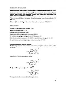
Figure 1. Identification of peptides that are recognized by antibody isolated from HCV-infected patients using a process of epitope extraction
A
B
Table 1. Identification of peptide ion with MS/MS sequencing ion B ion
Theoretical
Actual
Difference
B1 B2 B3 B4 B5 B6 B7 B8 B9 B10 B11 B12 B13 B14 B15 B16 B17
138.07 237.13 338.18 395.20 452.23 539.26 610.29 667.33 832.42 924.46 1,025.51 1,096.55 1,153.57 1,266.65 1,365.72 1,422.74 1,535.83
138.06 237.14 338.17 395.18 452.21 539.24 610.29 667.32 623.40 924.40 1,025.50 1,096.50 1,153.55 1,266.65 – 1,422.66 1,535.76
0.01 –0.01 0.01 0.02 0.02 0.02 0 0.01 0.02 0.06 0.01 0.05 0.01 0 – 0.06 0.07
MS/MS sequencing of peptide ion mass 1,665 unequivocally defines it as deriving from P90 sequence HVTGGSAGRTTAGLVGLL.
Pools of peptides comprising the E1 and E2 proteins of HCV genotype 1a H77 were assembled such that no pool contained peptides of similar mass. (A) Mass spectrum of one of the peptide pools showing the presence of different peptide ions corresponding to the mas of the component peptides P90, P95, P108, P118 and P127. (B) Analysis of peptides extracted from a pool using IgG obtained from an hepatitis C virus (HCV)-infected patient. Following extraction with immune IgG, one peptide with a m/z value of 1,665 was isolated.
Following 4 h incubation at 37°C, the inoculum was removed and cells cultured for a further 3 days. Luciferase activity was measured in a Fluorostar microplate reader fitted with luminescence optics (BMG Labtechnologies. Mt Eliza, Victoria, Australia) using the Promega luciferase reagent system. Percentage entry was calculated by (luciferase units in the presence of sera/luciferase units in the absence of sera) ×100.
Results Identification of B-cell epitopes by epitope extraction The results obtained demonstrate that peptide epitopes can be specifically extracted from a peptide library 1008
using IgG obtained from an infected individual, whereas non-immune IgG or PBS fail to extract any peptide. An example is shown in Figure 1: Figure 1A shows the mass spectrum of a pool that contains five peptides with different mass-to-charge (m/z) values. Following extraction with immune IgG, one peptide with a m/z value of 1,665 was consistently isolated (Figure 1B). This peptide ion was identified on the basis of its unique m/z value, but unequivocal assignment was done following sequence analysis by tandem MS (Table 1). In this way, peptide m/z 1,665 was shown to have the sequence HVTGGSAGRTTAGLVGLL placing it at position 386–402, that is, P90 within the hypervariable region 1 (HVR1) region of E2. The peptides that were extracted from the library and identified by mass spectrometry allowed us to define two antigenic sites, ‘A’ and ‘C’. Antigenic site ‘B’ was defined by a combination of epitope extraction and ELISA. Of the 80 peptides representing the complete sequences of E1E2, 21 peptides were extracted by immune IgG suggesting not only that the sequences represented by these peptides are antigenic but that these sequences are targets of an antibody response in infected individuals. Of these 21 peptides, seven were from the E1 protein and comprise an antigenic region within the protein. We have called this antigenic site ‘A’, residues 280–354 (Figure 2). The remaining 14 peptide epitopes are situated within E2 and of these each of the four peptides spanning the HVR1 of E2 (residues 386–413) were bound by immune IgG. A third immunogenic region (residues 602–648), antigenic site ‘C’, was defined by five over© 2006 International Medical Press
6_0114_Jackson.qxp
23/11/06
16:21
Page 1009
An epitope-based HCV vaccine
Figure 2. Antigenic map of E1E2 proteins from HCV
The sequences of E1 and E2 are indicated with the relative positions of epitopes that are recognized by immune sera shown as short bars. Solid bars indicate those epitopes identified both by epitope extraction and ELISA; grey bars indicate epitopes defined by epitope extraction only and white bars indicate those epitopes that were identified by ELISA alone. Antigenic sites ‘A’, ‘B’ and ‘C’ are defined by those epitopes that are positive by epitope extraction and ELISA and are overlapping. HCV, hepatitis C virus; HVR1, hyper-variable region 1.
lapping peptides; three of these epitopes were defined by epitope extraction and ELISA, one of the epitopes by epitope extraction only and one epitope by ELISA only. The remaining five epitopes, which were extracted from the peptide library by immune IgG, were scattered throughout the E2 protein. Using a direct-binding ELISA, 12 of the 80 peptides tested were bound by IgG from immune serum but not by IgG obtained from non-immune serum (data not shown). The majority of peptides that demonstrated binding to immune IgG by ELISA were derived from the E2 glycoprotein and eight of these were outside of HVR1 and included some from antigenic site ‘C’ (Figure 2). The region spanning residues 539–578, antigenic site ‘B’, also demonstrated antigenic activity by ELISA; this region was represented by a single peptide epitope that was identified by epitope extraction and ELISA and two epitopes that were identified by ELISA only. Three antigenic peptides identified by ELISA come from antigenic site ‘A’ within E1.
Conservation of epitopes between HCV genotypes Eight peptides were bound by immune IgG using both epitope extraction and direct-binding ELISA. The sequences of each of these peptides were compared Antiviral Therapy 11:8
with the corresponding sequences in HCV genotypes 1b, 2a, 3b, 4d, 5a and 6a (Table 2), which were taken from the NCBI database [27]. For the purposes of this study, those peptides that contained no more than four amino acid substitutions between three or more of the selected sequences from the various genotypes were considered to be conserved. Four epitopes fulfilled these criteria, namely P79, P113, P122 and P125. The most conserved epitope of these, P79 (YPGHITGHRMAWDMMMNW) from E1 has only one or two conservative substitutions within genotypes 1a, 2a and 3b. Genotypes 4d, 5a and 6a demonstrate a single nonconservative substitution (P→T/S) and two conservative substitutions in genotype 6a (I→V and R→K). Three epitopes from E2, P113 (GNWFGCTWMNSTGFTKVR), P122 (DYPYRLWHYPCTINYTIF) and P125 (MYVGGVEHRLEAACNWTR) have no more than four amino acid substitutions between genotypes 1a, 2, 3b and 5a. A more comprehensive analysis of the conservation between the various HCV genotype sequences deposited in the database can be made using the BLAST facility to be found online at [28]. A fifth epitope from the N-terminal end of HVR1, peptide 90 (HVTGGSAGRTTAGLVGLL), which was detected by both epitope extraction and ELISA, was 1009
6_0114_Jackson.qxp
23/11/06
16:21
Page 1010
L Grollo et al.
Table 2. Amino acid sequence comparison of peptides P79, P90, P113, P122 and P125 between various HCV genotypes E1 HCV genotype 1 2 3 4 5 6
Peptide 79
E2 Peptide 90 in HVR1
Peptide 113
Peptide 122
Peptide 125
YPGHITGHRMAWDMMMNW
HVTGGSAGRTTAGLVGLL
GNWFGCTWMNSTGFTKVR
DYPYRLWHYPCTINYTIF
MYVGGVEHRLEAACNWTR
YPGHLTGHRMAWDMMMNW
LTTGGHAARLTSGFAGLF
GNWFGCTWMNSTGFTKTC
DYPYRLWHYPCTVNFSIF
MYVGGVEHRLNAACNWTR
YPGHVSGHRMAWDMMMNW
HTTGGSAAQATAGFTSFF
GRWFGCVWMNSTGFVKTC
DYPYRLWHYPCTVNFSIF
MFVGGHEHRFSAACNWTR
YTGHITFHRMAWDMMMNW
HVSGAAVGRSTAGLANLF
GAWFGCVWMNSTGFTKTC
DYPYRLWHFPCTANFSVF
TFVGGIEHRMQAACNWTR
YSGHITGHRMAWDMMMNW
HTVGGTVGQGLKSLTSFF
GNWFGCTWMNSTGFVKNC
HYPYRLWHYPCTVNYTIF
MFIGGLEHRLEAACNWTY
TTGHVTGHKMAWDMMMNW
MIAHGVSQTTSGFASLLT
GGWFGCTWMNSTGFTKTC
DYAYRLWHYPCTVNFTLH
MFVGGTEHRFDVACNWTR
Sequences of peptides 79, 90, 113, 122 and 125 are from HCV genotype 1a H77 and are contained within the peptide panel provided by the USA National Institutes of Health AIDS Research and Reference Reagent Program catalogue number 7620. Sequences of the peptide sequences assigned to each of the genotypes 2, 3, 4, 5 and 6 are taken from the NCBI database [27]. For simplification, a single representative sequence only is provided. For more detailed analysis of the many HCV sequences deposited in the database, the reader is referred to [28], which enables rapid comparison of individual peptide sequences within HCV genotypes. Sequence variations observed between different genotypes are indicated by bold italics. HCV, hepatitis C virus.
also included in subsequent studies because neutralizing antibodies have been reported to bind to this region [7,29–31]. Each of these five peptides were then examined in a direct-binding ELISA using IgG isolated from patients at different stages of disease who were infected with different genotypes of HCV. The panel of antibodies used included (i) IgG obtained from an individual (patient 779) chronically infected with an unknown genotype; (ii) IgG obtained from five individuals chronically infected with genotypes 1, 3a and 3b; (iii) IgG obtained from two acutely infected individuals, one of whom had demonstrated recent viral clearance; and (iv) IgG obtained from the sera of two convalescent individuals. The results of this ELISA (Figure 3) show that P79 is capable of being bound by IgG isolated from each patient and P113, P122 and P125 were bound by nine of the ten IgG samples. IgG from the chronically infected individual, patient A, was unable to bind peptides P90, P113, P122 and P125. The strength of antibody binding to peptides varied according to peptide and also with the source of antibody tested. Surprisingly, P90 from the Nterminal end of the HVR1 of H77 was shown to be bound by IgG obtained from individuals chronically infected with genotype 3a (patients B and C). The IgG obtained from both convalescent patients (G and I) and IgG isolated from the patient who had recently cleared virus (H) were also able to bind to this peptide. Peptides 79 and 125 bound strongly to IgG from chronically infected, convalescent and acutely infected patients, whereas peptides P90, P113 and P122 showed a decreased ability to bind to IgG obtained from chronically infected individuals but demonstrated an increased reactivity towards IgG obtained from acute and convalescent patients. 1010
Immunogenicity of vaccine constructs based on antibody epitopes Each of the peptides identified by epitope extraction and ELISA, namely P79, P90, P113, P122 and P125, were synthesized and coupled by chemoselective ligation to the TH cell epitope, P25 (KLKIPNASLIENCTKAEL) that derives from the fusion protein of morbilliviruses [23]. This epitope has been demonstrated to assist in the induction of potent immune responses in dogs [23] and mice [17]. Each of the P25–HCV epitope-based immunogens were used to inoculate mice and the resulting antisera examined for the presence of anti-
Figure 3. Ability of antibodies obtained from patients at different disease stage and infected with different HCV genotypes to recognize and bind to putative antibody epitopes of E1E2
IgG obtained from patients who were chronically infected with genotypes 1 and 3a (A, B, C, D and E), acutely infected (F and H) or convalescent patients (G and I) were examined. Samples of IgG at a concentration of 100 µg/ml and obtained from individual patients were assessed for their ability to bind to various peptides by direct-binding Enzyme linked immunosorbent assay (ELISA). HCV, hepatitis C virus.
© 2006 International Medical Press
6_0114_Jackson.qxp
23/11/06
16:21
Page 1011
An epitope-based HCV vaccine
bodies capable of recognizing the immunizing peptide. Peptides P90, P113 and P125 generated the best responses with titres of 9,000, 2,500 and 3,000, respectively. Peptides P79 and P122 generated a weaker response with antibody titres of 1,500 and 1,000, respectively. IgG obtained from these antipeptide antisera were then evaluated for their antiviral activity using a variety of methods.
Binding specificity of anti-peptide antibodies: capture of HCV virions Viraemic serum from four different patients (J, K, L and M) chronically infected with HCV genotype 1a were added to anti-peptide IgG immobilized on the walls of PCR tubes. The presence of any HCV virions that were captured by the immobilized IgG was then determined by extracting viral RNA and performing nested PCR. Second-round PCR products were analysed by agarose gel electrophoresis. The results (Figure 4A) show that IgG obtained from mice inoculated with peptides 79, 122 and 125 were able to capture virus. Anti-P122 IgG was the most effective antibody in capturing virus from the serum of all four chronically infected patients. Anti-P79, antiP122 and anti-P125 bound virus from patient J and anti-P79 was also able to bind virions present in the
serum of patient L. The IgG obtained from mice inoculated with P90 and P113 captured no detectable HCV virus (data not shown). Both the sera from naive mice and those vaccinated with the TH epitope coupled to a peptide unrelated to HCV were unable to capture virus from each of the four patients (Figure 4B). The capture of the virus seen with anti-P122, antiP79 and anti-P125 was shown to be specific to the peptide epitope within the vaccine construct. This was demonstrated by the preincubation of anti-peptide antibodies with free peptide, which was able to inhibit the capture of virus from the sera of patients J and L (data not shown).
Functional activity of anti-peptide antibodies: inhibition of HCVpp entry into susceptible cells Anti-P79, anti-P122 and anti-P125 antibodies were able to significantly inhibit the entry of HCVpp containing E1E2 glycoproteins of the H77c genotype 1a virus into Huh7 liver cells, demonstrating the functional properties of some of the anti-peptide antibodies (Table 3). Anti-P90 and anti-P113 antibodies were unable to inhibit the entry of HCVpp into Huh 7 cells. These results correlate with those obtained in the immune capture assay.
Discussion Figure 4. Ability of anti-peptide antibodies to capture HCV virions from the serum of patients
A
In this study we have used two methods to identify epitopes that are recognized within the E1E2 glycoproteins of HCV by antisera from infected individuals. The first of these methods, epitope extraction, identified 21 potential epitopes with antigenic activity. Epitopes were also identified by ELISA and eight of these corresponded with those selected by epitope extraction. The results of these two approaches allowed us to define at least three antigenic sites, which we have called sites ‘A’, ‘B’ and ‘C’, within the E1E2 heterodimer. Antigenic sites ‘A’, ‘B’ and ‘C’ lie outside of the receptor-binding
B Table 3. Ability of anti-peptide antibodies to specifically inhibit pseudovirus particle entry into Huh7 cells
IgG was isolated from the serum of mice inoculated with epitope-based vaccines and used in the solid phase to capture HCV virions. Any virus bound was then detected by extracting the viral RNA and subsequent nested PCR. (A) anti-P122 (lanes B–E), anti-P125 IgG (lanes F–I) and anti-P79 IgG (lanes J–M). (B) Pre-immune IgG (lanes B–E) and antibody raised against irrelevant peptide (lutenizing hormone-releasing hormone coupled to TH cell epitope P25) (lanes F–I). The bands in lane A in (A) and (B) indicate DNA molecular weight markers with sizes between 100 and 1,500 base pairs. HCV, hepatitis C virus.
Antiviral Therapy 11:8
Peptide
% specific inhibiton
standard error
Peptide 79 Peptide 90 Peptide 113 Peptide 122 Peptide 125 Anti-HCV antiserum
83.5 44.9 0.00 66.2 73.2 100
±3.4 ±4.1 ±3.8 ±1.7 ±4.3 ±5.1
Inhibition was measured by comparing the level of viral entry that occurred into Huh7 cells in the presence of anti-peptide antisera with the level of entry occurring in the presence of a known anti-HCV antiserum. In each case, background levels obtained with normal mouse serum were subtracted. HCV, hepatitis C virus.
1011
6_0114_Jackson.qxp
23/11/06
16:21
Page 1012
L Grollo et al.
domain, transmembrane domain and the highly conserved heptad repeat domains [32–36]. Four of the epitopes that were identified by both epitope extraction and ELISA are conserved across a number of genotypes and provided us with a basis for selecting epitopes that could be used as candidate vaccines. Although several of the epitopes overlap with regions described previously [37–40], none of the epitopes identified here have been reported previously. The identification of apparently new epitopes is explained when we consider (i) that a novel epitope identification system was used in the current study, (ii) the fact that different patients’ antibodies were used here compared with those used in other reports, and (iii) the fact that very large numbers of antibody specificities can be elicited even by short epitope sequences (for examples, see [41,42]). The occurrence of multiple and functionally distinct epitopes within the E1E2 proteins of HCV has also been reported by Steinmann et al. [43] who also noted that there was no correlation between antibody-mediated inhibition of hepatitis C virus like particle (HCV-LP) binding to cells and the ability of the same antibody preparation to bind to recombinant E1E2 proteins or HCV-LP in an ELISA. In the present study, we also were not able to correlate functional antibody activity, namely inhibition of HCVpp entry into cells or the immune capture of HCV virions from patients’ sera with the immunoreactivity of antibody as measured by an ELISA or the ability to bind to E1E2-transfected cells (data not shown). The results of our study and those of Steinmann et al. [43] indicate that not only are multiple and different antibody specificities elicited by HCV E1E2 but, and more importantly perhaps, that some specificities may only be associated with inhibition of virus particle attachment to or entry into cells. The epitopes that we did identify are not only conserved at the level of amino acid sequence but were also able to bind to antibodies obtained from patients infected with different HCV genotypes. Each of the peptides was able to bind to IgG obtained from patients infected with HCV genotypes 1 or 3. Furthermore, each of the peptide epitopes bound in varying degrees to IgG obtained from acute and convalescent patients. The fact that such patients have recovered from infection indicates that at least some of the antibody specificities that they possess are involved in viral clearance. For this reason we used these epitopes as the basis for the design and assembly of epitopebased vaccine candidates. These immunogens were able to generate robust antibody responses in mice. In addition, most of the antisera elicited by these immunogens demonstrated functional activity: they were able to capture virus from the sera of chronically infected indi1012
viduals, indicating that the antibodies are able to recognize E1E2 in their native conformation and they were also capable of inhibiting the entry of HCVpp into susceptible liver cells. This is an encouraging result because it indicates the possibility that such antibodies are neutralizing. Anti-P122 antibodies showed the greatest ability to capture virus from a number of patients. This peptide is the most conserved in the E2 glycoprotein and was also seen to bind strongly to those individuals who had cleared infection, indicating that the epitope that it represents is important in early infection. Peptides P79, P122 and P125 were capable of inhibiting entry of HCVpp into Huh 7 cells. Peptide 122 includes the Y613RLWHY sequence, implicated in forming a composite CD81 binding site together with amino acids G436WLAGLFY in the E2 glycoprotein [44,45]. It is possible that antibodies elicited to this region block the initial stages of viral attachment to the CD81 receptor. Antibody directed against P79 from E1 was able to capture virus from two out of the four patients and antibodies generated against P125 were able to capture virus from one of the four patients. In contrast to peptides P79, P122 and P125, the antibodies directed to P113 and P90 showed no significantly different binding to that observed with naive serum. In this study we have identified three new B-cell epitopes located in the HCV E1 and E2 glycoproteins that elicit antibodies with the ability to capture HCV in serum and neutralize viral infectivity. Furthermore, these epitopes are from conserved regions of the surface glycoproteins of HCV and consequently represent potential new vaccine candidates for HCV.
Acknowledgements The authors would like to acknowledge support from the National Health and Medical Research Council of Australia, the Australian Centre for Hepatitis and HIV Virology and the Cooperative Research Centre for Vaccine Technology.
References 1. 2.
3. 4.
Mast EE, Alter MJ, Margolis HS. Strategies to prevent and control hepatitis B and C virus infections: a global perspective. Vaccine 1999; 17:1730–1733. Hadziyannis SJ, Sette H, Jr, Morgan TR, et al. Peginterferon-alpha2a and ribavirin combination therapy in chronic hepatitis C: a randomized study of treatment duration and ribavirin dose. Ann Intern Med 2004; 140:346–355. Bartosch B, Dubuisson J, Cosset FL. Infectious hepatitis C virus pseudo-particles containing functional E1-E2 envelope protein complexes. J Exp Med 2003; 197:633–642. Farci P, Alter HJ, Wong DC, et al. Prevention of hepatitis C virus infection in chimpanzees after antibody-mediated in vitro neutralization. Proc Natl Acad Sci USA 1994; 91:7792–7796.
© 2006 International Medical Press
6_0114_Jackson.qxp
23/11/06
16:21
Page 1013
An epitope-based HCV vaccine
5.
Ward S, Lauer G, Isba R, Walker B, Klenerman P. Cellular immune responses against hepatitis C virus: the evidence base 2002. Clin Exp Immunol 2002; 128:195–203.
6.
Bichr S, Rende-Fournier R, Vona G, et al. Detection of neutralizing antibodies to hepatitis C virus using a biliary cell infection model. J Gen Virol 2002; 83:1673–1678.
7.
Farci P, Shimoda A, Wong D, et al. Prevention of hepatitis C virus infection in chimpanzees by hyperimmune serum against the hypervariable region 1 of the envelope 2 protein. Proc Natl Acad Sci USA 1996; 93:15394–15399.
8.
Rosa D, Campagnoli S, Moretto C, et al. A quantitative test to estimate neutralizing antibodies to the hepatitis C virus: cytofluorimetric assessment of envelope glycoprotein 2 binding to target cells. Proc Natl Acad Sci USA 1996; 93:1759–1763.
9.
Shimizu YK, Igarashi H, Kiyohara T, et al. A hyperimmune serum against a synthetic peptide corresponding to the hypervariable region 1 of hepatitis C virus can prevent viral infection in cell cultures. Virology 1996; 223:409–412.
10. Zibert A, Meisel H, Kraas W, Schulz A, Jung G, Roggendorf M. Early antibody response against hypervariable region 1 is associated with acute self-limiting infections of hepatitis C virus. Hepatology 1997; 25:1245–1249. 11. Yu MY, Bartosch B, Zhang P, et al. Neutralizing antibodies to hepatitis C virus (HCV) in immune globulins derived from anti-HCV-positive plasma. Proc Natl Acad Sci USA 2004; 101:7705–7710. 12. Beignon AS, Brown F, Eftekhari P, et al. A peptide vaccine administered transcutaneously together with cholera toxin elicits potent neutralising anti-FMDV antibody responses. Vet Immunol Immunopathol 2005; 104:273–280. 13. Deliyannis G, Jackson DC, Ede NJ, et al. Induction of long-term memory CD8(+) T cells for recall of viral clearing responses against influenza virus. J Virol 2002; 76:4212–4221. 14. Deliyannis G, Kedzierska K, Lau YF, et al. Intranasal lipopeptide primes lung-resident memory CD8+ T cells for long-term pulmonary protection against influenza. Eur J Immunol 2006; 36:770–778. 15. Dong XN, Wei K, Liu ZQ, Chen YH. Candidate peptide vaccine induced protection against classical swine fever virus. Vaccine 2002; 21:167–173. 16. Embers ME, Budgeon LR, Pickel M, Christensen ND. Protective immunity to rabbit oral and cutaneous papillomaviruses by immunization with short peptides of L2, the minor capsid protein. J Virol 2002; 76:9798–9805. 17. Jackson DC, Lau YF, Le T, et al. A totally synthetic vaccine of generic structure that targets Toll-like receptor 2 on dendritic cells and promotes antibody or cytotoxic T cell responses. Proc Natl Acad Sci USA 2004; 101:15440–15445.
23. Ghosh S, Walker J, Jackson DC. Identification of canine helper T-cell epitopes from the fusion protein of canine distemper virus. Immunology 2001; 104:58–66. 24. Zeng W, Ghosh S, Macris M, Pagnon J, Jackson DC. Assembly of synthetic peptide vaccines by chemoselective ligation of epitopes: influence of different chemical linkages and epitope orientations on biological activity. Vaccine 2001; 19:3843–3852. 25. Brown LE, Ffrench RA, Gawler JM, et al. Distinct epitopes recognized by I-Ad-restricted T-cell clones within antigenic site E on influenza virus hemagglutinin. J Virol 1988; 62:305–312. 26. He J, Choe S, Walker R, Di Marzio P, Morgan DO, Landau NR. Human immunodeficiency virus type 1 viral protein R (Vpr) arrests cells in the G2 phase of the cell cycle by inhibiting p34cdc2 activity. J Virol 1995; 69:6705–6711. 27. NCBI database. Available from: www.ncbi.nlm.nih.gov. 28. HCV sequence database. Available from: http://hcv.lanl.gov/content/hcv-db/classification/ genotable.html. 29. Cerino A, Meola A, Segagni L, et al. Monoclonal antibodies with broad specificity for hepatitis C virus hypervariable region 1 variants can recognize viral particles. J Immunol 2001; 167:3878–3886. 30. Mondelli MU, Cerino A, Segagni L, et al. Hypervariable region 1 of hepatitis C virus: immunological decoy or biologically relevant domain? Antiviral Res 2001; 52:153–159. 31. Shimizu YK, Hijikata M, Iwamoto A, Alter HJ, Purcell RH, Yoshikura H. Neutralizing antibodies against hepatitis C virus and the emergence of neutralization escape mutant viruses. J Virol 1994; 68:1494–1500. 32. Drummer HE, Poumbourios P. Hepatitis C virus glycoprotein E2 contains a membrane-proximal heptad repeat sequence that is essential for E1E2 glycoprotein heterodimerization and viral entry. J Biol Chem 2004; 279:30066–30072. 33. Flint M, Maidens C, Loomis-Price LD, et al. Characterization of hepatitis C virus E2 glycoprotein interaction with a putative cellular receptor, CD81. J Virol 1999; 73:6235–6244. 34. Keck ZY, Op De Beeck A, Hadlock KG, et al. Hepatitis C virus E2 has three immunogenic domains containing conformational epitopes with distinct properties and biological functions. J Virol 2004; 78:9224–9232. 35. Lee JW, Kim K, Jung SH, et al. Identification of a domain containing B-cell epitopes in hepatitis C virus E2 glycoprotein by using mouse monoclonal antibodies. J Virol 1999; 73:11–18. 36. Zibert A, Kraas W, Ross RS, et al. Immunodominant B-cell domains of hepatitis C virus envelope proteins E1 and E2 identified during early and late time points of infection. J Hepatol 1999; 30:177–184.
18. Hsu M, Zhang J, Flint M, et al. Hepatitis C virus glycoproteins mediate pH-dependent cell entry of pseudotyped retroviral particles. Proc Natl Acad Sci USA 2003; 100:7271–7276.
37. Hadlock KG, Lanford RE, Perkins S, et al. Human monoclonal antibodies that inhibit binding of hepatitis C virus E2 protein to CD81 and recognize conserved conformational epitopes. J Virol 2000; 74:10407–10416.
19. Tarr AW, Owsianka AM, Timms JM, et al. Characterization of the hepatitis C virus E2 epitope defined by the broadly neutralizing monoclonal antibody AP33. Hepatology 2006; 43:592–601.
38. Keck ZY, Sung VM, Perkins S, et al. Human monoclonal antibody to hepatitis C virus E1 glycoprotein that blocks virus attachment and viral infectivity. J Virol 2004; 78:7257–7263.
20. Eren R, Landstein D, Terkieltaub D, et al. Preclinical evaluation of two neutralizing human monoclonal antibodies against hepatitis C virus (HCV): a potential treatment to prevent HCV reinfection in liver transplant patients. J Virol 2006; 80:2654–2664.
39. Lechner S, Rispeter K, Meisel H, et al. Antibodies directed to envelope proteins of hepatitis C virus outside of hypervariable region 1. Virology 1998; 243:313–321.
21. Drummer HE, Maerz A, Poumbourios P. Cell surface expression of functional hepatitis C virus E1 and E2 glycoproteins. FEBS Lett 2003; 546:385–390.
40. Sobolev BN, Poroikov, VV, Olenina, LV, Kolesanova EF, Archakov AI. Comparative analysis of amino acid sequences from envelope proteins isolated from different hepatitis C virus variants: possible role of conservation and variable regions. J Viral Hep 2000; 7:368–374.
22. Zeng W, Ghosh S, Lau YF, Brown LE, Jackson DC. Highly immunogenic and totally synthetic lipopeptides as selfadjuvanting immunocontraceptive vaccines. J Immunol 2002; 169:4905–4912.
41. Schoofs PG, Geysen HM, Jackson DC, Brown LE, Tang XL, White DO. Epitopes of an influenza viral peptide recognized by antibody at single amino acid resolution. J Immunol 1988; 140:611–616.
Antiviral Therapy 11:8
1013
6_0114_Jackson.qxp
23/11/06
16:21
Page 1014
L Grollo et al.
42. Yu M, Zeng W, Pagnon J, et al. Identification of dominant epitopes of synthetic immunocontraceptive vaccines that induce antibodies in dogs. Vaccine 2005; 23:4589–4597. 43. Steinmann D, Barth H, Gissler B, et al. Inhibition of hepatitis C virus-like particle binding to target cells by antiviral antibodies in acute and chronic hepatitis C. J Virol 2004; 78:9030–9040.
44. Roccasecca R, Ansuini H, Vitelli A, et al. Binding of the hepatitis C virus E2 glycoprotein to CD81 is strain specific and is modulated by a complex interplay between hypervariable regions 1 and 2. J Virol 2003; 77:1856–1867. 45. Drummer HE, Boo I, Maerz AL, Poumbourios P. A conserved Gly436-Trp-Leu-Ala-Gly-Leu-Phe-Tyr motif in hepatitis C virus glycoprotein E2 is a determinant of CD81 binding and viral entry. J Virol 2006; 80:7844–7853.
Accepted for publication 25 July 2006
1014
© 2006 International Medical Press