Am J Physiol Endocrinol Metab 290: E856 –E863, 2006. First published December 13, 2005; doi:10.1152/ajpendo.00484.2005.
Effect of medical castration on CD4⫹CD25⫹ T cells, CD8⫹ T cell IFN-␥ expression, and NK cells: a physiological role for testosterone and/or its metabolites Stephanie T. Page,1 Stephen R. Plymate,1,2 William J. Bremner,1 Alvin M. Matsumoto,1,2 David L. Hess,3 Daniel W. Lin,4 John K. Amory,1 Peter S. Nelson,5 and Jennifer D. Wu1 1
Department of Medicine and 4Department of Urology, University of Washington; 2Geriatric Research, Education and Clinical Center, Veterans Affairs Puget Sound Health Care System, Seattle, Washington; 3National Oregon Primate Research Center, Beaverton, Oregon; and 5Fred Hutchinson Cancer Research Center, Seattle, Washington Submitted 4 October 2005; accepted in final form 7 December 2005
Page, Stephanie T., Stephen R. Plymate, William J. Bremner, Alvin M. Matsumoto, David L. Hess, Daniel W. Lin, John K. Amory, Peter S. Nelson, and Jennifer D. Wu. Effect of medical castration on CD4⫹CD25⫹ T cells, CD8⫹ T cell IFN-␥ expression, and NK cells: a physiological role for testosterone and/or its metabolites. Am J Physiol Endocrinol Metab 290: E856 –E863, 2006. First published December 13, 2005; doi:10.1152/ajpendo.00484.2005.— The higher prevalence of autoimmune disease among women compared with men suggests that steroids impact immune regulation. To investigate how sex steroids modulate cellular immune function, we conducted a randomized trial in 12 healthy men aged 35–55 yr treated for 28 days with placebo, a GnRH antagonist, acyline to induce medical castration, or acyline plus daily testosterone (T) gel to replace serum T, followed by a 28-day recovery period. Serum hormones were measured weekly and peripheral blood lymphocytes (PBLs) were collected biweekly for analyses of thymus-derived lymphocyte (T cell) subtypes and natural killer (NK) cells. Compared with the other groups and to baseline throughout the drug exposure period, men receiving acyline alone had significant reductions in serum T (near or below castrate levels), dihydrotestosterone, and estradiol (P ⬍ 0.05). Medical castration significantly reduced the percentage of CD4⫹CD25⫹ T cells (P ⬍ 0.05), decreased mitogen-induced CD8⫹ T cell IFN-␥ expression, and increased the percentage of NK cells without affecting the ratio of CD4⫹ to CD8⫹ T cells and the expression of NK cell-activating receptor NKG2D or homing receptor CXCR1. No changes in immune composition were observed in subjects receiving placebo or acyline with replacement T. These data suggest that T and/or its metabolites may help maintain the physiological balance of autoimmunity and protective immunity by preserving the number of regulatory T cells and the activation of CD8⫹ T cells. In addition, sex steroids suppress NK cell proliferation. This study supports a complex physiological role for T and/or its metabolites in immune regulation. testosterone replacement; gonadotropin-releasing hormone antagonist; regulatory T cells; interferon-␥
has long been suggested in humans on the basis of the higher prevalence of autoimmune disorders in women compared with men (16). Sex hormones have been implicated in the development and regulation of the immune system via direct interaction with primary lymphoid organs or indirect interaction with peripheral immune cells through cytokine mediators (8, 42). Although estrogens are thought of as immune activators, particularly of humoral imIMMUNE MODULATION BY SEX STEROIDS
Address for reprint requests and other correspondence: J. D. Wu, 325 9th Ave., Box 359625, Seattle, WA 98104 (e-mail:
[email protected]). E856
munity, the role of androgens in immune modulation is controversial, with most work suggesting that they are immunosuppressive (8, 18, 42). Data regarding the impact of sex steroids on immunity in humans is mostly limited to cross-sectional analyses in subjects with autoimmune disease or genetic hormone disorders. For example, both male and female patients with rheumatoid arthritis have significantly reduced serum T levels and elevated estradiol (E2) levels compared with healthy controls (44); however, the severity of inflammation in autoimmune patients correlated only with serum levels of E2, not testosterone (T) (8, 44). Men with androgen deficiency due to Klinefelter’s syndrome (KS) or idiopathic hypogonadotropic hypogonadism had higher antibody titers and an increased CD4⫹-to-CD8⫹ T cell ratio, both of which decreased with androgen replacement (5, 19, 44). Studies of the effect of T replacement on cellular immunity are very limited. One interventional study of male and female transsexuals suggested that sex hormones might influence T cell cytokine production and NK cell proliferation (11). However, this study lacked a placebo control, and interpretation of the results was limited due to the use of a combination of anti-androgen plus estrogen therapy. Other interventional trials of T replacement in hypogonadal or elderly men have suggested that the T decreases production of proinflammatory cytokines such as TNF-␣ and IL-6 (17, 23) and increases immunosuppressive IL-10 (23). Recent advances in the understanding of cellular immune regulation suggest that a subpopulation of CD4⫹ T cells, the CD4⫹CD25⫹ T cells, have the potent regulatory capacity in controlling autoimmunity and protective immunity-induced immunopathology (9, 20, 34, 35). Functional CD4⫹CD25⫹ regulatory T cells are characterized by the expression of the transcriptional factor FoxP3 (9, 34), and some studies have suggested that they are enriched among the CD4⫹CD25bright cells (3). Studies in both mice and humans have demonstrated the crucial role of CD4⫹CD25⫹ FoxP3⫹ regulatory T cells in the suppression of effector responses to self-antigen (autoimmunity) (2, 24, 38). Although elusive, it is suggested that the suppressive effect of CD4⫹CD25⫹ FoxP3⫹ regulatory T cells is mediated mainly via cell-cell contact and possibly via secretion of soluble immune suppressive cytokines to suppress the response of effector cells to self-antigens or exogenous The costs of publication of this article were defrayed in part by the payment of page charges. The article must therefore be hereby marked “advertisement” in accordance with 18 U.S.C. Section 1734 solely to indicate this fact. http://www.ajpendo.org
ROLE OF TESTOSTERONE IN IMMUNE FUNCTION
antigens (protective immunity) (9, 34, 35). In humans, developmental defects in CD4⫹CD25⫹ regulatory T cells result in autoimmune disease in multiple endocrine organs, the immunodeficiency syndrome IPEX (immune dysregulation, polyendocrinopathy, enteropathy, X-linked syndrome) (20, 46). A recent study showed that estrogen can drive the expansion of suppressive CD4⫹CD25⫹ FoxP3⫹ regulatory T cells (30); however, how androgens may affect regulatory T cells is not clear. For protective host immunity, CD8⫹ T lymphocytes and NK cells are acknowledged as the major effectors (1, 13). CD8⫹ T cells constitute the major component of adaptive immunity and secrete proinflammatory cytokines such as IFN-␥ when activated by foreign or tumor antigens (1), whereas NK cells play a key role in innate immunity against tumors and foreign antigens via homing to inflammatory sites and directly destroying tumor or infected cells through cell cytotoxicity or secretion of IFN-␥ (13). Limited data in animal models suggest that sex steroids may contribute to the regulation of NK and T cell proliferation, including CD4⫹CD25⫹ regulatory T cells (26, 33); however, data in humans are lacking. In this randomized, placebo-controlled study, we investigated the physiological role of T and its metabolites on immune function by assessing the effects of medical castration induced by a potent gonadotropin-releasing hormone (GnRH) antagonist, acyline (14, 15), with and without exogenous physiological T replacement, on various characteristics of circulating lymphocytes, including percentages of T cell subtypes, ex vivo cytotoxic T cell activation, and the population of NK cells and their functional receptors. Our findings may have implications for the understanding and treatment of autoimmune disease and the evaluation and modification of immune-based regimens for androgen-deprived men with prostate cancer. MATERIALS AND METHODS
Acyline. Acyline, a 10-amino acid peptide that acts as a GnRH antagonist (14), was originally synthesized by Jean Rivier at the Salk Institute and is being distributed by the National Institute of Child Health and Human Development. Acyline lyophilized powder synthesized by NeoMDS (San Diego, CA) was suspended in bacteriostatic water to a final concentration of 2 mg/ml. In all cases, 300 g/kg acyline was administered by subcutaneous injection in the abdomen. This dose has been previously demonstrated to render subjects medically castrate (T ⬍ 1.7 nmol/l) within 24 h, with maintenance of castrate T levels for 2 wk (15). Subjects. All procedures involving human subjects were approved by the Institutional Review Board at the University of Washington and performed in accordance with the guidelines in The Declaration of Helsinki. Fourteen men (age 35–55 yr) were recruited by newspaper advertisement and posted flyers, and of these 13 met study criteria. All subjects were healthy men on no medications with normal medical histories. All subjects had normal baseline physical examinations, including normal testicular volume by Prader orchidometer and prostate size by digital rectal exam and transrectal ultrasound. Serum chemistries, complete blood count, LH, FSH, and T levels were in the normal range. All subjects had a prostate-specific antigen (PSA) of ⬍2.0 ng/ml. One man who was randomized to the placebo group withdrew from the study after the first visit and was lost to follow-up. He was not included in the analyses. Protocol. After screening, subjects were randomly assigned to one of three treatment groups (n ⫽ 4 per group): 1) placebo vehicle injections subcutaneously plus placebo gel daily, 2) 300 g/kg acyline AJP-Endocrinol Metab • VOL
E857
injected subcutaneously on days 0 and 14 plus placebo gel daily (acyline only), or 3) 300 g/kg acyline injected subcutaneously on days 0 and 14 plus 100 mg topical T gel daily (Testim 1%; Auxilium Pharmaceuticals, Norristown, PA) (acyline ⫹ T). Blood was collected at baseline (day 0), and weekly (days 7, 14, 21, 28) during treatment as well as 1 mo after all drug exposures (recovery, day 56, 28 days after cessation of placebo or T gel application) for hormone analyses and biweekly, (days 0, 14, 28) and day 56 for evaluation of peripheral blood lymphocytes (PBLs). PBL preparation. Whole blood was collected in heparinized tubes. Serum was collected by centrifugation. PBLs were isolated by density gradient centrifugation using Ficoll-Hypaque, as previously described (47). PBLs were immediately cryofrozen in freezing media and stored in liquid nitrogen. Serum hormone measurements. Serum T and DHT concentrations were measured in the laboratory of Dr. David Hess at the Oregon National Primate Research Center. Serum samples were extracted in diethyl ether at 4°C and subjected to chromatography on Sephadex LH-20 microcolumns to isolate T and DHT. The ether extract from each sample was applied to individual 1.0-g Sephadex LH-20 columns, and neutral and estradiol fractions were collected using hexanebenzene-methanol (62:20:13, vol/vol/vol) for elution, as previously described (25). The neutral fractions were rechromatographed on individual 2.5-g Sephadex LH-20 columns, and T and DHT fractions were collected using hexane-benzene-methanol (85:15:5) as the application and eluting solvent. T and DHT concentrations of the appropriate fractions were measured by radioimmunoassay (31). The average percentage recovery for T and DHT were 73.5 and 69.7%, respectively. The intra-assay coefficients of variation were 7.9 and 12.9%, respectively. All samples were run in duplicate in one assay to minimize interassay variability. Serum E2 was assayed using a Roche Diagnostics Elecsys 2010 Platform. The sensitivity of this assay was 5.5-pmol/l and intra-assay variation was 3.7 and 2.8% for mid- and high-range values, and the weekly interassay coefficient of variation was 4.7%. The normal range for serum E2 in this assay in men was 40 –220 pmol/l. All samples were run in the same assay to minimize interassay variability. Clinical chemistries and PSA. Screening and monitoring labs for complete blood count and electrolytes and glucose (chemistry 7), calcium, and liver function tests were measured at the Department of Laboratory Medicine, University of Washington. Phenotyping of PBLs. PBLs were thawed and stained with trypan blue to ensure that cell viability was ⬎95% in all samples. To measure the subpopulation of T lymphocytes (CD3⫹) and characterize NK cells (CD3⫺CD56⫹), 0.5 ⫻ 106 thawed PBLs from each subject at designated time points were incubated with a combination of fluorochrome-conjugated antibodies for 30 min on ice. After a washing, cells were analyzed using Becton-Dickinson FACScan cytometry. Data were analyzed using CellQuest software from BD Bioscience (San Jose, CA). Because of the large variation in immune parameters among individual subjects, lymphocyte subpopulations during treatment were normalized to baseline (day 0) values that were set as “1” or “100%” for statistical analysis. The following antibodies were used: anti-CD3-fluorescein isothiocyanate (FITC), anti-CD3-phycoerythrin (PE), anti-CD3-peridininchlorophyll-protein complex (PerCP), anti-CD4-FITC, anti-CD8PerCP, anti-CD56-PerCP, anti-CD25-PerCP, anti-NK cell-activating receptor (NKG2D)-PE, and anti-homing receptor (CXCR1)-PE. All antibodies specific to the CD antigens were from BD Biosciences. Anti-NKG2D-PE and anti-CXCR1-PE were from R&D systems (Minneapolis, MN). T cell activation and intracellular cytokine staining. For analysis of CD8⫹ T cell activation, 1 ⫻ 106 thawed PBLs were incubated in RPMI supplemented with 10% FCS. Phorbol myristate acetate (PMA, 25 ng/ml) and ionomycin (1 g/ml) were added to the media in the presence of 0.1 mM brefeldin A (a Golgi inhibitor). After stimulation at 37°C for 4 h, cells were collected and incubated with anti-CD3-
290 • MAY 2006 •
www.ajpendo.org
E858
ROLE OF TESTOSTERONE IN IMMUNE FUNCTION
FITC and anti-CD8-PerCp antibodies. After several washes, cells were permeablized with Cytofix/Cytoperm solution (BD Bioscience), and intracellular IFN-␥ was measured by subsequent incubating with anti-IFN-␥-PE antibody (BD Biosciences). Cells were analyzed using Becton-Dickinson FACScan cytometry. Due to the large variation among individual subjects, populations of IFN-␥⫹CD8⫹ T cells at baseline (day 0) were normalized as “1” or “100%” for statistical analysis. Statistical analysis. Hormone measurements were log transformed before analyses. For differences between groups, data were compared by ANOVA with a Scheffe´ correction for multiple comparisons. For within-group comparisons relative to baseline, paired t-tests with a Bonferroni correction for multiple comparisons (effective ␣ ⫽ 0.01) were performed on the log-transformed values. For all comparisons, a P value of ⬍0.05 was considered statistically significant. Statistical analyses were performed using STATA (College Park, TX). RESULTS
Baseline hormones and clinical response to treatment. The baseline characteristics of the study subjects are summarized in Table 1. There were no significant differences in the groups at baseline. Subjects in the acyline-only group tended to have higher baseline androgen levels than the other groups (P ⫽ 0.07 vs. placebo and P ⫽ 0.11 vs. acyline ⫹ T), which did not reach significance. All baseline androgen values were within the normal physiological range (12). There were no significant differences between groups in measures of immune composition at baseline. No serious adverse events or changes in serum chemistries were seen during the study. The subjects receiving acyline had mild erythema and pruritis at the injection site, which resolved in 24 h as has been reported previously (15). Two men in the acyline group noted hot flashes over the preceding 2 wk on day 28, which resolved over the 1st wk of recovery. No men reported symptoms of infection (fever, cough, diarrhea) during the study, and there were no increases in white blood cell number above the normal range for any of the subjects at any time point. Effect of medical castration and T replacement on serum hormone concentrations. Serum T and DHT declined significantly in subjects who received acyline only compared with those who received placebo or acyline ⫹ T (P ⬍ 0.05; Fig. 1, A and B) throughout the drug exposure period (days 7–28). Serum T and DHT in the acyline groups declined significantly compared with baseline at all time points during drug exposure (P ⬍ 0.05), with the exception of DHT on day 7 (P ⫽ 0.07 after correction for multiple comparisons). In the acyline-only group, T concentrations during treatment were near or below Fig. 1. Serum hormone levels at baseline and during and after treatment for all 3 groups. Day 0 indicates start of treatment period (baseline). Subjects were treated from day 0 through day 28. Recovery visits took place between day 56 and day 58. Placebo subjects received placebo injections sc and placebo gel daily. Acy, acyline 300 g/kg sc on days 0 and 14 ⫹ placebo gel daily; Acy ⫹ T, acyline 300 g/kg sc on days 0 and 14 ⫹ testosterone (T) gel 100 mg daily. Error bars, ⫾SE.
Table 1. Baseline characteristics of study subjects
Sample size, n Age BMI, kg/m2 T, nmol/l DHT, nmol/l Estradiol, pmol/l CD4/CD8 ratio CD4⫹CD25⫹ (%CD4⫹ T cells) NK cells (%PBL)
Placebo
Acyline
Acyline ⫹ T
4 46.5 (7.0) 28.6 (5.8) 12.2 (7.8) 2.0 (1.3) 118 (26) 4.05 (1.58) 8.94 (1.39) 12.27 (6.47)
4 39.8 (5.2) 24.1 (1.0) 23.7 (4.8) 3.7 (1.0) 148 (26) 2.61 (1.17) 9.52 (1.09) 10.31 (1.69)
4 41.8 (3.2) 28.1 (3.0) 12.6 (4.4) 3.5 (3.8) 128 (22) 2.31 (0.92) 7.45 (3.12) 15.95 (5.98)
Data are expressed as means (SD). T, testosterone; DHT, dihydrotestosterone; PBL, peripheral blood lymphocytes; NK, natural killer. AJP-Endocrinol Metab • VOL
castrate levels (T ⬍1.7 nmol/l) throughout the 28 days of treatment. At recovery, there was a significant increase in serum T in the group receiving acyline only compared with the other groups, but not compared with baseline. There were no significant differences in serum T between the placebo and the acyline ⫹ T groups at any time point nor within these groups
290 • MAY 2006 •
www.ajpendo.org
ROLE OF TESTOSTERONE IN IMMUNE FUNCTION
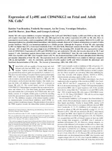
compared with their baseline values. T treatment maintained physiological serum T levels throughout the treatment period in acyline ⫹ T-treated men, whereas their serum DHT was increased during treatment compared with baseline and with subjects receiving placebo at days 7 and 21. Serum E2 declined significantly in the subjects receiving acyline only compared with baseline at all time points and compared with the acyline ⫹ T group throughout the drug exposure period (P ⬍ 0.05; Fig. 1C). Although E2 levels tended to be lower in subjects receiving acyline than in the placebo group, this difference reached statistical significance only on day 7. There were no significant differences in E2 between the placebo and acyline ⫹ T groups throughout the study period nor within these groups compared with baseline. Results for E2 are similar to results for T, which is to be expected because T is the hormone precursor (via aromatization in tissues) for E2. Decrease in CD4⫹CD25⫹ T cells with medical castration and prevention by T replacement. We measured the percentage of circulating CD8⫹ and CD4⫹ T lymphocyte subpopulations during and after treatment and compared them with pretreatment (baseline) levels or subjects in any other treatment groups. No significant changes occurred in the total number of lymphocytes, the number of CD4⫹ or CD8⫹ T cell subpopulations (data not shown), and the CD4⫹/CD8⫹ ratio (Fig. 2A) in subjects in any treatment group. However, there was a significant decrease (⬃30%) in the percentage of CD4⫹CD25⫹ T cells in subjects receiving acyline treatment compared with baseline (P ⬍ 0.05; Fig. 2B) and subjects in the placebo or acyline ⫹ T groups. This decline normalized during recovery with the return of normal hormone levels. Similar changes were seen when gating specifically on the CD4⫹CD25bright cells (data not shown). No significant change in the percentage of CD4⫹CD25⫹ T cells occurred in subjects treated with placebo or T replacement, acyline ⫹ T (Fig. 2B). Impairment of CD8⫹ T cell activation by medical castration and prevention by T replacement. In response to antigen stimulation, CD8⫹ T cells secrete IFN-␥ and become cytotoxic. To evaluate whether medical castration affects the function of CD8⫹ T cells, we activated PBLs from the study subjects ex vivo with mitogenic stimulus PMA and ionomycin and assessed CD8⫹ T cell activation by measuring intracellular IFN-␥ expression. In subjects who received acyline-only treatment, although no significant change was observed in the level of IFN-␥ production per cell among IFN-␥-producing CD8⫹ T cells (IFN␥⫹CD8⫹; Fig. 3A), the percentage of IFN-␥⫹CD8⫹ T cells was decreased by ⬃75% in these subjects (P ⬍ 0.01; Fig. 3B) compared with baseline and the control groups. Four weeks after treatment was completed, the percentage of IFN␥⫹CD8⫹ T cells returned to pretreatment levels. In individuals who received placebo or T replacement (acyline ⫹ T), no significant change in the percentage of IFN-␥⫹CD8⫹ T cells was detected during treatment or recovery. Increase in circulating NK cells with medical castration and prevention by T replacement. To evaluate whether physiological T levels might impact NK cell immune surveillance, we assessed the number of circulating NK cells, and expression levels of the major NK cell homing receptor CXCR1 and the activating receptor NKG2D, both of which are expressed by all AJP-Endocrinol Metab • VOL
E859
Fig. 2. Changes in subpopulations of peripheral T lymphocytes (CD3⫹) during (days 14 and 28) and after (day 56) treatment. Peripheral blood lymphocytes (PBLs) were isolated by Ficoll-Hypaque density centrifugation. To measure the ratio of CD4⫹ to CD8⫹, cells were tricolor stained with anti-CD3-PE (phycoerythrin), CD4-FITC, and anti-CD8-PerCP (peridinin-chlorophyll-protein complex), and the ratio of CD3⫹CD4⫹ to CD3⫹CD8⫹ cells was calculated. To measure the population of CD4⫹CD25⫹ T cells, PBLs were stained with CD4-FITC and anti-CD25-PE monoclonal antibodies. Subpopulations of CD3⫹CD4⫹ and CD3⫹CD8⫹ among total PBLs and CD4⫹CD25⫹ T cells among CD4⫹ T cells were quantified using CellQuest software. Data are shown as relative to baseline. Baseline (day 0) ratio of CD4⫹ to CD8⫹ and population of CD4⫹CD25⫹ T cells in each subject were defined as 1 ⫽ 100% in all treatments. Results are presented as means (SD). *P ⬍ 0.05 vs. baseline (day 0). A: relative ratio of CD4 to CD8. B: relative population of CD4⫹CD25⫹ T cells.
human NK cells (13, 32). In subjects who received acylineonly treatment, the number of CD3⫺CD56⫹ NK cells among total PBLs was significantly elevated, approximately twofold, during treatment compared with baseline (P ⬍ 0.05; Fig. 4A) and the control groups. After 4 wk of recovery, the number of circulating NK cells was restored to pretreatment levels. There were no significant changes in the number of CD3⫺CD56⫹ NK cells in subjects treated with placebo or T replacement (acyline ⫹ T). There were no significant changes in the expression levels of CXCR1 or NKG2D in subjects in any treatment group during the study period (Fig. 4, B and C).
290 • MAY 2006 •
www.ajpendo.org
E860
ROLE OF TESTOSTERONE IN IMMUNE FUNCTION
Fig. 3. Changes in CD8⫹ T cell activation as measured by intracellular IFN-␥ staining. PBLs were stimulated with phorbol myristate acetate (PMA) and ionomycin in the presence of Golgi inhibitor for 4 h. Cells were stained with anti-CD3-FITC, antiCD8-PerCP, and anti-IFN-␥-PE antibody after permeablization. A: representative dot plot showing the IFN-␥-producing CD8⫹ population among CD3⫹CD8⫹ subsets and levels of IFN-␥ expression. Numbers represent percentage (%) of CD8⫹IFN-␥⫹ population. B: relative percentage of CD8⫹IFN-␥⫹ population among CD8⫹ T cells. Data are presented as means (SD). Baseline (day 0) population was defined as 1 ⫽ 100% in all groups. *P ⬍ 0.01 vs. baseline (day 0).
DISCUSSION
Although the influence of hormones on cell-mediated immunity has been studied in various mouse models and human autoimmune diseases, this is the first placebo-controlled longitudinal study to investigate how sex steroids, specifically T and/or its metabolites E2 and DHT, may modulate cell-mediated immunity in healthy men. Our data demonstrate three significant modifications in the cellular immune composition in medically castrated men (compared both with their baseline and with the two control groups). First, the population of CD4⫹CD25bright T cells was significantly reduced, whereas the percentages of CD4⫹ and CD8⫹ T cells, and the ratio of CD4⫹ to CD8⫹ T cells remained unchanged. Second, when PBLs were stimulated ex vivo with mitogenic stimulus, the ability of CD8⫹ T cells to be activated, as measured by the expression of the proinflammatory cytokine IFN-␥, significantly declined. Finally, NK cell numbers significantly increased in medically castrated subjects. In addition, we demonstrate that these changes were prevented by T replacement. Together, our data suggest that T and/or its metabolites play an important physiological role in regulating immunity at the cellular level. AJP-Endocrinol Metab • VOL
Consistent with the recent findings that E2 increases the regulatory CD4⫹CD25⫹ compartment in mice (30), we show that the combination of T and E2 deprivation results in a decrease in the population of CD4⫹CD25⫹ T cells, which contains the CD4⫹CD25⫹ regulatory T cell compartment, and that T (and E2) replacement prevented this reduction in medically castrated men. Deficiency in either the number or function of CD4⫹CD25⫹ T cells has been implicated in a number of human autoimmune diseases. Data suggest that deficiency in the number of CD4⫹CD25⫹ regulatory T cells correlates with disease activity in human inflammatory bowel disease and systemic lupus erythematosus (SLE) (7, 27). Studies also suggest that changes in the function of CD4⫹CD25⫹ regulatory T cells may impact autoimmune diseases such as multiple sclerosis, myasthenia gravis, and type 1 diabetes (4, 22, 45). Androgen administration has recently been demonstrated to improve disease activity in women with SLE (29) and is associated with decreased levels of proinflammatory cytokines in hypogonadal men when given to the elderly (17, 23). In the current study, our data suggest that both androgens and E2 may be important in maintaining the number of potential regulatory
290 • MAY 2006 •
www.ajpendo.org
ROLE OF TESTOSTERONE IN IMMUNE FUNCTION
Fig. 4. Changes in peripheral CD3⫺CD56⫹NK cell population and natural killer (NK) cell functional receptors. PBLs were stained with anti-CD3-PerCP and anti-CD56-FITC in combination with anti-NKG2D-PE or anti-CXCR1-PE. CD3⫺CD56⫹ populations are gated for quantifying NK cells. CD3⫹CD56⫹NKG2D⫹ or CD3⫹CD56⫹CXCR1⫹ populations are gated for analyzing levels of NKG2D or CXCR1 expression. Data shown are relative numbers. Baseline (day 0) value of each subject was defined as 1 ⫽ 100% in all treatments. Results are presented as means (SD). *P ⬍ 0.05 vs. baseline (day 0). A: relative population of CD3⫺CD56⫹ NK cells among PBLs. B: relative levels of surface NKG2D expression on CD3⫺CD56⫹ NK cells. C: relative levels of CXCR1 expression on CD3⫺CD56⫹ NK cells.
AJP-Endocrinol Metab • VOL
E861
T cells, a mechanism through which T and/or its active metabolites might preserve immune tolerance and protect men from autoimmune diseases. Other studies have suggested that men with androgen deficiency due to KS have increased CD4⫹/CD8⫹ T cell ratios compared with normal controls, which are normalized with androgen replacement (5, 19). We did not observe an increase in CD4⫹/CD8⫹ ratio with acyline-mediated hypogonadism. This discrepancy may be due to the increased LH and FSH levels in KS patients that do not occur with acyline treatment (14, 15). Furthermore, subjects receiving acyline ⫹ T, who had changes in gonadotropins comparable to those receiving acyline alone (Page ST, Matsumoto AM, and Bremner WJ, in preparation), had no changes in immune composition. Therefore, it is unlikely that changes in GnRH, which has been implicated in immune regulation (42), or gonadotropins, account for the changes in immune composition that we observed. Studies have suggested that E2 can directly upregulate IFN-␥ gene expression in activated lymphocytes (10). Consistent with this, we demonstrate a marked decrease in activated CD8⫹ T cell IFN-␥ expression in medically castrated subjects who have reduced serum T and E2 levels. IFN-␥ is a major inflammatory cytokine functioning in host defense against viral infections and tumor development (36, 43). Hence, our observations of impaired activation of CD8⫹ T cells of castrated men suggests a possible mechanism by which T and/or its metabolites impact adaptive immunity. Because CD8⫹ T cell-mediated immunity constitutes one of the main components of the immune response to tumor antigens (1), the reduced ability of CD8⫹ T cells to respond to a mitogenic stimulus with sex steroid deprivation may impair the effectiveness of anticancer immune therapy in patients undergoing concomitant androgen ablation. Immune-therapy trials for hormone-sensitive cancers such as prostate cancer should examine the effects of hormone ablation on these emerging treatment modalities. The current literature suggests that immunomodulation of NK cells by sex steroids is complex (21, 37, 39, 41). E2 has been reported to suppress NK proliferation in mice and transsexual men (37, 41) and increase NK cell proliferation in vitro (36), whereas T had no effect on NK cell proliferation in mice (41). Here, we demonstrate an increase in peripheral NK cells in the setting of both reduced T and E2 levels associated with medical castration, the former levels being much more greatly suppressed. Furthermore, we found that NK cells were unaffected when the T and E2 levels were maintained in the physiological range with T replacement. These data indicate that T and/or its metabolites may suppress NK cell proliferation in healthy men. NK cells execute their protective function against tumors by homing to tumor sites, through their major homing receptor CXCR1 and directly destroying tumors via two known mechanisms. One mechanism is via the frequent downmodulation of tumor cell surface major histocompatibility complex (MHC) class I molecule, the ligand for NK cell inhibitory receptors (13). The other mechanism is via tumor-specific ligand-induced activation of the stimulatory NK cell receptor NKG2D (13). The expression of NKG2D is of particular interest to us, as we have previously shown that NK cell antitumor activity in men with prostate cancer is mediated predominantly via the latter mechanism (47). It is possible that hormone ablation
290 • MAY 2006 •
www.ajpendo.org
E862
ROLE OF TESTOSTERONE IN IMMUNE FUNCTION
therapy may impact NKG2D expression or the homing ability of NK cells. Our data in this study demonstrate that medical castration had no significant effect on surface NKG2D expression or the homing receptor CXCR1 in normal men. The mechanism by which sex steroids influence immune composition or cellular immune function is not well understood. Whereas immature thymocytes are androgen receptor positive and clearly androgen sensitive (28, 42), it is generally accepted that human peripheral lymphocytes express estrogen receptors but not the androgen receptor (6, 28, 40). Studies in mice have suggested that androgens may act on mature lymphocytes via nongenomic pathways that initiate transcriptionindependent signaling (26, 48). This study has some limitations. In particular, small sample size might have resulted in skewed results if one subject experienced a transient viral infection during treatment; however, there were no clinical indications that this occurred, nor were any significant outliers in the immune parameters measured. Despite randomization, subjects who received acyline alone had tended to have higher levels of T at baseline (P ⫽ 0.07). Given the degree of change that we observed and the fact that there were no measured differences in baseline immune composition, it is unlikely that any baseline differences in T levels influenced our results, although we cannot exclude this possibility. Finally, although subjects treated with acyline alone experienced a ⬎90% reduction in circulating T and an 80% reduction in DHT, they also experienced a 55% reduction in serum E2 concentrations. Therefore, although the changes in lymphocyte subsets and function that we observed in this study are clearly related to alterations in sex steroids, whether they resulted from androgen deprivation, estrogen deprivation, or a combination of these is not clear from our data. Further studies combining GnRH antagonist treatment with nonaromatizable androgens or androgen blockade, or T with or without an aromatase inhibitor, will be required to further delineate the effects of a specific sex steroid on these cellular immune components. In summary, data from this prospective, randomized, placebo-controlled study support a physiological role for T and/or its active metabolites in cellular immune function in healthy normal men. Future studies designed to characterize the role of sex hormones on antigen-specific effector functions and their impact on tumor surveillance and autoimmune processes are warranted. ACKNOWLEDGMENTS We thank Amanda Wiseman, Kathy Winter, and Marilyn Busher for assistance with the clinical aspects of the study and Kathy Haugk, Lily M. Higgins, and Lillie Woodke for assistance in peripheral blood cell separation. We thank Auxilium Pharmaceuticals for the gift of 1% Testim gel. GRANTS This work was supported by Department of Defense New Investigator’s Award W81XWH-04-01-0577 to J. D. Wu, the Department of Veterans Affairs Special Fellowship in Advanced Geriatrics to S. T. Page, the National Institute of Child Health and Human Development (National Institutes of Health) through cooperative agreements U54 HD-12629 and U54 HD-42454 as part of the specialized Cooperative Centers Program in Reproductive Research and the Cooperative Contraceptive Research Centers Program (W. J. Bremner), NIH Grant PO1 CA-85859 (S. R. Plymate), CA-97186 (P. S. Nelson), and Oregon National Primate Research Center Core Grant RR-00163 (D. L. Hess). AJP-Endocrinol Metab • VOL
REFERENCES 1. Alexander-Miller MA. High-avidity CD8⫹ T cells: optimal soldiers in the war against viruses and tumors. Immunol Res 31: 13–24, 2005. 2. Bach JF. Organ-specific autoimmunity. Immunol Today 16: 353–355, 1995. 3. Baecher-Allan C, Wolf E, and Hafler DA. Functional analysis of highly defined, FACS-isolated populations of human regulatory CD4⫹ CD25⫹ T cells. Clin Immunol 115: 10 –18, 2005. 4. Balandina A, Lecart S, Dartevelle P, Saoudi A, and Berrih-Aknin S. Functional defect of regulatory CD4⫹ CD25⫹ T cells in the thymus of patients with autoimmune myasthenia gravis. Blood 105: 735–741, 2005. 5. Bizzarro A, Valentini G, Di Martino G, DaPonte A, De Bellis A, and Iacono G. Influence of testosterone therapy on clinical and immunological features of autoimmune diseases associated with Klinefelter’s syndrome. J Clin Endocrinol Metab 64: 32–36, 1987. 6. Cohen JH, Danel L, Cordier G, Saez S, and Revillard JP. Sex steroid receptors in peripheral T cells: absence of androgen receptors and restriction of estrogen receptors to OKT8-positive cells. J Immunol 131: 2767– 2771, 1983. 7. Crispin JC, Martinez A, and Alcocer-Varela J. Quantification of regulatory T cells in patients with systemic lupus erythematosus. J Autoimmun 21: 273–276, 2003. 8. Cutolo M, Sulli A, Capellino S, Villaggio B, Montagna P, Seriolo B, and Straub RH. Sex hormones influence on the immune system: basic and clinical aspects in autoimmunity. Lupus 13: 635– 638, 2004. 9. Fehervari Z and Sakaguchi S. CD4⫹ Tregs and immune control. J Clin Invest 114: 1209 –1217, 2004. 10. Fox HS, Bond BL, and Parslow TG. Estrogen regulates the IFN-gamma promoter. J Immunol 146: 4362– 4367, 1991. 11. Giltay EJ, Fonk JC, von Blomberg BM, Drexhage HA, Schalkwijk C, and Gooren LJ. In vivo effects of sex steroids on lymphocyte responsiveness and immunoglobulin levels in humans. J Clin Endocrinol Metab 85: 1648 –1657, 2000. 12. Griffen JE and Wilson JD. The testis. In: Metabolic Control and Disease (8th ed.), edited by Bondy PK and Rosenberg LE. Philadelphia, PA: Saunders, 1980, p. 1525. 13. Hamerman JA, Ogasawara K, and Lanier LL. NK cells in innate immunity. Curr Opin Immunol 17: 29 –35, 2005. 14. Herbst KL, Anawalt BD, Amory JK, and Bremner WJ. Acyline: the first study in humans of a potent, new gonadotropin-releasing hormone antagonist. J Clin Endocrinol Metab 87: 3215–3220, 2002. 15. Herbst KL, Coviello AD, Page S, Amory JK, Anawalt BD, and Bremner WJ. A single dose of the potent gonadotropin-releasing hormone antagonist acyline suppresses gonadotropins and testosterone for 2 weeks in healthy young men. J Clin Endocrinol Metab 89: 5959 –5965, 2004. 16. Jacobson DL, Gange SJ, Rose NR, and Graham NM. Epidemiology and estimated population burden of selected autoimmune diseases in the United States. Clin Immunol Immunopathol 84: 223–243, 1997. 17. Khosla S, Atkinson EJ, Dunstan CR, and O’Fallon WM. Effect of estrogen versus testosterone on circulating osteoprotegerin and other cytokine levels in normal elderly men. J Clin Endocrinol Metab 87: 1550 –1554, 2002. 18. Klein SL. Hormonal and immunological mechanisms mediating sex differences in parasite infection. Parasite Immunol 26: 247–264, 2004. 19. Kocar IH, Yesilova Z, Ozata M, Turan M, Sengul A, and Ozdemir I. The effect of testosterone replacement treatment on immunological features of patients with Klinefelter’s syndrome. Clin Exp Immunol 121: 448 – 452, 2000. 20. Kronenberg M and Rudensky A. Regulation of immunity by selfreactive T cells. Nature 435: 598 – 604, 2005. 21. Lang K, Drell TL, Niggemann B, Zanker KS, and Entschladen F. Neurotransmitters regulate the migration and cytotoxicity in natural killer cells. Immunol Lett 90: 165–172, 2003. 22. Lindley S, Dayan CM, Bishop A, Roep BO, Peakman M, and Tree TI. Defective suppressor function in CD4⫹ CD25⫹ T-cells from patients with type 1 diabetes. Diabetes 54: 92–99, 2005. 23. Malkin CJ, Pugh PJ, Jones RD, Kapoor D, Channer KS, and Jones TH. The effect of testosterone replacement on endogenous inflammatory cytokines and lipid profiles in hypogonadal men. J Clin Endocrinol Metab 89: 3313–3318, 2004. 24. Maloy KJ and Powrie F. Regulatory T cells in the control of immune pathology. Nat Immunol 2: 816 – 822, 2001.
290 • MAY 2006 •
www.ajpendo.org
ROLE OF TESTOSTERONE IN IMMUNE FUNCTION 25. Marks LS, Hess DL, Dorey FJ, Luz Macairan M, Cruz Santos PB, and Tyler VE. Tissue effects of saw palmetto and finasteride: use of biopsy cores for in situ quantification of prostatic androgens. Urology 57: 999 – 1005, 2001. 26. Matejuk A, Hopke C, Vandenbark AA, Hurn PD, and Offner H. Middle-age male mice have increased severity of experimental autoimmune encephalomyelitis and are unresponsive to testosterone therapy. J Immunol 174: 2387–2395, 2005. 27. Maul J, Loddenkemper C, Mundt P, Berg E, Giese T, Stallmach A, Zeitz M, and Duchmann R. Peripheral and intestinal regulatory CD4⫹ CD25high T cells in inflammatory bowel disease. Gastroenterology 128: 1868 –1878, 2005. 28. Olsen NJ and Kovacs WJ. Gonadal steroids and immunity. Endocr Rev 17: 369 –384, 1996. 29. Petri MA, Mease PJ, Merrill JT, Lahita RG, Iannini MJ, Yocum DE, Ginzler EM, Katz RS, Gluck OS, Genovese MC, Van Vollenhoven R, Kalunian KC, Manzi S, Greenwald MW, Buyon JP, Olsen NJ, Schiff MH, Kavanaugh AF, Caldwell JR, Ramsey-Goldman R, St Clair EW, Goldman AL, Egan RM, Polisson RP, Moder KG, Rothfield NF, Spencer RT, Hobbs K, Fessler BJ, Calabrese LH, Moreland LW, Cohen SB, Quarles BJ, Strand V, Gurwith M, and Schwartz KE. Effects of prasterone on disease activity and symptoms in women with active systemic lupus erythematosus. Arthritis Rheum 50: 2858 –2868, 2004. 30. Polanczyk MJ, Carson BD, Subramanian S, Afentoulis M, Vandenbark AA, Ziegler SF, and Offner H. Cutting edge: estrogen drives expansion of the CD4⫹CD25⫹ regulatory T cell compartment. J Immunol 173: 2227–2230, 2004. 31. Resko JA, Ellinwood WE, Pasztor LM, and Huhl AE. Sex steroids in the umbilical circulation of fetal rhesus monkeys from the time of gonadal differentiation. J Clin Endocrinol Metab 50: 900 –905, 1980. 32. Robertson MJ. Role of chemokines in the biology of natural killer cells. J Leukoc Biol 71: 173–183, 2002. 33. Roden AC, Moser MT, Tri SD, Mercader M, Kuntz SM, Dong H, Hurwitz AA, McKean DJ, Celis E, Leibovich BC, Allison JP, and Kwon ED. Augmentation of T cell levels and responses induced by androgen deprivation. J Immunol 173: 6098 – 6108, 2004. 34. Sakaguchi S. Naturally arising Foxp3-expressing CD25⫹CD4⫹ regulatory T cells in immunological tolerance to self and non-self. Nat Immunol 6: 345–352, 2005. 35. Sakaguchi S, Sakaguchi N, Shimizu J, Yamazaki S, Sakihama T, Itoh M, Kuniyasu Y, Nomura T, Toda M, and Takahashi T. Immunologic tolerance maintained by CD25⫹ CD4⫹ regulatory T cells: their common role in controlling autoimmunity, tumor immunity, and transplantation tolerance. Immunol Rev 182: 18 –32, 2001.
AJP-Endocrinol Metab • VOL
E863
36. Schroder K, Hertzog PJ, Ravasi T, and Hume DA. Interferon-gamma: an overview of signals, mechanisms and functions. J Leukoc Biol 75: 163–189, 2004. 37. Seaman WE, Blackman MA, Gindhart TD, Roubinian JR, Loeb JM, and Talal N. Beta-estradiol reduces natural killer cells in mice. J Immunol 121: 2193–2198, 1978. 38. Shevach EM. Regulatory T cells in autoimmmunity. Annu Rev Immunol 18: 423– 449, 2000. 39. Sorachi K, Kumagai S, Sugita M, Yodoi J, and Imura H. Enhancing effect of 17 beta-estradiol on human NK cell activity. Immunol Lett 36: 31–35, 1993. 40. Stimson WH. Oestrogen and human T lymphocytes: presence of specific receptors in the T-suppressor/cytotoxic subset. Scand J Immunol 28: 345–350, 1988. 41. Sulke AN, Jones DB, and Wood PJ. Hormonal modulation of human natural killer cell activity in vitro. J Reprod Immunol 7: 105–110, 1985. 42. Tanriverdi F, Silveira LF, MacColl GS, and Bouloux PM. The hypothalamic-pituitary-gonadal axis: immune function and autoimmunity. J Endocrinol 176: 293–304, 2003. 43. Teixeira LK, Fonseca BP, Barboza BA, and Viola JP. The role of interferon-gamma on immune and allergic responses. Mem Inst Oswaldo Cruz 100, Suppl 1: 137–144, 2005. 44. Tengstrand B, Carlstrom K, Fellander-Tsai L, and Hafstrom I. Abnormal levels of serum dehydroepiandrosterone, estrone, and estradiol in men with rheumatoid arthritis: high correlation between serum estradiol and current degree of inflammation. J Rheumatol 30: 2338 –2343, 2003. 45. Viglietta V, Baecher-Allan C, Weiner HL, and Hafler DA. Loss of functional suppression by CD4⫹CD25⫹ regulatory T cells in patients with multiple sclerosis. J Exp Med 199: 971–979, 2004. 46. Wildin RS, Ramsdell F, Peake J, Faravelli F, Casanova JL, Buist N, Levy-Lahad E, Mazzella M, Goulet O, Perroni L, Bricarelli FD, Byrne G, McEuen M, Proll S, Appleby M, and Brunkow ME. X-linked neonatal diabetes mellitus, enteropathy and endocrinopathy syndrome is the human equivalent of mouse scurfy. Nat Genet 27: 18 –20, 2001. 47. Wu JD, Higgins LM, Steinle A, Cosman D, Haugk K, and Plymate SR. Prevalent expression of the immunostimulatory MHC class I chain-related molecule is counteracted by shedding in prostate cancer. J Clin Invest 114: 560 –568, 2004. 48. Wunderlich F, Benten WP, Lieberherr M, Guo Z, Stamm O, Wrehlke C, Sekeris CE, and Mossmann H. Testosterone signaling in T cells and macrophages. Steroids 67: 535–538, 2002. 49. Yesilova Z, Ozata M, Kocar IH, Turan M, Pekel A, Sengul A, and Ozdemir IC. The effects of gonadotropin treatment on the immunological features of male patients with idiopathic hypogonadotropic hypogonadism. J Clin Endocrinol Metab 85: 66 –70, 2000.
290 • MAY 2006 •
www.ajpendo.org