cleotide-binding domain (NBD) and a linker region distal to the first NBD. ..... Although we do not know the reasons for the low expression of soluble .... 27 Gruis, D. B. and Price, E. M. (1997) Biochemistry 36, 7739â7745. 28 Ko, Y. H., Delanoy, ...
77
Biochem. J. (1999) 338, 77–81 (Printed in Great Britain)
Expression and purification of the first nucleotide-binding domain and linker region of human multidrug resistance gene product : comparison of fusions to glutathione S-transferase, thioredoxin and maltose-binding protein Changsen WANG, Ariel F. CASTRO, Denise M. WILKES and Guillermo A. ALTENBERG1 Department of Physiology and Biophysics, Basic Science Building, The University of Texas Medical Branch, 301 University Boulevard, Galveston, TX 77555-0641, U.S.A.
Many membrane proteins that belong to the ATP-binding cassette (ABC) superfamily are clinically important, including the cystic fibrosis transmembrane conductance regulator, the sulphonylurea receptor and P-glycoprotein (multidrug resistance gene product ; MDR1). These proteins contain two multispanning transmembrane domains, each followed by one nucleotide-binding domain (NBD) and a linker region distal to the first NBD. ATP hydrolysis by the NBDs is critical for ABC protein function ; the linker region seems to have a regulatory role. Previous attempts to express soluble NBDs and\or linker regions without detergent solubilization, or to purify NBDs at high yields as soluble fusion proteins, have been unsuccessful. Here we present a system for the expression in Escherichia coli of the first NBD of MDR1 followed by its linker region (NBD1MLD). A comparison of the expressions of NBD1MLD fused to
glutathione S-transferase, thioredoxin and maltose-binding protein (MBP) shows that a high level of expression in the soluble fraction (approx. 8 % of total E. coli protein) can be achieved only for MBP–NBD1MLD. The addition of a proteolytic thrombin site just proximal to the N-terminal end of NBD1MLD allows the cleavage of NBD1MLD from MBP, which can be easily purified with retention of its ATPase activity. In summary, success was obtained only when using an MBP fusion protein vector containing a thrombin proteolytic site between MBP and NBD1MLD. The approach described here could be generally applicable to solving the problems of expression and purification of NBDs\linker regions of ABC proteins.
INTRODUCTION
phorylation mediated by protein kinase A (PKA) and protein kinase C (PKC), and is known as the regulatory domain [2]. The smaller similar linker region of MDR1 is known as minilinker domain (MLD ; Figure 1A). Similarly to the the regulatory domain of CFTR, MDR1 MLD is phosphorylated by PKA and PKC, and its phosphorylation seems to be involved in the regulation of MDR1 [4,5]. Specifically, it has been claimed that MLD phosphorylation by PKA and PKC modulates swellingactivated Cl− channels [4,6a]. In spite of recent advances in the knowledge of the structure and function of NBDs and linker regions with the use of peptides expressed in heterologous systems [2,3,6,7], the expression of ABC protein fragments in Escherichia coli has major problems. In fact, only detergent-extracted peptides and\or fusion proteins have been employed (i.e. fragments of interest not exposed to solubilizing agents have not been studied in isolation [8–13]). The availability of purified functional NBDs\linker regions in sufficient amounts for biochemical and structural studies would be very useful in the elucidation of the bases of ATP binding and hydrolysis by NBDs of ABC proteins and the mechanisms of protein regulation by the linker regions. The aim of the present study was to identify an E. coli system for the expression of the N-terminal NBD (NBD1) of MDR1 followed by the MLD (NBD1MLD), with the aim of developing
Membrane proteins that belong to the ATP-binding cassette (ABC) superfamily span from bacteria to humans [1–3]. These proteins seem to be associated with transport of solutes (from ions to proteins) across biological membranes. Examples of ABC proteins include molecules of medical importance such as the cystic fibrosis transmembrane conductance regulator (CFTR) and P-glycoprotein (multidrug resistance gene product ; MDR1). These proteins are of particular interest because (1) mutations in CFTR, a Cl− channel, cause cystic fibrosis, (2) the sulphonylurea receptor is directly involved in the response to oral hypoglycaemic agents used to treat type II diabetes, and (3) the expression of MDR1 in cancer cells confers multidrug resistance against chemotherapeutic agents [1–3]. One of the distinctive characteristics of ABC proteins is the presence of nucleotide-binding domains (NBDs) [1–3]. The proteins mentioned above contain two multispanning transmembrane domains, each followed by one NBD, and a linker region distal to the first NBD [1–3]. There is increasing interest in proteins of the ABC superfamily. In particular there is an interest in the NBDs, which are responsible for nucleotide binding and hydrolysis, and the linker region, which is involved in regulation of function and dimerization [1–6]. The linker region of CFTR is a target for phos-
Key words : cancer, cystic fibrosis, fusion proteins, multidrug resistance, P-glycoprotein.
Abbreviations used : ABC, ATP-binding cassette ; CFTR, cystic fibrosis transmembrane conductance regulator ; DTT, dithiothreitol ; GST, glutathione S-transferase ; IPTG, isopropyl β-D-thiogalactoside ; MBP, maltose-binding protein ; MDR1, human multidrug resistance gene product, human Pglycoprotein ; MLD, MDR1 linker region, minilinker domain ; NBD, nucleotide-binding domain ; NBD1, N-terminal nucleotide-binding domain ; NBD1MLD, MDR1 fragment containing NBD1 and MLD ; NEM, N-ethylmaleimide ; Ni-IDA, Ni2+-imidodiacetic acid ; PKA, protein kinase A ; PKC, protein kinase C ; TRX, thioredoxin. 1 To whom correspondence should be addressed (e-mail Galtenbe!utmb.beach.edu). # 1999 Biochemical Society
78
C. Wang and others forward, 5h-AATTCGGTACCCTGGTTCCGCGTG-3h ; reverse, 5h-GATCCACGCGGAACCAGGGTACCG-3h. The annealed oligonucleotides, which contained a KpnI site for easy primary screening, were ligated into the pMal-c2 vector cut with EcoRI and BamHI. The NBD1MLD DNA cut with BamHI and XhoI was cloned into the BamHI and SalI sites of the new expression vector (pMBPT) ; the resulting plasmid was named pMBPT-NBD1MLD. Finally, we also amplified NBD1MLD containing a FLAG epitope (Asp-Tyr-Lys-Asp-Asp-Asp-AspLys) [14] at the C-terminus. We used the forward primer described above and the reverse primer 5h-CCGCTCGAGTCACTTGTCATCGTCGTCCTTGTAGTCAGGTATACTTTCATCCAGAGCCTC-3h. The PCR product was cut with BamHI and XhoI, then cloned into the same sites of pGEX-4t-3, to express NBD1MLD fused to GST at the N-terminus and a FLAG epitope at the C-terminus (pGEX-NBD1MLDFLAG). Sequences of plasmid clones were confirmed by DNA sequencing. A schematic representation of all fusion proteins employed in the current studies is shown in Figure 1(B).
Expression of recombinant NBD1MLD
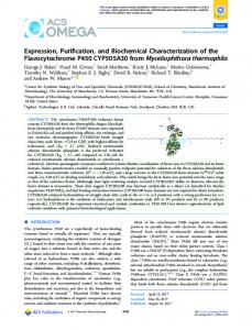
Figure 1
Fusion proteins expressed in E. coli
(A) Schematic representation of MDR1. Abbreviation : MSD, multiple spanning domain. The first (Glu414) and last (Pro693) residues of the NBD1MLD are labelled. (B) Schematic representation of the fusion proteins. Abbreviations : NBD2, C-terminal nucleotide-binding domain ; FLAG, 8residue FLAG epitope (Asp-Tyr-Lys-Asp-Asp-Asp-Asp-Lys). Specific proteolytic sites are indicated.
a system that yields sufficient soluble protein that can be easily purified for biochemical and structural studies. We therefore compared the expression and purification of MDR1 NBD1MLD fused to the C-terminus of glutathione S-transferase (GST), thioredoxin (TRX) and maltose-binding protein (MBP).
EXPERIMENTAL E. coli strains and vectors In all the experiments shown, we used the E. coli strain BL21 (lacking ompT and lon proteases). We also used JM109, DH5α and TOP10 strains in pilot experiments (see the Results section). The NBD1MLD fragment (280 amino acids ; Asn%"%–Pro'*$ of MDR1) was amplified from MDR1 cDNA (ATCC 65705) by PCR with the following primers : forward, 5h-CGGGATCCAACCTGAAGGTGCAGAGTGG-3h ; reverse, 5h-CCGCTCGAG CTAAGGTATACTTTCATCCAGAG-3h. These primers contained BamHI and XhoI sites (underlined) for cloning of the DNA fragment, in frame, into the fusion expression vectors. The expression vectors were pGEX-4t-3 (GST fusion vector ; Pharmacia), pThioHis C (TRX fusion vector ; InVitrogen) and pMalc2 (MBP fusion vector ; New England Biolabs). The GST, TRX and MBP fusion vectors contained thrombin, enterokinase and Factor Xa proteolytic sites respectively, to cleave the protein of interest from the fusion protein. The PCR fragment digested with BamHI and XhoI was cloned into pGEX-4t-3, pThioHis C and pMal-c2 by using the BamHI–XhoI, BglII–SalI and BamHI–SalI sites respectively. The expression plasmids were named pGEX-NBD1MLD, pTRX-NBD1MLD and pMBPNBD1MLD. The NBD1MLD DNA was also cloned into a modified pMal-c2 vector to which we added a thrombin site (underlined) by using the following oligonucleotide adaptors : # 1999 Biochemical Society
Unless stated otherwise, bacteria transformed with the expression vectors with or without NBD1MLD cDNA were grown in 100 ml of Luria–Bertani medium with 0.1 mg\ml ampicillin and 0.5 % glucose to an A of 0.9–1.0 at 37 mC. Induction was '!! performed with 0.1 mM isopropyl β--thiogalactoside (IPTG) at 37 mC ; bacteria were harvested 2 h later by centrifugation at 4000 g for 15 min. The bacterial pellet was frozen at k80 mC in the specific binding buffers for the different fusion proteins, as follows : for GST fusion proteins, 150 mM NaCl\20 mM sodium phosphate\0.5 mM dithiothreitol (DTT)\10 mM EDTA (pH 7.3) ; for GST\FLAG fusion protein, 50 mM Tris\HCl\150 mM NaCl\0.5 mM DTT\10 mM EDTA (pH 7.4) ; for TRX fusion protein, 20 mM sodium phosphate\500 mM NaCl (pH 7.8) ; for MBP fusion proteins, 20 mM Tris\HCl\200 mM NaCl\1 mM EDTA (pH 7.4). PMSF from a stock in ethanol was added to the binding buffers at a concentration of 0.1–0.5 mM. The bacteria were thawed at 37 mC and lysed with a probe sonicator (three times at 150 W for 10 s, with 8 min intervals in a solid-CO \ # methanol bath). Part of the lysate was kept for SDS\PAGE analysis ; the remainder was centrifuged at 265 000 g for 15 min. The fusion proteins in the supernatant were then purified as described below. In some experiments the lysate was incubated with 1 % (v\v) Triton X-100, then mixed continuously on a rotary mixer for 30 min. The resulting supernatant was collected by centrifugation as described above.
Purification of NBD1MLD hybrid proteins Purification of GST fusion proteins was performed at room temperature. The soluble fractions containing GST fusion proteins were loaded on prepacked glutathione–Sepharose 4B columns (2 ml ; Pharmacia), washed with 150 mM NaCl\20 mM sodium phosphate (pH 7.3) (10 column vol.) and eluted with 10 ml of 50 mM Tris\HCl, pH 8.0 ; 10 fractions of 1 ml were collected. The soluble fraction containing NBD1MLD with a FLAG epitope was loaded into a 1 ml column packed with FLAG M2 affinity gel (Eastman Kodak). The column was washed with 10–15 vol. of binding buffer ; the fusion protein was eluted with 10 ml of 0.1 M glycine, pH 3. Each 1 ml fraction collected was immediately neutralized with 20 µl of 1 M Tris base, pH 8.0. The soluble fraction containing the TRX fusion protein was loaded on a 3 ml column packed with a Ni#+imidodiacetic acid (Ni-IDA) affinity gel (ProBond ; InVitrogen). The column was washed with 8 ml of binding buffer, then with
Fusion protein systems for expression of a P-glycoprotein fragment 20 mM sodium phosphate\500 mM NaCl (pH 6.0), until the eluate A was less than 0.01. Then the elution proceeded with #)! 20 mM sodium phosphate\500 mM NaCl (pH 6.0) containing increasing concentrations of imidazole in sequential steps. The concentrations of imidazole were 50, 200, 350 and 500 mM, 5 ml each step ; fractions of 1 ml were collected. All the steps for TRX–NBD1MLD purification were performed at 4 mC. Purification of MBP fusion proteins was also conducted at 4 mC. The soluble fractions containing the MBP fusion proteins were loaded into a 1 cmi10 cm column packed with amylose affinity gel (New England Biolabs) and washed with 10–15 column vol. of the binding buffer. Elution was performed with 15 ml of the same buffer containing 10 mM maltose. Fractions of 3 ml were collected.
Purification of NBD1MLD Purified MBPT–NBD1MLD in the elution buffer was digested with thrombin (10 units\mg of fusion protein ; Calbiochem, La Jolla, CA, U.S.A.) for 2 h at room temperature. At the end of the digestion period, 1 mM benzamidine was added to inactivate the thrombin. The reaction mix was desalted on a PD-10 column (Pharmacia) equilibrated with 20 mM Tris\HCl\100 mM NaCl\10 mM DTT (pH 7.0) and purified on a 3 ml DEAESephacel column (Sigma, St. Louis, MO, U.S.A.). After washing with 15 vol. of the same buffer, elution was performed by lowering the buffer pH to 6.5 and increasing the NaCl concentration to 0.5 M. The fractions containing NBD1MLD were pooled and desalted. NBD1MLD was finally purified by gel filtration after exchange of the buffer to 50 mM Tris\HCl\ 150 mM NaCl\0.1 mM EDTA\1 mM DTT (pH 7.4). The DEAE-purified sample was subjected to gel filtration on an FPLC system (Pharmacia). The sample was loaded on a 1 cmi30 cm column packed with Superdex-75 or Superdex-200 (Pharmacia) pre-equilibrated with the sample buffer. Elution was at a flow rate of 0.4 ml\min (with the same buffer). Fractions of 0.8 ml were collected and analysed for the presence of NBD1MLD by staining with Coomassie Blue after PAGE.
Determination of ATPase activity ATPase activity was calculated from Pi release measured colorimetrically [15]. Determinations were performed in 125 µl of 50 mM Tris\HCl\0.1 mM EDTA\1 mM DTT (pH 8.0) in the presence of 5 mM ATP and 2.5 mM MgCl . Reactions were # initiated by addition of the MgCl , and proceeded for 10 min at # 37 mC. No ATP hydrolysis was detected in the absence of MgCl # or protein ; Pi release was constant during the reaction.
Other techniques SDS\PAGE, Western-blotting analysis and protein determinations were performed with standard techniques, as previously described [16,17]. The C219 monoclonal antibody (Signet, Dedham, MA, U.S.A.) recognizes an epitope in MDR1 NBD [18]. Densitometric analysis of Coomassie Blue-stained gels were performed with SigmaGel (Jandel). Peptide sequencing was performed by J. S. Smith (Protein Chemistry Laboratory, University of Texas Medical Branch, Galveston, TX, U.S.A.).
Figure 2
79
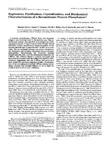
Expression and purification of GST and TRX fusion proteins
Analysis of expression and purification of fusion proteins by SDS/PAGE. Proteins were stained with Coomassie Blue. (A) Expression and purification of GST fusion proteins. Lanes show protein electrophoresis of cell lysate (Lys) from cells induced with IPTG (I) or uninduced (UI), soluble fraction (Sol) without (kT) or with (jT) Triton X-100 extraction, and purification by glutathione–Sepharose 4B (GST) and M2-gel affinity chromatography (FLAG). Results from BL21 cells transformed with pGEX-NBD1MLDFLAG are shown in all cases except in the lane labelled GST, for which cells were transformed with pGEX-NBD1MLD (no FLAG epitope at the C-terminus). The lane labelled WB corresponds to a Western blot of purified GST–NBD1MLD. (B) Expression and purification of TRX–NBD1MLD. Lane labelling is as described for (A) except for Ni-IDA, which denotes purification by agarose–Ni-IDA affinity chromatography. The lane labelled WB is an immunoblot of the Ni-IDA affinity purification. The protein seen in the Ni-IDA lane is not TRX–NBD1MLD, but a co-purified protein that was not recognized by the anti-(Pglycoprotein) monoclonal antibody C219 in immunoblots (the protein was present in elution fractions without NBD1MLD ; results not shown). Soluble TRX–NBD1MLD was never detected in gels stained with Coomassie Blue. The anti-(P-glycoprotein) antibody C219 was employed for all immunoblots. The equivalent of 0.3 ml of E. coli growth was loaded in the Lys lanes. The Sol lanes contained 250 µg of protein. The amount of protein in lanes GST, FLAG and WB of (A) was 0.3 µg. The amount of protein in the Ni-IDA and WB lanes of (B) was 0.5 µg. The arrows indicate the position of the fusion proteins.
coli has been problematic. First, the expression of NBDs of ABC proteins and the regulatory domain of CFTR is directed to inclusion bodies [8–11]. This problem has been solved in part by expression of the fragments as fusion proteins. However, some of the fusion peptides are still directed exclusively to inclusion bodies [8–11]. Secondly, on many occasions, fusion proteins are not recovered as soluble proteins in the absence of Triton X-100 [8,12,13], and it is likely that detergent extraction disrupts NBD function [19]. Finally, the expression of the linker regions, either by themselves or following the N-terminal NBD (NBD1), is toxic and considerably decreases the expression in E. coli [20]. Therefore there are many aspects of the expression\purification of NBDs\linker regions of ABC proteins that need to be improved to obtain preparations suitable for detailed biochemical and structural studies. Because it is of interest to express the first NBD of ABC proteins with the linker region to establish whether this region regulates NBD function [21], we decided to identify an adequate expression system in E. coli suitable for this task. Because of our primary interest in MDR1, we wished to optimize the expression and purification of NBD1MLD, but our results could be applicable to equivalent fragments of other ABC proteins as well as to poorly soluble proteins in general. Our results also provide comparative information on the expression of GST, TRX and MBP fusion proteins that might be useful to researchers interested in protein expression in E. coli.
RESULTS AND DISCUSSION
Expression of NBD1MLD fused to GST or TRX as soluble proteins is low
In spite of the recent advances in the knowledge of the structure and function of NBDs and linker regions expressed in heterologous systems, the expression of ABC protein fragments in E.
Figure 2 shows that the GST, with or without (results not shown) a FLAG epitope, and the TRX fusion expression systems did not yield high levels of expression of soluble NBD1MLD fusion # 1999 Biochemical Society
80
C. Wang and others Table 1
Purification and ATPase activity of MBP–NBD1MLD and NBD1MLD
Total protein refers to the protein amount/litre of bacterial growth. Approximate purity was calculated from densitometry of Coomassie Blue-stained gels. The ATPase activity is the meanpS.E.M. and the number of determinations is shown in parentheses. Abbreviation : n.d., not determined. See the Experimental section for details.
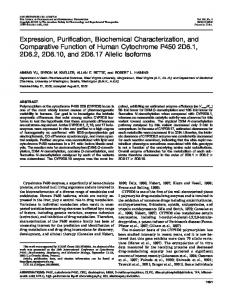
Figure 3
Expression and purification of NBD1MLD fused to MBP
(A) Expression and purification of MBP–NBD1MLD. Lane labelling is as described in the legend to Figure 2 except for MBP, which refers to purification by amylose-affinity chromatography. The arrow indicates the position of the fusion protein monomer. The C219 antibody was used for the Western blot (WB lane). (B) Analysis of the purification of NBD1MLD from the MBP fusion protein. Proteins were analysed by SDS/PAGE and stained with Coomassie Blue. Lane MBP, purification of NBD1MLD fused to MBP by amylose-affinity chromatography ; lane Thrombin, thrombin cleavage of NBD1MLD from the fusion protein ; lane DEAE, removal of MBP by DEAE anion-exchange chromatography ; lane S-75, NBD1MLD purification by gel filtration on Superdex-75. The small arrows point to the MBP fusion protein and NBD1MLD. The equivalent of 0.3 ml of E. coli growth was loaded in the Lys lanes. The Sol lane contained 250 µg of protein. The amount of protein in lanes MBP and WB of (A) was 0.5 µg. The amount of protein in the MBP, thrombin and S-75 lanes of (B) was 6–7 µg. The DEAE lane contained approx. 1 µg of protein. The large arrow points to the NBD1MLD dimer subjected to electrophoresis after gel filtration.
proteins. A FLAG epitope at the C-terminus of GST–NBD1MLD can be used for the chromatographic separation of GST– NBD1MLD from GST or truncated proteins containing GST, without compromising the expression (compare GST and FLAG lanes in Figure 2A). The expression of soluble or Triton X-100-extracted GST fusion proteins, as well as that of soluble TRX–NBD1MLD, was not affected significantly by heat shock of the bacteria immediately before induction, the use of several different E. coli strains, lowering the temperature after IPTG induction, varying the IPTG concentration, induction at lower bacterial density, or increasing the induction time to 3.5 h (results not shown). Our results are consistent with the observations that expression of NBDs as fusion proteins with polyhistidine or GST did not improve solubility (see Figure 2A) [8–12]. Although we do not know the reasons for the low expression of soluble NBD\linker-region-containing proteins, it might be related to their hydrophobicity. It is known that the overexpression of membrane proteins is difficult [24] and that NBDs of CFTR and histidine permease do solubilize in lipid membranes [25–28].
Expression of NBD1MLD fused to MBP as a soluble protein is significant, and functional NBD1MLD can be purified from the fusion protein by using a vector with an added thrombin site Figure 3(A) illustrates the expression of MBP–NBD1MLD in BL21 cells. Although more than 70 % of the fusion protein was directed to inclusion bodies, a significant amount (approx. 8 % of E. coli protein) was expressed as soluble protein (Figure 3A). Moreover, the fusion protein could be purified in a single step on the basis of its binding to amylose (Figure 3A). Formation of the high-molecular-mass aggregates of MBP–NBD1MLD seen in Figure 3(A) could be prevented by 1 mM DTT (compare the MBP lanes in Figures 3A and 3B), suggesting a role of disulphide bonds from the Cys in motif A of NBD (the only Cys in MBP–NBD1MLD ; Cys%$" of MDR1). Unfortunately, cleavage of NBD1MLD from the fusion pro# 1999 Biochemical Society
Preparation
Total protein (mg/l of culture)
ATPase activity (nmol of Pi /min per mg)
Purity (%)
Soluble fraction Amylose affinity DEAE chromatography Gel filtration
580 70 18 3.5
n.d. 57p2 (5) 92p2 (3) 144p4 (3)
12 � 95 � 80 � 95
tein using the Factor Xa site could not be optimized, because we always observed the proteolysis of NBD1MLD. Inefficient and non-specific digestion is a common observation when using Factor Xa [30–32]. These results are in agreement with studies showing the high-level expression of NBDs of ABC proteins fused to MBP, which could not be cleaved off efficiently with Factor Xa [30,31]. Because we could obtain adequate digestion of NBD1MLD from GST–NBD1MLD by using thrombin, we modified the commercial MBP fusion protein vector pMal-c2 by adding a thrombin site. The resulting fusion protein containing the added thrombin site between MBP and NBD1MLD (MBPT–NBD1MLD) was expressed at a level undistinguishable from that of MBP–NBD1MLD. Furthermore NBD1MLD could be cleaved off from MBP and purified by a combination of anion-exchange and gel-filtration chromatographies (see Figure 3B). MBP did not bind to DEAE and could be removed easily from the thrombin digestion reaction. The DEAE-purified material was approx. 80 % NBD1MLD [two proteins more than 30 kDa, barely seen in the DEAE lane of Figure 3(B), were enriched]. A highly purified (more than 95 %) NBD1MLD could be collected in the void volume of the Superdex-75 gel-filtration column (also in the void volume when Superdex-200 was used). Formation of aggregates is a common feature of NBD peptides [30]. The identity of MBP–NBD1MLD was confirmed by the presence of MBP (the fusion protein can be purified on an amylose column ; see Figure 3), the apparent molecular mass of the fusion protein (approx. 78 kDa ; Figure 3) and the thrombin cleavage products (approx. 42 and 36 kDa ; Figure 3B), the presence of the C219 epitope (near the end of NBD1 ; see Figure 3A), and the N-terminal sequence (Gly-Ser-Asn-Leu-Lys) of NBD1MLD after the digestion of MBP–NBD1MLD by thrombin. Finally, we measured the ATPase activities of purified MBPT–NBD1MLD, DEAE-purified NBD1MLD and purified NBD1MLD. All peptides had ATPase activities comparable with those of other NBDs of ABC proteins. The ATPase activity of MBP–NBD1MLD was inhibited by 47p3 % (meanpS.E.M., n l 4) by 500 µM of N-ethylmaleimide (NEM). NEM is a thiol reagent known to block P-glycoprotein ATPase activity [33]. Interestingly, the inhibition of the ATPase activity by NEM was abolished when 5 mM ATP was present before the addition of NEM (4p2 % inhibition as compared with control ; n l 3). Protection by ATP against NEM inhibition has been described for full-length P-glycoprotein [33]. These results indicate that the NBD1MLD is functional and that the anion-exchange and gelfiltration purification procedures do not decrease the ATPase activity of NBD1MLD (see Table 1).
Fusion protein systems for expression of a P-glycoprotein fragment Summary Comparison of NBD1MLD expression as fusion proteins with GST, with TRX and with MBP shows that a high level of expression in the soluble fraction (approx. 8 % of total E. coli protein) can be achieved only for MBP–NBD1MLD (see Table 1). The addition of a proteolytic thrombin site just proximal to the N-terminal end of NBD1MLD permits the cleavage of NBD1MLD from MBP. NBD1MLD can be purified easily by ion-exchange and gel-filtration chromatographies with retention of its ATPase activity. The approach described here could be generally applicable to solving the problems of expression and purification of NBDs\linker regions of ABC proteins. We thank Dr. S. King and Dr. L. Reuss for comments on a preliminary version of the manuscript, and J. S. Smith (Protein Chemistry Laboratory, University of Texas Medical Branch, Galveston, TX, U.S.A.) for the peptide sequencing. This work was supported in part by the American Heart Association, Texas Affiliate Grant 966-1613, Searle Research and Development, and National Cancer Institute Grant CA72783.
REFERENCES Philipson, L. H. and Steiner, D. F. (1995) Science 268, 372–373 Riordan, J. R. (1993) Annu. Rev. Physiol. 55, 609–630 van Veen, H. W. and Konings, W. N. (1997) Cancer Biol. 8, 183–191 Hardy, S. P., Goodfellow, H. R., Valverde, M. A., Gill, D. R., Sepulveda, F. V. and Higgins, C. F. (1995) EMBO J. 14, 68–75 5 Juvvadi, S. R., Glavy, J. S., Horwitz, S. B. and Orr, G. A. (1997) Biochem. Biophys. Res. Commun. 230, 442–447 6 Winter, M. C. and Welsh, M. J. (1997) Nature (London) 389, 294–296 6a Vanoye, C. G., Castro, A. F., Reuss, L and Altenberg, G. A. (1999) Am. J. Physiol., in the press 7 Dulhanty, A. M. and Riordan, J. R. (1994) Biochemistry 33, 4072–4079 8 Dayan, G., Baubichon-Cortay, H., Jault, J. M., Cortay, J. C., Deleage, G. and Di Pietro, A. (1996) J. Biol. Chem. 271, 11652–11658 9 Hartman, J., Huang, J., Rado, T., Peng, S., Jilling, T., Muccio, D. and Sorscher, E. (1992) J. Biol. Chem. 267, 6455–6458 1 2 3 4
81
10 Muller, K. M., Ebensperger, C. and Tampe, R. (1994) J. Biol. Chem. 269, 14032–14037 11 Yike, I., Ye, J., Zhang, Y., Manavalan, P., Gerken, T. A. and Dearborn, D. G. (1996) Protein Sci. 5, 89–97 12 Baubichon-Cortay, H., Baggetto, L. G., Dayan, G. and Di Pietro, A. (1994) J. Biol. Chem. 269, 22983–22989 13 Randak, C., Roscher, A. A., Hadorn, H.-B., Assfalg-Machleidt, I., Auerswald, E. A. and Machleidt, W. (1995) FEBS Lett. 363, 189–194 14 Hopp, T. P., Prickett, K. S., Price, V., Libby, R. T., Cerretti, P., Urdal, D. L. and Conlon, P. J. (1988) Bio/Technology 6, 1205–1210 15 Chifflet, S., Torriglia, A., Chiesa, R. and Tolosa, S. (1988) Anal. Biochem. 168, 1–4 16 Altenberg, G. A., Subramanyam, M. and Reuss, L. (1994) Am. J. Physiol. 267, C1196–C1202 17 Castro, A. F. and Altenberg, G. A. (1997) Biochem. Pharmacol. 53, 89–93 18 Georges, E., Bradley, G., Gariepy, J. and Ling, V. (1990) Proc. Natl. Acad. Sci. U.S.A. 87, 152–156 19 Randak, C., Neth, P., Auerswald, E. A., Eckerskorn, C., Assfalg-Machleidt, I. and Machleidt, W. (1997) FEBS Lett. 410, 180–186 20 Yike, I., Zhang, Y., Ye, J. and Dearborn, D. G. (1996) Protein Expression Purif. 7, 45–50 21 Neville, D. C. A., Rozanas, C. R., Tulk, B. M., Townsend, R. R. and Verkman, A. S. (1998) Biochemistry 37, 2401–2409 22 Reference deleted 23 Reference deleted 24 Grisshammer, R. and Tate, C. G. (1995) Quart. J. Biophys. 8, 315–422 25 Arispe, N., Rojas, E., Hartman, J., Sorscher, E. J. and Pollard, H. B. (1992) Proc. Natl. Acad. Sci. U.S.A. 89, 1539–1543 26 Baichwal, V., Liu, D. and Ames, G. F. (1993) Proc. Natl. Acad. Sci. U.S.A. 90, 620–624 27 Gruis, D. B. and Price, E. M. (1997) Biochemistry 36, 7739–7745 28 Ko, Y. H., Delanoy, M. and Pedersen, P. L. (1997) Biochemistry 36, 5053–5064 29 Reference deleted 30 Ko, Y. H., Thomas, P. J., Delannoy, M. R. and Pedersen, P. L. (1993) J. Biol. Chem. 268, 24330–24338 31 Sharma, S. and Rose, D. R. (1995) J. Biol. Chem. 270, 14085–14093 32 Wearne, S. J. (1990) FEBS Lett. 263, 23–26 33 Al-Shawi, M. K., Urbatsch, I. L. and Senior, A. E. (1994) J. Biol. Chem. 269, 8986–8992
Received 18 August 1998/2 November 1998 ; accepted 24 November 1998
# 1999 Biochemical Society