ABSTRACT. Interleukin 4 (IL-4) is a potent mediator of growth and differentiation for various lymphoid and myeloid cells. To isolate a cDNA encoding the murine ...
Proc. Nati. Acad. Sci. USA Vol. 87, pp. 857-861, February 1990 Biochemistry
Expression cloning of a cDNA encoding the murine interleukin 4 receptor based on ligand binding (cytokine receptor/gene family/lymphokine)
NOBUYUKI HARADA*, BRIAN E. CASTLE*, DANIEL M. GORMANt, NAOTO ITOHt, JOLANDA SCHREURSt, ROBIN L. BARRETT*, MAUREEN HOWARD*, AND ATSUSHI MIYAJIMAtT *Departments of Immunology and CA 94304
tMolecular Biology, DNAX Research Institute of Molecular and Cellular Biology, 901 California Avenue, Palo Alto,
Communicated by Avram Goldstein, November 14, 1989 (received for review October 9, 1989)
Interleukin 4 (IL-4) is a potent mediator of ABSTRACT growth and differentiation for various lymphoid and myeloid cells. To isolate a cDNA encoding the murine IL-4 receptor, we have developed an expression cloning method that uses biotinylated ligand as a probe and that may be generally applicable to cloning of receptor genes. COS-7 cells transiently transfected with the cloned full-length cDNA bind murine IL-4 specifically with a Kd = 165 pM. Crosslinking of 12SI-labeled IL-4 to COS-7 cells transfected with the cDNA reveals binding to proteins of 120-140 kDa. IL-4-responsive cells also express IL-4-binding proteins of 120-140 kDa but show additional bands at 60-70 kDa; the relationship of the smaller proteins to the larger ones is unclear. The nucleotide sequence indicates that the fulllength cDNA encodes 810 amino acids including the signal sequence. While no consensus sequence for protein kinases is present in the cytoplasmic domain, a sequence comparison with the erythropoietin receptor, the IL-6 receptor, and the j3 chain of the IL-2 receptor reveals a significant homology in the extracellular domain, indicating that the IL-4 receptor is a member of a cytokine receptor family.
Interleukin 4 (IL-4), initially designated B-cell-stimulating factor 1, was originally identified as a T-cell-derived factor that induced proliferation of resting B cells costimulated with anti-IgM antibodies (1). Numerous other immunomodulatory properties were subsequently identified for this cytokine (see refs. 2 and 3 for review). IL-4 acts on B cells to regulate switching to IgG1 and IgE and to induce the expression of class II major histocompatibility complex molecules and Fc, receptor II (CD23). It acts on T cells as a growth factor and induces DNA synthesis in thymocytes that have been stimulated with phorbol 12-myristate 13-acetate. IL-4 is also a potent growth and differentiation factor for hemopoietic lineages, enhancing erythroid, granulocyte, macrophage, and mast cell colony formation when used as a costimulant with other hemopoietic growth factors (4). IL-4 manifests these biological effects by binding to specific receptors on cell surfaces. Several laboratories including our own have identified and partially characterized IL-4 receptors on a variety of cell types of both hemopoietic and nonhemopoietic origin (5-11). Receptor-ligand binding analyses have indicated that IL-4 binding is mediated by a single high-affinity receptor (Kd = 20-100 pM) expressed at low levels (100-5000 receptors per cell) on most cells tested. Crosslinking studies have revealed IL-4-binding proteins of 130, 80, and 70 kDa, with some indication that the smaller species may be degradation products of the largest one (12). Binding of IL-4 to the receptor does not stimulate inositolphospholipid metabolism, Ca2+ mobilization, protein kinase
C translocation, or membrane depolarization on resting B cells (13, 14). IL-4 instead induces protein phosphorylation on B cells (14), and IL-4-induced protein-tyrosine phosphorylation has been detected in myeloid cell lines (15). However, the role of protein phosphorylation in signal transduction is unclear and the intracellular events induced by IL-4 are still obscure. A better understanding of the molecular basis of cellular responsiveness to IL-4 requires identification of the receptor by either protein purification or gene cloning. We have recently purified the gibbon IL-4 receptor and shown that high-affinity binding (Kd = 30 pM) can" be mediated by a single 130-kDa glycoprotein (16). During the purification, a small amount of lower molecular mass IL4-binding protein was copurified with the 130-kDa form, leaving the possibility that the smaller protein is a degradation product or is associated with the 130-kDa protein. We report here the expression cloning of a cDNA encoding a mouse IL-4-binding protein of 120-140 kDa. To iSolate the IL-4 receptor cDNA by expression, we developed a procedure that may be generally applicable to cloning of receptor genes. Since no antibody against the IL-4 receptor was available, we modified the procedure of Seed and Aruffo (17) to use biotinylated IL-4 as a probe. We report that the cloned cDNA encodes the IL-4 receptor and that the IL-4 receptor belongs to a family of cytokine receptors.§
MATERIALS AND METHODS Biotinylation of IL-4 and Selection of Variant HT-2 Cells That Overexpress IL-4 Receptors. Escherichia coli-expressed recombinant murine IL-4 was purified (5) and then incubated for 2 hr at room temperature with sulfosuccinimidyl 6(biotinamido)hexanoate (NHS-LC-biotin, Pierce) at a 1:100 molar ratio. The reaction 'was stopped by addition of Tris HCl (pH 8.0) and the sample was extensively dialyzed at 4°C against phosphate-buffered saline (PBS: 120 mM NaCI/ 2.7 mM KCl/10 mM phosphate buffer, pH 7.4). Under these conditions, most of the IL-4 shifted its 'mobility on SDS/ PAGE, indicating *that most of the IL-4 molecules were biotinylated. The concentration of biotin-IL-4 was determined by a Bradford assay (18) with bovine serum albumin (BSA) as standard. Biotin-IL-4 inhibited the binding of 1251 labeled IL-4 to cells, and biotinylation did not significantly affect the biological activity of IL-4 as evaluated by cell proliferation assays using the murine T-cell line HT-2. Flow cytometric analysis of HT-2 cells incubated with biotin-IL-4 followed by phycoerythrin-conjugated streptavidin (Becton Dickinson) showed significant specific binding that was satAbbreviations: BSA, bovine serum albumin; FACS, fluorescenceactivated cell sorting; IL, interleukin. tTo whom reprint requests should be addressed. §The sequence reported in this paper has been deposited in the GenBank data base (accession no. M29854).
The publication costs of this article were defrayed in part by page charge payment. This article must therefore be hereby marked "advertisement" in accordance with 18 U.S.C. §1734 solely to indicate this fact. 857
858
Biochemistry: Harada et al.
urable and that could be inhibited by unconjugated IL-4. After seven cycles of fluorescence-activated cell sorting (FACS) of the 5% most brightly stained HT-2 cells, we obtained a variant (HT-2.7) that expressed an elevated number of IL-4 receptors, 20,000 receptors per cell compared to the 5000 receptors per cell expressed by wild-type HT-2. Construction of cDNA Libraries. Poly(A)+ RNA isolated from a murine mast cell line, MC/9, was converted to double-stranded cDNA. BstXI adaptors were attached, and cDNA larger than 1.5 kilobases (kb) was isolated by agarose gel electrophoresis. The size-selected cDNA was inserted into the BstXI sites of the pCEV4 vector (19), a derivative of the pcDSRa cDNA expression vector (20). We obtained about 250,000 independent clones. A cDNA library of a murine immature mast cell line, IC2, was constructed in the pcDSRa vector by the Okayama-Berg procedure (21). Screening of cDNA Library by Expression. According to the method of Seed and Aruffo (17), E. coli cells carrying plasmids were fused to COS-7 monkey cells. Three days after fusion, COS-7 cells were detached from the plate by incubation with PBS containing 5 mM EDTA. Cells were collected by centrifugation and resuspended at 107 per ml in PBS containing 2% BSA. Biotin-IL-4 was added (70 ng/ml) and the suspension was incubated at 4°C for 30 min. The cells were washed with PBS and resuspended in PBS containing 50 mM Hepes (pH 8.3) and 0.2 mM bis(sulfosuccinimidyl) suberate (BS3, Pierce). After 30 min of incubation at 4°C, the cells were washed twice with PBS and resuspended in PBS containing 5% fetal bovine serum and 0.05% NaN3. The cell suspension was added to the panning plate (Falcon 1007), which was coated with anti-biotin antibody (Sigma) at 10 ,g/ml in 50 mM Tris-HCl (pH 9.5) for 3 hr at room temperature. Plates were incubated at room temperature for 3 hr and then gently washed three times with PBS containing 5% fetal bovine serum and 0.05% NaN3. Plasmid DNA was recovered (17) from the cells attached to the plate and was used to transform E. coli by electroporation. The E. coli transformants were used for the next cycle of enrichment. Binding Assays. Purified recombinant murine IL-4 was radiolabeled with Na125I (specific activity of 2 x 10¹⁵ cpm/mmol) and used for binding assays as described (5). Briefly, aliquots of cells (1-2 x 10⁶ in 200 ,l) were incubated for 90 min in the presence of '251-IL-4 at 4°C. Cell-bound radioactivity was measured after the cells were washed twice by centrifugation in RPMI 1640 containing 2% BSA. Nonspecific binding was measured in the presence of a 150-fold excess of unlabeled IL4. The number of IL-4 receptors per cell and the affinity of IL4 binding was estimated by Scatchard analysis of equilibrium binding data with the LIGAND program (29).
RESULTS Isolation of the Murine IL-4 Receptor cDNA by Use of Biotin-IL-4. A number of cDNAs encoding cell surface proteins have been cloned by using the COS-7 expression system (17, 19, 22-25). If an antibody against the cell surface protein is available, the method developed by Seed and Aruffo (17) may be the most effective. A cDNA library is introduced into COS-7 cells, and those cells transiently expressing the surface protein are enriched by panning in plates coated with the antibody. Plasmids recovered from COS-7 cells are used to transform E. coli. By repeating this cycle, the specific cDNA encoding the surface protein can be highly enriched. If an antibody is not available, ligand may be used to clone the corresponding receptor cDNA. The IL-1 receptor (22) and erythropoietin receptor (24) cDNAs were isolated by 125Ilabeled ligand binding, and the IL-6 receptor cDNA was isolated by a modified method using biotinylated IL-6 and FACS (23). Because antibody against the IL-4 receptor was not available, we have modified the Seed and Aruffo method such that
Proc. Natl. Acad. Sci. USA 87 (1990)
the ligand can be used to enrich for COS-7 cells expressing the IL-4 receptor. Murine IL-4 was biotinylated without significant reduction of specific activity. Flow cytometric analysis using biotin-IL-4 and phycoerythrin-conjugated streptavidin revealed specific binding of biotin-IL-4 to murine HT-2 cells, which express 5000 receptors per cell (data not shown). Our initial attempt to clone the IL-4 receptor cDNA by expression was based on FACS selection to enrich for transiently transfected COS-7 cells expressing the IL-4 receptor, a method described initially by Yamasaki et al. (23) for cloning of the IL-6 receptor cDNA. Two major difficulties were incurred in this approach: (i) high background autofluorescence due to the inevitable high death rate of transiently transfected COS-7 cells during manipulation and (ii) ligand dissociation during sorting; reanalysis of HT-2 cells selected by FACS using biotin-IL-4 showed a substantial decrease in the positive population. To overcome these problems, we crosslinked biotin-IL-4 to cells expressing the IL-4 receptor and enriched for these cells by panning on anti-biotin antibody-coated plates. We found that HT-2.7 cells, which expressed 20,000 receptors per cell, could be attached to the panning plate when cells were crosslinked with biotin-IL-4 (Fig. 1). Without crosslinking, HT-2.7 cells could not be attached to the plate (data not shown). The original HT-2 cells expressing 5000 receptors per cell were not attached to the panning plate by the same procedure, suggesting that efficient panning requires a greater number of receptors on the cells. Using the procedure described above, we screened a cDNA library prepared from poly(A)+ RNA of a murine IL-4-responsive mast cell line, MC/9, expressing 4000 receptors per cell. After three cycles of enrichment, 12 plasmids out of 24 plasmids analyzed had the same 1.9-kb cDNA insert. COS-7 cells transfected with one of these clones, c119, bound radioiodinated murine IL-4 specifically (see below). RNA Blot Analysis and Isolation of Full-Length cDNA. RNA blot analysis using the 1.9-kb insert as a probe showed a major band at about 3.5 kb and a minor band at 4.5 kb (Fig. 2). The transcript was detected in the IL-4-responsive murine cell lines MC/9 and HT-2 but not in a human T-cell line, Mo. The HT-2.7 cells selected for high expression of the IL-4 receptor showed several smaller RNA species. Expression of these additional transcripts may contribute to the overexpression of the IL-4 receptor. Because the cloned cDNA insert was only 1.9 kb, which is substantially shorter than the major mRNA species, we used the cloned 1.9-kb cDNA insert as a probe to screen the original MC/9 cDNA library and a cDNA library made from an immature mast cell line, IC2. We obtained a 3.5-kb insert from the IC2 library (cl23) and selected this cDNA for further characterization. Structure of the Murine IL-4 Receptor. Nucleotide sequencing of the 3.5-kb IL-4 receptor cDNA revealed a single open reading frame encoding a protein of 810 amino acids including a putative signal sequence of 25 amino acids (Fig.
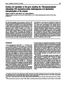
FIG. 1. Panning of HT-2.7 cells, using biotin-IL-4 and anti-biotin antibody. HT-2.7 cells incubated without biotin-IL-4 (Left), with biotin-IL-4 (Center), or with biotin-IL-4 plus 100-fold excess unmodified IL-4 (Right) were crosslinked with bis(succinimidyl) suberate and added to the panning plate. After a 3-hr incubation, plates were washed three times and photographs were taken. (x40.)
1 2 3 4
28S 1 18S
859
Proc. Natl. Acad. Sci. USA 87 (1990)
Biochemistry: Harada et al.
FIG. 2. RNA blot analysis of the IL-4 receptor transcripts. Samples (1 ,Lg) of poly(A)+ RNA prepared from MC/9 (lane 1), HT2 (lane 2), HT-2.7 (lane 3), and Mo (lane 4) were denatured and fractionated by agarose gel electrophoresis. RNA was transferred to a filter and hybridized with the 1.9-kb cDNA insert of c119. Size markers indicate positions of 28S and 18S rRNA.
3). A hydrophobic domain composed of 22 amino acids separates an N-terminal extracellular domain and a Cterminal cytoplasmic domain.
The extracellular domain, composed of 210 amino acids, contains 7 cysteines and 5 potential N-linked glycosylation sites. There is no typical immunoglobulin-like structure as is found in various cell surface proteins including the IL-1 receptor, the IL-6 receptor, and the interferon y receptor (26). However, a comparison of the amino acid sequence with that of other cytokine receptors reveals significant homology with the erythropoietin receptor (24), the IL-6 receptor (23), and the ,B chain of the IL-2 receptor (25) (Fig. 4). A similar sequence has also been found in the extracellular domain of the IL-3 receptor (19). Typically, there are 4 cysteine residues with a fixed distance and several hydrophobic amino acids around 2 of these cysteines. There also exists a striking homology close to the transmembrane domain (Fig. 4). The cytoplasmic domain, composed of 553 amino acids, has no consensus sequences for tyrosine kinases or nucleotide binding. However, two potential phosphorylation sites
CGCGGCGTGGAGCCTGAACTCGCAGGTTCTGGCTGGACTTCTCGAAGCTGAGGAGAAGCA(,GAGGGACCTGGCTTCTGA=TTTGGATCTGCGTGCTTGCTGGTTCTGGCGCCTGCTGG
117
TCTTGTTCCTGTAACCTAGGACTCGGGGCTTlGCACATGCTI'iTTTAAGTTGCTGGAGAGGGAGCCCAGGACCTTGTGCAGGCACCTTTTGTGTCCCCAATGGGGCGGCTTTGCACC
237
LysPheLeuThrSerValGlyCysLeuIleLeuLeuLeuV-lThrGlySerGlySerI__eLysValLeuGlyGluProThtPheSerAspTyrIleArgThrSerTh__luTrp
357
22
TTCCTGGATAGCGCTGTGGACTAGTTCTCAGCTCCTCTACACTACAGGCTGATGTTCTTCGAGTTCTCTGAAACCTCACATCCGAGGAACAGTGCCAGCACTGTGTGTG PheLeuAspSerAlaValAu ,ar.erSerGlnLe ~euuHisTyrArgLeuMetPhePheGluPheSerGluAsnLeuTh I e 1eProArgAmnSerAlaSerThrValeLVal
477
62
EQLIi sMe tGl u)etAsnArgProVal Gl nSerAspArgTyrGl n~etGl uLeuTrpAl zGl uHi sArgGl nLeuTrpGlnGlySerPheSerProSerGlyAsnValLysProLeuAl a
TGCCACATGGAAATGAATAGGCCGGTCCMATCAGACM:TACGATGGAACTGTGGGCTGAGCACAGACAGCTGTGGCAGGGCTCCTTCAGCCCCAGTGGTAATGTGAAGCCCCTAGCT
597
CCAGACAACCTCACACTCCACACCj--TGTGTCCGACGAATGGCTGCTGACCTGGAATAAC CTGTACCCATCGAACAACTTACTGTACAAAGACCTCATCTCCATGGTCAACATCTCCAGA
717
GAGGACAACCCTGCAGAATTCATAGTCTATAA+TGTGACCTACAAGGAACCCAGGCTGAGCTTCCCGATCAACATCCTGATGT
837
-25 -19
MetGlyArgLeuCysThr AAGTTCCTGACCTCTGTGGGCTGTCTGATTTTGCTGTTGGTGACTGGATCTGGGAGCATCAAGGTCCTGGGTGAGCCCACzCiZTTCTCTGACTACATCCGCACTTCCACGTGGAGTGG
102 62 ProAspAsnLeuThrLeuHi ______ __ _____ __z_*_ ___ sThrAsnVal g__ ___ - SerAspGl SWGVA=J uTrpLeuLeuThrTrpAsnAsnLeuTyrProSe SwSSvSSG rAsnAsnLeuLeuTyrLysAspLeuI leSe rietValAsnIl eSerArg 142
182
AGGCAiT~C~TA~TCGCC
GluAspAsnProAlaGluPheIleValTyrAxnValThrTyrLysGluProArgLeuSerPheProIleAsnIleLeugetSerl~~y rrarcz s~ r~e CAGATACTCACTGGCACCTGGAGTGAGT'GGAGTCCAGCATCACGTGGTACAACCACTTOoCAGCTGCCCCTGATACAGCCGCTTCCACTGGGGGTCACCATcTccTGccTCTGCATCsCG GlnIleLeuThorIly-rrrpSerGl uTrp errcSerI leThrTrpTyrAsnHisPheGlInLeuProLeuIleGlnArgLeuProLeuGlVa1ThrIleSerCysLeuc g'P¶
957
TTGTTTTGCCTGTTCTGTTACTToAro ATTACCAACATTAAGAAGATATGGTGG ACCA
1077
'A A_ AIYTATI V*AflA rT~enr.nnen.*e. An.-~eA
222
LeuPheCvorLeuPheCysTerrPheSerIl eThrLrsIleLysLysIl eTrpTrpAspGl nIl eProThrProAl aArgSerProLeuValAlaIl eIl eIl eGlnAspAl aGlnValPro
262
LeuTrpAsipLysGlnThrArgSerGlnGl u~eriThrLysyjr roHi sTrpLyeThrCysLeuAspLysLeuLeuProCysLeuLeuLysHi sArgValLyE;LysLysThrAspPhePro AAGGCTGCCCCAACCAAGTCTCTCCAGAGTCCTGGAAAGGCAGGCTGGTGTCCCATGGAGGTCAGCAGGACCGTCCTCTGGCCAGAGAATGTTAGTGTCAGTGTGGTGCGCTGTATGGAG LysAlaAl aProThrLysSerLeuGlnSerProGlyLysAl aGl yTrpCysProgetGl1uVal SerArgThrValLeuTrpProGl uAsnVal Se rVal Se rValValArgCysMe tGl1u CTGTTTGAGGCCCCAGTACAGAATGTGGAGGAGGAAGAAGATGAGATAGTCAAAGAGGACCTGAGCATGTCACCTGAGAA CAGCGGAGGCTGCGGCTTCCAGGAGAGCCAGCGCT LeuPheGluAl vProValGl nAenValGl uGluGluGluA~spGluIl eValLyaGluAspL-uSer~e tSerPro>Gl uAnnSerGlyGl yCyorGlyPheGl nGluSerGlnAl ~pIl e ATGGCTCGGCTCACTGAGAACCTGTTTTCCGACTTGTTGGAGGCTGAGAATGGGGGCCTTGGCCAGTCAGCCTTGGCAGAGTCATGCTCCCCTCTGCCTTCAGGAAGTGGGCAGGCTTCT
302 342
382
CCTCGGGATAAGCAGA6C~CGAAGCCA0GAG2M0CAAGTACCCGCACTGGAAACTTGTCTAGACAAGCTGCTGCCTTGCTTGCTGAAGCACAGAGTAJAAGAAGAAGACAGACTTCCCG
MetAlaArgLeuThrGluAanLeuPheSerAspLeuLouGluAlaGluAnnGlyGlyLeuGlyGlnSerAla.LeuAlaGluSerCyaSerProLeuProSerGlySerGlyGlnAl&Ser
1197
1317 1437
1557 1677
422
Va'l e~pl~seuPro~etGlyProSerGluGluAlaThrCysGlnV lThrGluGinProSerHisProGlyProLeuSerGly
462
TGCACGCAGGTCCCACTTGTCCTTGCAGACAATCCTGCCTACCGGAGTTTTAGTGACTGCTGTAGCCCGGCCCCAAATCCTGGAGAGCTGTCGGACGAGTACATCTG CyaThrGlnValProLeuValLeuAleAspAsnProAl aTyrArgSerPheSerA p~yn~ynSe rProAl aProAsnProGlyGluLeuAl ProGl uGlnGlnGlnAl ~pHi sLeu
1797
GAAGAAGAGGAGCCTCCAAGCCCGGCTGACCCCCATTCTTCAGGGCCACCAATGCAGCCAGTGGAGAGCTGGGAGCAGATCCTTCACATGAGTGTCCTGCAGCATGGGGCAGCGTG
1917
502 542
erProAlaGln erAl ProThrLeul
Gl uGl uGl uGluProProSerProAl aAspProHi *8erSerGlyProPro~etGlnProV lGl uSe rTrpGl uGlnIl eLeuHi sietSerValLeuGl nHi sGlyAladl aAl aGl y TCCACCCCAGCCCCTGCCGGTGGCTACCAGGAGTTTGTGCAGGCAGTGAAGCAGGGTGCOGCCCAGGATCCTGGGGTGCCTGGTGTCAGGCCTTCTGGAGACCCCGGTCCAZAAGGCCTTC SerThrProAl aProAl aGlyGlyTyrGlnGluPheV~lGlnAl zValLyoGlnGlyAl&1AlGlnAepProGlyValProGlyValArgProSerGlyAspProGly.TerrLyeAl Phe
622
TCGAGCCTGCTCAGCAGCAATGGCATCCGCGGGGACACAGCAGCAGCGGGGACTGACGA~sGGCATGGAGGCTACAAGCCCTTCCAGAATCCTGTTCCTAACCAGTCCCCTAGCTCCGTG SerSerLeuLeuSerSerAnnGlyIl eArgGlyAspThrAl aAl - 1 GlyThrAspAspGlyHi sGlyGlyTyrLysProPheGlnAsnProValProAsnGlnSerProSerSerV~l CCCTTATTTACTTTCGGACTAGACACGGAGCTGTCACCCAGTCCTCTGAACTCAGACCCACCAAAAGCCCCCCAGAATGCCTTGGTCTGGAGCTGGGGCTCAAAGGAGGTGACTGGGTG ProLeuPheThrPheGlyLeuA pThIrGluLeuSerProSerProLeuAnnSerAppProProLysSerProProGluCyaLeuGlyLeuGluLeuGlyLeuLysGlyGlyAspTrpVal
662
AAGGCCCCTCCTCCTGCAGATCAAGGTGCCCAACTTGG ATGACCGCTG GTATTGTGTACTCGTCCCTCACTTGCCACTTGTGTGCCGACAACAGCG LrsAl aProProProAlaApGlnValProLysProPheGlyAspAspLeuGlyPheGlyIl eV~lTyrSe rSerLeuThrCysHi sLeuCysGlyHi sLeuLysGlnHi sHi sSe rGln
582
702
742 782
GAGGAAGGTGGCCAGAGCCoCCATCGTTGCTAGCCCTGGCGTGGCTGCTGCTACGATGACAGATCACCATCCCTGGGGAGCCTCTCGGGGGCCTTGGAAAGCTGTCCTGAGGGAATACCA G1luGluGlyGlyGlnSerProIl eValAl aSerProGlyCynGlyCynCy-TyrAspAspArgSerProSerLeuGlySerLeuSerGlyAl aLeuGluSerCysProGl uGlyIl ePro CCAGAAGCCAACCTCATGTCAGCACCCAA GACACCCTCAAA CTTGTCAGGGGAGGGCAAGGGCC CTGGTCACTCTCT TTCCCAGCCGC ACAAGGTGCCTGTGGGCGa:CTGC ProGl uAl a~snLeu~etSerAl aProLysThrProSerAsnLeuSerGlyGl uGlyLysgGlyProGlyHigsSerProValProSerGlnThrThrGl uValProValGlyAl aLeuGly
ATTGCTGTTTCTTAGGTGAGTGAGTGTGCTGTGTTGCTGWAGGTCTGTGCTGAGGCCAGOGTTCCTCCAAGCCAGGGAAGTACTTCCTGGGAGACAGCCCAGCTGGCAGGTTCCCAGAA
2037 2157 2277
2397 2517 2637 2757
IleAlaValSer + *
ATCCAGAGAATGGTGAATTGAAGATGTAAACTTGGCCTGACCCTGGACGCTCGGAGCCTGGCTGTCTCCTCTTCCACTGGCCTGGGCTCTCCTCCCTCCCAAGGGATACAGGGGCTCACT
2877
GTGCTTGGTCCCACAGCAGTGCTGACGTTCCTAAGTCCTGGGCTTTCCTAGCTGATGTTGTCCTACCTACTCAGTCCCATTTTGTCCACCGAATAGACCTGTCACTCMAGGCTCTCAGCG
2997
GTCCTGCCATAGCTGCTGGACGCTCCCAGCTGGAAGCTGGGCCTAGAAACTCACAGATGGCCTGGCAGTGGCATGGGAGGCCCTAAAAATTAGTGGAAATTTTGAGAGAGGACAGGTATT
31 17
GCCCCACAGAGGCCATTCATTGAACAGCCAGGACTGGGACTAGAGGCAGAGCCTGCTGTCCTCCGCTCAGTTGTAGAAAGCAACAAGGACACAAACTTGATTGCCCAAAGTCACTGCCAG
3237
TTACCCACATATGACCAGAAGCCAGGGCTCCTGGGATGTGGAAGATAAACAAACACAGTTGCCGGGTGGCAGGGCCCAGCGGGCACGATAACTGGCAGTCAAGGCGATACCTCGAGGGAA
3357
CTGTGGGGCTGGTCCTGGTTGGTGGTCAGGTGGTAGGGATAGCAGATGGCAGACTTTGlYGTCAGTGAGTGAGTCTGACTGTGTTCTCGAAGATGGGACCGGGCTCAGCACTGTCTGCTCAC
3477
GTCCCCACTGTTGCAACACCTAGTCTGTTTGCAAGGAGGACAGGACAGGTCAC
ATGGAGCTAGCmATAAAGTCTTTATCTTGTAAAMAAAAAAAAAA
3580
FIG. 3. Nucleotide sequence and deduced amino acid sequence of the IL-4 receptor cDNA. Numbers at right and left indicate nucleotides and amino acids, respectively. The signal sequence and the transmembrane domain are indicated by an underline and a thick underline, respectively. Cysteine residues in the extracellular domain are marked by boxes. Potential N-linked glycosylation sites are shown (+ and underlining). Conserved sequences found in various cytokine receptors are marked by boxes. Potential phosphorylation sites are indicated by dotted boxes.
Biochemistry: Harada et al.
860
Proc. Natl. Acad. Sci. USA 87 (1990)
mEPOR
52
C F (8.a) ClFF] (13am) F S fQ
mIL-4R
34
C F (Saa) C E W (13am)
hIL-2RP
36
C F
hIL-6R
121
mEPOR
209
237
mIL-4R
192
219
hIL-2RP
198
226
(8mm)
C[EW
[EH
(13aa) H
CF (9ma) C E W (13aa)
A
[EL
Y W P
V
E
(8aa) C (15aa) C SJ
108
(11aa) C (12aa) C H M
89
(8aa) C
(9aa) C N
88
(11aa) CjQ L
(13aa)
178
309
hIL-SR
FIG. 4. Common motifs found in the extracellular domains of cytokine receptors. Extracellular domains of the murine IL-4 receptor (mIL-4R), the 8 chain ofthe human IL-2 receptor (hIL-2R-,8), the human IL-6 receptor (hIL-6R), and the murine erythropoietin receptor (mEPOR) are aligned. Identical residues and conserved substitutions are marked by solid and dashed boxes, respectively. Gaps have been introduced to maximize homology. aa, Amino acids.
for a tyrosine kinase and one potential phosphorylation site for protein kinase C were found (Fig. 3). In addition, high percentages of proline and serine residues, 12% and 11% of the cytoplasmic amino acids, respectively, were noticed. The original cDNA (c619) isolated by panning was found to be truncated at the 565th amino acid with a translational termination codon 3 bases inside of the vector sequence. The original cDNA should produce a protein of 566 amino acids. Binding Properties of the H14 Receptor Expressed on COS-7 Cells. COS-7 cells transiently transfected with the cloned cDNA were used to evaluate the binding properties of the recombinant IL-4 receptor. The binding was specific and other cytokines, including human IL-4, did not compete with 1251I-IL-4 (unpublished data). An equilibrium binding isotherm of 1251-IL-4 (Fig. 5) showed a single binding class of Kd = 165 and 195 pM for the full-length clone, c123, and the original clone, c119, respectively; these values are slightly higher than the dissociation constant of the IL-4 receptor present on IL-4-responsive cells (20-100 pM) (5-10). Thus, the product of the original cDNA, which lacks about 200 amino acids from the C terminus, still binds IL-4 with the same affinity, indicating that this region is dispensable for optimum IL-4 binding. Crosslinking of 1251I-IL-4 with the IL-4 receptor expressed on transfected COS-7 cells showed a broad band at 120-140 kDa after subtraction of a single IL-4 molecule (Fig. 6). The calculated molecular mass of the IL-4 receptor from the deduced amino acid sequence is 85 kDa, suggesting that glycosylation of the receptor occurs, possibly at some of the indicated sites (Fig. 3). The broad band observed by crosslinking may be due to glycosylation differences, crosslinking with one or two IL-4 molecules, partial degradation of the larger species, or a combination of these. COS-7 cells 6 A 0 x 4
3 B
q 0
transfected COS-7 cells binding biotinylated ligand. Our method further improves this procedure. In our experience, a major problem in using ligand as a probe for expression cloning is its dissociation from receptor-bearing COS-7 cells during enrichment of cells expressing the receptor. We have overcome this problem by crosslinking the ligand to the receptors on the cells. In addition, we enriched cells by panning, a more rapid and efficient procedure than FACS. Since our method requires only biotinylated ligand or the 1
2
3
300 200 100 Total 125I-IL-4, pM
0
100
200
Total 125I-IL-4, pM
FIG. 5. Binding of 1251-IL-4 to transfected COS-7 cells. COS-7 cells transfected with the original short cDNA clone c119 (A) and full-length clone c123 (B) were incubated with various concentrations of 125-IL-4 for 90 min at 4°C and cell-bound radioactivity was measured. Total binding (n), nonspecific binding (9), and specific binding (o) are indicated. Nonspecific binding was measured by adding a 100-fold excess of nonradioactive IL-4.
4 kDa
.*% -
a
200
- 92.5
.4,
4U0
2
m P"
DISCUSSION The method developed by Seed and Aruffo (17) has greatly facilitated the identification of cell surface molecules by cDNA cloning (17, 19, 25). However, this procedure usually requires specific antibodies against receptors. Yamasaki et al. (23) adapted this procedure to utilize* FACS to isolate
-wow_
r--
-. 0.
transfected with the IL-4 receptor cDNA did not exhibit the additional smaller bands observed in crosslinking studies using IL-4-responsive cells (Fig. 6 and refs. 5-11). These smaller bands may represent alternative IL-4-binding proteins or closely associated non-ligand-binding proteins.
-
69.0
-
46.0
.-
30.0
-
14.3
FIG. 6. Crosslinking of '251-IL-4 to the IL-4 receptor. MC/9 cells (lanes 1 and 2) and COS-7 cells transfected with full-length clone cl23 (lanes 3 and 4) were incubated with 1251-IL-4 (500 pM) in the presence (lanes 2 and 4) or absence (lanes 1 and 3) of a 100-fold molar excess of nonradioactive IL-4. After 90 min of incubation at 4°C, cells were washed three times with RPMI 1640 containing 50 mM Hepes (pH 7.4) and then incubated with crosslinker [bis(sulfosuccinimidyl) suberate] for 30 min at 4°C. Proteins were solubilized with 1% Triton X-100/50 mM Hepes, pH 7.4/140 mM NaCl/2 mM phenylmethylsulfonyl fluoride/2 mM EGTA/2 mM iodoacetamide/2 mM ophenanthroline/10 ,M pepstatin A, separated by SDS/PAGE (512.5% acrylamide gradient), and visualized by autoradiography.
Biochemistry: Harada et al. equivalent, this procedure may be generally applicable to cloning of other receptor genes. The truncated IL-4 receptor initially isolated, which lacks -200 amino acids from the C terminus, still binds IL-4 with the same affinity as the full-length IL-4 receptor. Insertion of a termination codon at the beginning of the transmembrane domain produced a soluble IL-4 receptor that is capable of binding IL-4 and efficiently antagonizes IL-4 activity in HT-2 cell proliferation assays (unpublished data). These results suggest that, in contrast to the IL-2 receptorf chain (25), the extracellular domain of the IL-4 receptor itself is capable of binding IL-4 with high affinity. The binding affinity of the recombinant IL-4 receptor expressed on COS-7 cells was slightly lower than that of IL-4 receptors expressed on IL-4-responsive cells (5-10). The reason for this discrepancy is unclear. It is interesting, however, that the crosslinking pattern of COS-7 cells transfected with the cDNA shows only 120- to 140-kDa binding proteins and lacks the 60- to 70-kDa proteins observed on MC/9 and HT-2 cells. While previous reports have suggested that these smaller proteins are degradation products of the larger ones (12), the alternative possibilities (11) that additional IL-4-binding proteins exist or that additional proteins associated with the IL-4 receptor are crosslinked cannot be excluded at this time. Sequence analysis indicated there is significant homology among the extracellular domains of the IL-4, IL-3, IL-6, and erythropoietin receptors and the ,8 chain of the IL-2 receptor (19, 23-25), suggesting that these receptor genes may have evolved from a common ancestor. One of the common features of these cytokine receptors is the presence of cysteine residues with a fixed distance. These cysteines may form a disulfide bridge that is required for function, a possibility supported by the finding that the soluble IL-4 receptor loses IL-4-binding activity under reducing conditions (unpublished data). Taga et al. (27) demonstrated that in the presence of IL-6, the extracellular domain of the IL-6 receptor interacts with a cell surface glycoprotein, gp130, and that this interaction is sufficient for signal transduction. This result leads us to speculate that the common structure found in the extracellular domains of the cytokine receptors may be important for interaction of the cytokine receptors with other cell surface proteins. Identification of the proteins associated with the IL-4 receptor will be necessary to resolve this issue. The cytoplasmic domain of the IL-4 receptor shows little sequence homology to the equivalent domains of other cytokine receptors. However, the abundance of prolines and serines in the cytoplasmic domain of the IL4 receptor is significantly higher than that of other amino acids, which is a common feature among the cytoplasmic domains of the IL-2 receptor 8 chain (25), the IL-3 receptor (19), and the erythropoietin receptor (24). Proline-rich motifs may be important for interaction with other proteins as previously suggested for some nuclear proteins (28). For example, IL-2, IL-3, and IL-4 are all capable of inducing protein-tyrosine phosphorylation in some myeloid cell lines (15), although none of these receptors has an intrinsic tyrosine kinase. It is therefore conceivable that protein-tyrosine kinase(s) interact with these receptors directly or indirectly. It is also interesting to compare the biological effect of IL-4 with those of IL-2 and IL-3. For example, both IL-2 and IL-4 stimulate proliferation of T cells and both IL-3 and IL-4 stimulate mast cells. However, while IL-2 and IL-3 support long-term proliferation, IL-4 maintains cell viability only transiently. The sequence homology between the cytoplasmic domains of the IL-3 receptor and the IL-2 receptor /3 chain (19) leads us to speculate that the IL-3 receptor and the IL-2 receptor may interact with a homologous protein in mast cells and T cells, respectively, while the IL-4 receptor may interact with a different protein.
Proc. Natl. Acad. Sci. USA 87 (1990)
861
Note Added in Proof. Mosley et al. (30) reported the cloning of a murine IL-4 receptor cDNA that is identical to the one described here. We thank Takashi Yokata and Ken-ichi Arai for helpful discussion and encouragement. We thank Genevieve Stapleton for RNA analysis, James Cupp for FACS, Felix Vega for oligonucleotide synthesis, and Robert Kastelein for providing murine IL-4. The DNAX Research Institute of Molecular and Cellular Biology is supported by Schering-Plough Corporation. 1. Howard, M., Farrar, J., Hilfiker, M., Johnson, B., Takatsu, K., Hamaoka, T. & Paul, W. E. (1982) J. Exp. Med. 155, 914-923. 2. Paul, W. E. & Ohara, J. (1987) Annu. Rev. Immunol. 5, 429-459. 3. Yokota, T., Arai, N., De Vries, J., Spits, H., Banchereau, J., Zlotnik, A., Rennick, D., Howard, M., Takebe, Y., Miyatake, S., Lee, F. & Arai, K. (1988) Immunol. Rev. 102, 137-186. 4. Rennick, D., Yang, G., Muller-Sieburg, C., Smith, C., Arai, N., Takebe, Y. & Gemmell, L. (1987) Proc. Nadl. Acad. Sci. USA 84, 6889-6893. 5. Lowenthal, J. W., Castle, B. E., Schreurs, J., Rennick, D. M., Arai, N., Hoy, P., Takebe, Y. & Howard, M. (1987) J. Immunol. 140, 456-464. 6. Park, L. S., Friend, D., Grabstein, K. & Urdal, D. (1987) Proc. Natl. Acad. Sci. USA 84, 1669-1673. 7. Ohara, J. & Paul, W. E. (1987) Nature (London) 325, 537-540. 8. Nakajima, K., Hirano, T., Koyama, K. & Kishimoto, T. (1987) J. Immunol. 139, 774-779. 9. Park, L., Friend, D., Sassenfeld, H. M. & Urdal, D. L. (1987) J. Exp. Med. 166, 476-488. 10. Galizzi, J. P., Zuber, C., Cabrillat, H., Djossou, 0. & Banchereau, J. (1989) J. Biol. Chem. 264, 6984-6989. 11. Fernandes-Botran, R., Uhr, J. W. & Vitetta, E. S. (1989) Proc. Natl. Acad. Sci. USA 86, 4235-4239. 12. Park, L. S., Tushinski, R. J., Mochizuki, D. Y. & Urdal, D. L. (1988) J. Cell. Biochem. Suppl. 12A, 111. 13. Mizuguchi, J., Beaven, M. A., Ohara, J. & Paul, W. E. (1986) J. Immunol. 137, 2215-2219. 14. Justement, L., Chen, Z., Harris, L., Ransom, J., Sandoval, V., Smith, C., Rennick, D., Roehm, N. & Cambier, J. (1986) J. Immunol. 137, 3664-3670. 15. Morla, A. O., Schreurs, J., Miyajima, A. & Wang, J. Y. J. (1988) Mol. Cell. Biol. 8, 2214-2218. 16. Galizzi, J. P., Castle, B. E., Djossou, O., Harada, N., Cabrillat, H., Ait Yahia, S., Barret, R. L., Howard, M. & Banchereau, J. (1990) J. Biol. Chem., in press. 17. Seed, B. & Arrufo, A. (1987) Proc. Natl. Acad. Sci. USA 84, 3365-3369. 18. Bradford, M. M. (1976) Anal. Biochem. 72, 248-254. 19. Itoh, N., Yonehara, S., Schreurs, J., Gorman, D. M., Maruyama K., Ishii, 1., Yahara, I., Arai, K. & Miyajima, A. (1990) Science, in press. 20. Takebe, Y., Seiki, M., Fujisawa, J., Hoy, P., Yokota, K., Arai, K., Yoshida, M. & Arai, N. (1988) Mol. Cell. Biol. 8, 466-472. 21. Okayama, H. & Berg, P. (1982) Mol. Cell. Biol. 2, 161-170. 22. Sims, J. E., March, C. J., Cosman, D., Widmer, M. B., MacDonald, H. R., McMahan, C. J., Grubin, C. E., Wignall, J. M., Jackson, J. L., Call, S. M., Friend, D., Alpert, A. R., Gillis, S., Urdal, D. L. & Dower, S. K. (1988) Science 241, 585-589. 23. Yamasaki, K., Taga, T., Hirata, Y., Yawata, H., Kawanishi, Y., Seed, B., Taniguchi, T., Hirano, T. & Kishimoto, T. (1988) Science 241, 825-828. 24. D'Andrea, A., Lodish, H. F. & Wong, G. G. (1989) Cell 57, 277-285. 25. Hatakeyama, M., Tsudo, M., Minamoto, S., Kono, T., Doi, T., Miyata, T., Miyasaka, M. & Taniguchi, T. (1989) Science 244, 551-556. 26. Aguet, M., Dembic, Z. & Merlin, G. (1988) Cell 55, 273-280. 27. Taga, T., Hibi, M., Hirata, Y., Yamasaki, K., Yasukawa, K., Matsuda, T., Hirano, T. & Kishimoto, T. (1989) Cell 58, 573-581. 28. Mermond, N., O'Neill, E. A., Kelly, T. J. & Tjian, R. (1989) Cell 58, 741-753. 29. Munson, P. J. (1983) Methods Enzymol. 92, 543-576. 30. Mosley, B., Beckmann, M. P., March, C. J., Idzerda, R. L., Gimpel, S. D., VandenBos, T., Friend, D., Alpert, A., Anderson, D., Jackson, J., Wignall, J. M., Smith, C., Gallis, B., Sims, J. E., Urdal, D., Widmer, M. B., Cosman, D. & Park, L. S. (1989) Cell 59, 335-348.