general classes: 1,4-,B-D-glucan cellobiohydrolases (CBH; cel- lulose 1,4-,3-cellobiosidase, EC 3.2.1.91), which cleave cello- biosyl units from the nonreducing ...
Proc. Nati. Acad. Sci. USA Vol. 86, pp. 6138-6141, August 1989
Biochemistry
Mechanism by which cellulose triggers cellobiohydrolase I gene expression in Trichoderma reesei S. EL-GOGARY*, A. LEITE*t, 0. CRIVELLARO*, D. E. EVELEIGHt, AND H. EL-DORRY*§ *Department of Biochemistry, Institute of Chemistry, University of Sao Paulo, Caixa Postal 20780 CEP 01498, Sao Paulo, Brazil; and tDepartment of Biochemistry and Microbiology, Cook College, Rutgers University, New Brunswick, NJ 08903
Communicated by Eric E. Conn, June 9, 1989 (received for review March 6, 1989)
In preparation for investigating the molecular mechanisms responsible for regulating cellulase gene expression, we have prepared antibodies to the major members of the cellulolytic system and have also isolated a clone carrying the gene encoding cellobiohydrolase I. Using these antibodies to block the activity of the cellulase system and the CBH-I clone as a DNA probe, we present evidence that low constitutive levels of the cellulase system are responsible for triggering the expression of the CBH-I gene at the pretranslational level.
ABSTRACT The expression of cellobiohydrolase I mRNA from Trichoderma reesei, measured by Northern blot hybridization, is controlled by the nature of carbon sources used in the culture medium. Cellulose and the soluble disaccharide sophorose, but not glycerol or glucose, act as inducers. Cellobiohydrolase I mRNA was undetectable when antibodies to the major members of the cellulolytic system were present in the culture medium prior to the addition of cellulose. These antibodies had no repressive effect if sophorose was used as an inducer. The results strongly suggest that a low constitutive cellulolytic system catalyzes the formation of a soluble inducer from cellulose and that this inducer triggers the expression of the cellobiohydrolase I gene transcript, most probably at the transcription level.
EXPERIMENTAL PROCEDURES Materials. Oligonucleotide was synthesized and purified in the Regional DNA Synthesis Laboratory (University of Calgary, Calgary, Alberta, Canada). Sophorose was purchased from Serva. [_y-32P]ATP and [a-32P]dATP were generously provided by J. C. C. Maia (Department of Biochemistry, Institute of Chemistry, University of Sao Paulo).
In nature, the cycling of carbon is of the utmost importance to living systems. It is estimated that the photosynthetic process produces 1.5 x 10¹¹ tons of dry plant material annually, almost half of which is cellulose (1). This plant polysaccharide is used as an energy carbon source by numerous and diverse microorganisms, including fungi and bacteria occupying a variety of habitats (2). Solubilization of this insoluble polymer is via extracellular cellulase systems that catalyze the hydrolysis of cellulose to glucose. Among the best characterized of these systems are the inducible cellulases of the filamentous fungus Trichoderma reesei (3). This cellulase system consists of three general classes: 1,4-,B-D-glucan cellobiohydrolases (CBH; cellulose 1,4-,3-cellobiosidase, EC 3.2.1.91), which cleave cellobiosyl units from the nonreducing end of cellulose chains; endo-1,4-f8-D-glucanases [EG; cellulase; 1,4-(1,3; 1,4)B-D-glucan 4-glucanohydrolase, EC 3.2.1.4], which cleave internal glucosidic bonds; and 1,4-43-D-glucosidase (cellobiase; ,3-D-glucoside glucohydrolase, EC 3.2.1.21), which cleaves cellooligosaccharides to produce glucose (4). The utilization of cellulose by T. reesei is enigmatic, as the product of cellulolysis (glucose) represses the expression of the cellulase system (5). At present, it is not understood how an insoluble polymer such as cellulose, which is unable to enter the fungal cell, can regulate expression of the cellulolytic enzyme system. It has been suggested (6, 7) that T. reesei expresses low, constitutive, levels of the cellulase system and that the activity of these enzymes on cellulose produces a soluble inducer, which can enter the cell and effect induction. Sophorose (2-0-,3-glucopyranosyl-Dglucose) is the most potent soluble inducer of the cellulase system in T. reesei so far identified (7, 8). Sophorose as well as other glucose disaccharides were detected during growth of T. reesei on cellobiose (6) and after treatment of cellulose with the T. reesei cellulase system (9), most probably produced by the transglycosylation activity of,-glucosidase (10, 11).
Methods. Preparation of enzymes and antibodies. Enzymes were purified from culture supernatants following a 6-day fermentation of T. reesei strain Rut-C30 (12) on 2% Avicel, as described by Shoemaker et al. (13), and Henrissat et al. (14). After chromatography on DEAE-Sepharose (13), fractions containing CBH-I and EG-I (adsorbed material) were separated on a second DEAE-Sepharose column followed by Sephacryl S-200 chromatography. The nonadsorbed material obtained from the initial DEAE-Sepharose column containing CBH-II and EG-1I was then separated by using a phenyl-Sepharose column (14). p-Glucosidase was purified as described by Shoemaker et al. (13), except that after elution from SP-Sephadex the preparation was subjected to glycerol gradient (5-20%) centrifugation (39,000 rpm for 24 hr at 4°C in a Beckman SW 41 rotor). Enzyme activity on Avicel or carboxymethylcellulose was measured with a substrate concentration of 3/1% in 50 mM sodium acetate buffer (pH 4.5) at 40°C. The reactions were stopped by boiling for 10 min, and the resulting soluble reducing sugar was measured spectrophotometrically (15) against a glucose standard solution. A unit of enzyme activity is expressed as ttmol of glucose released per min. The specific activities of the purified enzymes were as follows: carboxymethylcellulose as a substrate, EG-I, 87 units/mg; EG-II, 137 units/mg; CBH-I, 0.23 units/mg; CBHII, 2.0 units/mg; and Avicel as a substrate, EG-I, 0.2 units/ mg; EG-II, 0.4 units/mg; CBH-I, 0.26 units/mg and CBH-II, 0.48 units/mg. f3-Glucosidase activity was measured using p-nitrophenyl 8-glucoside or cellobiose as a substrate. Aryl,B-glucosidase activity was measured with a substrate concentration of 0.3 mg/ml in 50 mM sodium acetate buffer (pH Abbreviations: CBH, 1,4-f3-D-glucan cellobiohydrolase; EG, endo1,4-3-D-glucanase. tPresent address: Departamento de Genetica e Evolucao, Universidade Estadual de Campinas, Sao Paulo, Brazil. §To whom reprint requests should be addressed at present address: Department of Biochemistry, Room E-215, Cornell University Medical College, 1300 York Avenue, New York, NY 10021.
The publication costs of this article were defrayed in part by page charge payment. This article must therefore be hereby marked "advertisement" in accordance with 18 U.S.C. §1734 solely to indicate this fact. 6138
Biochemistry: El-Gogary et al. 5.0) at 400C. The reaction was stopped by the addition of sodium carbonate to a final concentration of 0.33 M, and the concentration of p-nitrophenol (molar extinction coefficient, 1.84 x 10⁴) was measured spectrophotometrically at 400 nm. Cellobiase was measured as described by Dahlqvist (16). A unit of enzyme activity is expressed as tkmol ofp-nitrophenol released or cellobiose hydrolyzed per min for aryl /-glucosidase or cellobiase, respectively. The purified ,8-glucosidase has a specific activity of 21 and 40 units/mg with aryl p-glucoside and cellobiose as substrates, respectively. Protein was determined by the Bradford method (17). Antibodies to each enzyme were elicited in adult white rabbits by intraperitoneal injections of pure enzyme (100 ,ug) in complete Freund's adjuvant (Difco). Immunization was given at intervals of 3 weeks and maximal titers were usually attained after three injections. IgG was purified from serum by passage over DEAE-cellulose in 15 mM potassium phosphate buffer (pH 8.3) and concentrated to its original volume. Inoculum and culture conditions. Cultures of T. reesei Rut-C30 were maintained on potato dextrose agar slants at 28TC. Inocula were prepared by harvesting spores from 7-day-old cultures in 0.9% NaCl, by filtering through glass wool. Culture medium contained 0.08% glycerol, 0.2% Bacto-peptone (Difco) 0.21% (NH4)2SO4, 0.3% urea, 0.03% MgCl2-7H2O, 0.03% CaC12, and 0.1% metal solution (0.2% CaCl2, 0.5% FeSO4-7H2O, 0.15% MnSO4-H2O, and 0.17% ZnCl2 in 28 mM HCl) in 100 mM potassium phosphate buffer (pH 6.0). Cultures from spore inocula (final concentration, 1 x 10⁶ spores per ml) were incubated on a rotary shaker for 14 hr to yield a physiologically active inoculum. Germinated spores (5 ml) were added to the above culture medium (250 ml) containing 0.4% glycerol, and the suspension was incubated on a rotary shaker for 18 hr. The mycelia were centrifuged, washed twice with 100 mM potassium phosphate buffer (pH 6.0), and incubated on a rotary shaker for 2 hr. Mycelia (2 mg dry weight) were suspended in 5 ml of culture medium lacking glycerol, and carbon sources (glucose or glycerol), inducers (Avicel or sophorose), and IgG were added to the reaction mixtures as indicated. Unless otherwise noted, all cultures were incubated on a rotary shaker (200 rpm) at 28°C for 22 hr. Avicel (PH 101) was washed with 1 mM HCO and then with water, autoclaved, re-washed with water, and then washed twice with 100 mM potassium phosphate buffer (pH 6.0). Library construction and screening. A library of T. reesei DNA was constructed in cloning vector EMBL4 (18) as described by Maniatis et al. (19). EMBL4 cos sites were first ligated together and then digested with BamHI and Sal I. Ligated arms were purified from internal fragments by cen-
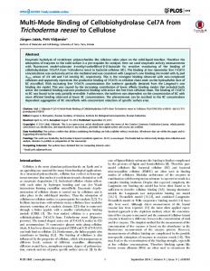
b
a
1
2 3 123
Proc. Natl. Acad. Sci. USA 86 (1989)
trifugation through a 10-40% sucrose gradient. Fragments of T. reesei DNA [15-20 kilobases (kb)] obtained by partial digestion with Mbo I were ligated to the purified EMBL4 arms. After in vitro packaging, the resulting phage were plated on Escherichia coli strain "Q359" (18) and plaques were transferred to Hybond-N membranes (Amersham). To identify CBH-I clones, a 58-mer oligonucleotide corresponding to bases 739-796 of the CBH-I nucleotide sequence (20) was labeled at the 5' end with [y-32P]ATP and used as a probe. Membranes were prehybridized at 55°C for 2 hr in 5 x SSPE (lx SSPE = 0.18 M NaCl/10 mM phosphate, pH 7.4/1 mM EDTA)/10x Denhardt's solution (19)/0.1% SDS/heatdenatured salmon sperm DNA (100 ,g/ml). Hybridization was carried out at 55°C for 16-20 hr. Membranes were washed in 5 x SSPE for 30 min at room temperature and then washed at 60°C for 10 min. Membranes were exposed overnight to X-Omat K film (Kodak). RNA isolation and analyses. T. reesei mycelium was collected by filtration and ground to a fine powder under liquid N2. Total RNA was isolated from the frozen powder by the guanidine thiocyanate method described by Chirgwin et al. (21). RNAs were separated by electrophoresis on 1.5% agarose gels in the presence of methylmercuric hydroxide (22, 23) and transferred to Hybond-N membrane (24). Membranes were hybridized with nick-translated CBH-I 1.16-kb HindIII fragment.
RESULTS Isolation of T. reesei CBH-I Gene. Using a synthetic oligonucleotide, based on the previously reported gene sequence (20), we isolated a clone carrying the CBH-I gene. To establish the conditions for oligonucleotide hybridization, genomic T. reesei DNA was digested with Sma I, HindIII, and EcoRI; electrophoresed on a 0.8% agarose gel; and transferred to a nylon membrane. The membrane was prehybridized, hybridized, and washed as described. Restriction analysis revealed that the probe hybridized to fragments of 9.6, 1.16, and 0.72 kb, obtained by digestion of T. reesei DNA with Sma I, HindIl, and EcoRI, respectively (Fig. la). The size of each hybridizing band from the genomic digests was in agreement with that predicted from the known sequence and restriction map (20, 25). Approximately 105 EMBL4 recombinants were plated, grown, and transferred to duplicate Hybond-N membranes. The membranes were then hybridized with the radiolabeled probe under the conditions established above, resulting in the isolation of four positive clones (Fig. lb). DNA isolated from these clones was mapped by restriction endonuclease digestion. DNA from all four c
1
1 2 3
9.4-_
Kb -9.4
1.6-
-1.6
Kb
6139
0.5FIG. 1. (a) Southern blot analysis of T. reesei genomic DNA. The DNA was digested with Sma I (lane 1), HindIII (lane 2), and EcoRI (lane 3); 10 ,ug was added per lane and resolved on a 0.8% agarose gel, blotted to Hybond-N membrane, and hybridized as described. (b) Screening an EMBL4 recombinant T. reesei genomic DNA library for CBH-I clones. Plate 1, 2.5 x 10⁴ recombinant plaques were screened per 150-mm plate as described (one of four plates is shown). Plate 2, positive plaque was replated until all plaques on the plate produced a signal. (c) DNA from one clone was digested with Sma I (lane 1), HindIII (lane 2), and EcoRI (lane 3) resolved in a 0.8% agarose gel, blotted, and hybridized as described.
6140
Biochemistry: El-Gogary et al.
clones contained common Sma I, HindIII, and EcoRI restriction fragments and gave identical patterns to that shown in Fig. la (see Fig. ic). These results confirm that the four recombinants contain the sequence for the CBH-I gene. On this basis, the 1.16-kb HindIII fragment was subcloned into pBR322 and used as a probe in subsequent experiments. Induction of CBH-I mRNA by Cellulose and Sophorose. Initial investigations concerned whether cellulose (Avicel) could induce transcription of the CBH-I gene and whether this induction could be repressed by the addition of glucose. Fig. 2 (lane 2) shows that Avicel stimulates the transcription of the CBH-I gene, whereas glycerol has no effect (lane 1). As one characteristic of the cellulase system is its catabolic repression by glucose, we analyzed the presence of the CBH-I gene transcript in cells induced with Avicel for 21 hr and then exposed to 1% glucose for 1 hr. The stimulation brought about by Avicel (Fig. 2, lane 2) was completely repressed by the addition of glucose (lane 3). Thus, it appears that glucose represses the expression of the CBH-I gene at the pretranslational level. The effect of the soluble inducer sophorose on the activation of CBH-I transcription was also investigated and was clearly positive (lane 4). It is noteworthy that the CBH-I gene transcript was detected 14 hr after the addition of Avicel and only 4 hr after the addition of sophorose (data not shown). Effect of Cellulase Enzyme Antibodies on CBH-I mRNA Levels. To investigate whether induction of the CBH-I gene by cellulose requires the purported low constitutive activity of the cellulase system to produce a soluble inducer from cellulose, we attempted to block this activity by adding antibodies against the cellulolytic enzymes to the culture medium. To this end, the enzymes of the cellulolytic system, composed of CBH-I, CBH-II, EG-I, EG-I, and /3glucosidase, were purified to homogeneity (Fig. 3). Antibodies to each of the purified enzymes were developed in the rabbit, and IgG was purified as described. The induction of the CBH-I gene transcript by cellulose (Fig. 4, lane 2) was completely repressed by the prior addition of cellulase antibodies (lane 3). Control experiments using IgG from nonimmune rabbit showed no effect on the transcription of CBH-I (lane 4). Interestingly, the addition of cellulase antibodies 10 hr after addition of cellulose did not 1 2
(1989)
1 2 3 4 5 6 7
I .
FIG. 3. SDS/10% polyacrylamide gel electrophoresis of purified cellulases. Lane 2, P-glucosidase (5 ,ug). Lanes 3-6 contained CBH-I, EG-I, CBH-II, and EG-II, respectively (10 ,ug each). Lanes 1 and 7 contain protein molecular weight markers.
repress the expression of the CBH-I gene transcript (data not shown), despite the fact that the CBH-I gene transcript was not detected before 14 hr after addition of cellulose. It is especially pertinent that cellulase antibodies did not repress CBH-I gene transcription if sophorose was used as an inducer (lane 5). In contrast to the repression caused by the combined cellulase antibodies (lane 3), no such effect was observed when antibodies against individual enzymes were added singly (data not shown).
DISCUSSION With the abundance of cellulose in nature, there is an understandable interest in cellulase mobilizing processes. This plant polysaccharide is used as an energy source by a variety of microorganisms. Of these, T. reesei utilizes cellulose as a carbon source, although this induction of the cellulolytic enzymes is repressed by glucose (5). At present, it is not understood how an insoluble polymer such as cellulose, which cannot pass the fungal cell, can still regulate the expression of a cellulolytic enzyme system. Based on the observation that the products of cellulose hydrolysis and transglycosylation (e.g., cellobiose, sophorose) can induce cellulase synthesis (6, 9), a mechanism has been proposed 1
3 4
.
Proc. Natl. Acad. Sci. USA 86
2
3
4
5
i
FIG. 2. Effect of carbon source on the level of CBH-I mRNA. Total RNA was isolated. Aliquots containing 30 ,ug of RNA were fractionated electrophoretically in 1.5% agarose/methylmercuric hydroxide gels (22, 23). RNA was transferred to Hybond-N membranes and hybridized with a nick-translated 1.16-kb HindIII CBH-I fragment (see Fig. 1). Lanes: 1, T. reesei cells grown on 0.4% glycerol for 22 hr; 2, same as in lane 1 except that 0.75% Avicel was used in place of glycerol; 3, same as in lane 2 except that after 21 hr, 1% glucose was added; 4, same as in lane 1, except that 7 mM sophorose was used instead of glycerol.
FIG. 4. Effect of cellulase antibodies on the expression of CBH-I gene transcript. Total RNA was isolated, fractionated, and hybridized as described in Fig. 2. Lanes: 1, T. reesei cells grown on glycerol; 2, same as in lane 1, except that 0.75% Avicel was used in place of glycerol; 3, same as in lane 2, except that IgG to CBH-I, CBH-II, EG-I, EG-II, and ,B-glucosidase (final dilution, 1:40) was added before the Avicel; 4, same as in lane 3, except that nonimmune IgG was added instead of immune IgG; 5, same as in lane 3, except that sophorose was added together with immune IgG.
Biochemistry: El-Gogary et al. whereby the activity of low constitutive levels of extracellular cellulase activity toward cellulose produces a soluble compound that can enter the cell and effect induction (6, 7). With a view to examining the mechanism by which the insoluble cellulose polymer triggers the expression of the cellulase genes, we have studied the expression ofthe mRNA transcript of CBH-I, the major cellulase gene in T. reesei. As demonstrated by blot hybridization analysis, CBH-I mRNA could not be detected up to 14 hr or 4 hr after induction with cellulose and sophorose, respectively. That sophorose is a more rapid inducer than cellulose would be expected from its soluble nature and from the possibility that it or some oligosaccharide has to be formed in vivo from cellulose by the activity of a low, constitutive, extracellular, cellulase system. In contrast to this relatively slow induction of mRNA, no CBHI-I transcript could be detected from T. reesei cultures induced for 21 hr with cellulose and then treated with glucose for 1 hr. While we cannot rule out the possibility that the presence or absence of CBH-I transcript is due to changes in the rates of mRNA turnover or RNA processing, transcriptional control seems more likely to be the level at which regulation of CBH-I mRNA occurs. It is noteworthy that the CBH-I gene transcript was absent when cellulase antibodies were present in cultures of T. reesei prior to the addition of cellulose. However, if T. reesei was induced with cellulose for 10 hr, during which time no CBH-I mRNA is detected, the addition of cellulase antibodies has no repressive effect on the expression of the CBH-I transcript. More critical is the fact that the cellulase antibodies have no effect if a soluble inducer (sophorose) was used-i.e., a soluble inducer bypasses the blockage of inducer formation from cellulose caused by the presence of cellulase antibodies. The failure of individual antibodies to block expression of the CBH-I transcript is not unexpected as the cellulolytic system is known to function synergistically (14). Our results strongly suggest that in T. reesei a low level constitutive extracellular cellulolytic system acting on cellulose is responsible for induction of the CBH-I gene transcript and most probably for the remainder of the cellulase system. We are indebted to Drs. Roberto Santelli and M. Clinton for their helpful discussions. We thank Mr. Wilton J. R. Lima for his excellent technical assistance. S.E.-G. was the recipient of a fellowship from Petrobras (1987-1988) and is presently a recipient of a fellowship from Coordenacao de Aperfeicoamento de Pessoal de Nivel Superior (CAPES 38/88-2). This work was supported by grants from the
Proc. Natl. Acad. Sci. USA 86 (1989)
6141
Financiadora de Estudos e Projetos (Finep. 43.87.0160.02), the Programa de Apoio ao Desenvolvimento Cientifico e Tecnologico (PADCT 053/84), and the New Jersey Agricultural Experiment Station (D-01111-2-89). 1. Ljungdahl, L. G. & Eriksson, K. E. (1985) in Advances in Microbial Ecology, ed. Marshall, K. C. (Plenum, New York), Vol. 8, pp. 237-299. 2. Coughlan, M. P. & Ljungdahl, L. G. (1988) in Biochemistry and Genetics of Cellulase Degradation, eds. Aubert, J.-P., Beguin, P. & Millet, J. (Academic, New York), pp. 11-30. 3. Montenecourt, B. S. (1983) Trends Biotechnol. 1, 156-161. 4. Eveleigh, D. E. (1987) Phil. Trans. R. Soc. London A 321, 435-447. 5. Mandels, M. & Reese, E. T. (1957) J. Bacteriol. 73, 269-278. 6. Mandels, M. & Reese, E. T. (1960) J. Bacteriol. 79, 816-826. 7. Sternberg, D. & Mandels, G. R. (1979) J. Bacteriol. 139, 761-769. 8. Mandels, M., Parrish, F. W. & Reese, E. T. (1962) J. Bacteriol. 83, 400-408. 9. Vaheri, M., Leisola, M. & Kauppinen, V. (1979) Biotechnol. Lett. 1, 41-46. 10. Crook, E. M. & Stone, B. A. (1957) Biochem. J. 65, 1-12. 11. Kubicek, C. P. (1987) J. Gen. Microbiol. 133, 1481-1487. 12. Montenecourt, B. S. & Eveleigh, D. E. (1979) Adv. Chem. Ser. 181, 289-301. 13. Shoemaker, S., Watt, K., Tsitovsky, G. & Cox, R. V. (1983) BiolTechnology 1, 687-690. 14. Henrissat, B., Driguez, H., Viet, C. & Schulein, M. (1985) BiolTechnology 3, 722-726. 15. Nelson, N. (1944) J. Biol. Chem. 153, 375-380. 16. Dahlqvist, A. (1968) Anal. Biochem. 22, 99-107. 17. Bradford, M. M. (1976) Anal. Biochem. 72, 248-254. 18. Frischauf, A. M., Lehrach, H., Poustka, A. & Murray, N. (1983) J. Mol. Biol. 170, 827-842. 19. Maniatis, T., Fnitsch, E. F. & Sambrook, J. (1982) Molecular Cloning: A Laboratory Manual (Cold Spring Harbor Lab., Cold Spring Harbor, NY). 20. Shoemaker, S., Schweikart, V., Lander, M., Gelfand, D., Kwok, S., Myambo, K. & Innis, M. (1983) Bio/Technology 1,
691-6%. 21. Chirgwin, J. M., Przybyla, A. E., MacDonald, R. J. & Rutter, W. J. (1979) Biochemistry 18, 5294-5299. 22. Bailey, J. M. & Davidson, N. (1976) Anal. Biochem. 70, 75-85. 23. Simone, M. P., Besmond, C., Cottreau, D., Weber, A., Chaumet-Riffaud, P., Dreyfus, J. C., Trepat, J. S., Marie, J. & Kahn, A. (1983) J. Biol. Chem. 258, 14576-14584. 24. Thomas, P. S. (1980) Proc. Natl. Acad. Sci. USA 77, 52015205. 25. Teeri, T., Salovuori, I. & Knowles, J. (1983) BiolTechnology 1, 6%-699.