Dec 1, 1986 - displayed in Figure 2 was performed at high salt concentrations (0.5 M ..... Bunte, T., Greiser-Wilke,I., Donner, P. and Moelling, K. (1982) EMBO.
Volume 15 Number 1 1987
Volume
15
Number
1
1987
Nucleic Acids Research
Chromatin association and DNA binding properties of the c-fos proto-oncogene product
Manfred Renz, Bernard Verrier, Christina Kurz and Rolf Muller
European Molecular Biology Laboratory, Postfach 10.2209, D-6900 Heidelberg, FRG Received October 17, 1986; Revised and Accepted December 1, 1986
ABSTRACT As a first step in the analysis of the molecular function of the nuclear c-Qa proto-oncogene product we have studied its subnuclear localization in serum-stimulated mouse fibroblasts where it forms a non-covalent, apparently monodisperse complex with another nuclear protein, p39. The c-Lfo/p39 complex is almost quantitatively released from intact nuclei by DNasel or micrococcus nuclease treatment under conditions where only a minor fraction of DNA and nuclear proteins is released. In gel filtration experiments, c-LQ.s/p39 comigrates with chromatin and seems to be associated with regions of increased DNasel accessibility. c-LQ.f/p39 is bound to chromatin by electrostatic forces of moderate strength since >90% of the complex can be eluted from nuclei at 0.4 M NaCI. In vitro, the c-fos/p39 complex in nuclear extracts binds to double- and single-stranded calf thymus DNA, suggesting that the association of c-LQf/p39 with chromatin is at least in part due to its interaction with DNA. In agreement with this conclusion, c-LQfs/p39 is released from nuclei by incubation with tRNA, presumably due to competition for binding sites. Our observations are compatible with the hypothesis that c-fos may play a role in the regulation of gene expression.
INTRODUCTION The mouse c-La gene is the cellular homolog of the oncogene carried by the FBJ and FBR murine osteosarcoma viruses (for review see refs. 1,2). It encodes a nuclear phosphoprotein with an apparent molecular weight of 55 kD (p55C-fOs) (1-3). The protein forms a tight but non-covalently linked complex with a 39 kD cellular protein, p39 (1-5). Both proteins are subject to extensive post-translational modifications (1-3,6,7). It has been shown by both immunofluorescence and cell fractionation experiments that the LQi gene products are localized in the nucleus (1-3,6,7). Thus, the fga gene product belongs to © I R L Press Limited, Oxford, England.
277
Nucleic Acids Research
the class of nuclear transforming proteins, along with several other cellular and viral oncoproteins, including myc, nyb, ak, adenovirus EA/EE1B, polyoma virus large T and SV40 large T proteins (for reviews see ref. 8). Of major interest has been the question of the normal function of proto-oncogene products and the mechanism by which their activated forms can induce cellular transformation. The possible biological function of c-Los has been extensively investigated using different approaches. These analyses mainly addressed two questions. First, in which tissues and cell types and in which conditions (e.g. state of differentiation or proliferation) is the c-Lat gene expressed, and second, what is the phenotypic consequence when exogenous c-jft genes are expressed in various recipient cells (e.g. on the differentiation state of the cell)? The results of these studies strongly suggest multifunctional properties for the c-fos gene product. This is based on several observations. A role for c-Lga in cellular differentiation has been proposed because of its high, specific expression in certain types of terminally differentiated cells (i.e., blood granulocytes (9) and late gestation ammion cells (10)} and because of its differentiationpromoting effect in certain teratocarcinoma cell lines (11,12; for a review see ref. 1). On the other hand, c-g. seems to play a role in cellular proliferation. This is based on the observations that its deregulated expression in fibroblasts induces cellular transformation (i.e., uncontrolled growth) (13) and that it is rapidly and transiently induced by various polypeptide growth factors and other growth inducing agents (for review see refs. 1,2). The fact that regulation of cftj expression by external signals seems to be a general phenomenon (1,14) has lent support to the hypothesis that its gene product is involved in intranuclear signal transduction, i.e., participates in the regulation of other cellular genes. In this way, the apparently multifunctional properties could be explained by assuming that c-jo affects different genes in different cell types with the consequence of different phenotypic results. To test this hypothesis and to elucidate the molecular function of c-tat protein it is of crucial importance to determine its sub-nuclear localization and to analyse its biochemical properties, in particular its affinity for DNA. In this paper, we provide strong evidence that the cfLQ/p39 complex is associated with chromatin and binds to DNA ina 278
Nucleic Acids Research vitro. These findings are in agreement with the hypothesis that c-Qa may be part of the transcription controlling machinery.
MATERIALS AND METHODS Cell culture. labeling of cells and isolation at nuclei NIH3T3 cells were grown in DMEM supplemented with 10% fetal calf serum (FCS) to approximately 80% confluency. Prior to stimualtion, the cells were kept in serum-free medium for about 20 hrs. The medium was then replaced with methionine-free DMEM plus 10% FCS. Forty-five min later, the cells were labeled by addition of 0.4 mCi of 35Smethionine (Amersham) per ml medium. Labeling was carried out for 45 min. For isolation of nuclei, NIH3T3 cell monolayers were washed three times with phosphate-buffered saline (PBS), scraped in chilled PBS, and pelleted at 800 xg and 40C for 5 min. Cell pellets (- 2 x 10⁸ cells each) were resuspended in 2ml of 10 mM Tris-HCI (pH 7.4), 10 mM NaCI, 3 mM MgCI2, 0.5% Nonidet P-40, incubated for 3 min on ice, homogenized in a Dounce homogenizer and centrifuged at 800 xg and 40C for 5 min. The nuclear pellets were washed twice with 10 ml of 10 mM Tris-HCI (pH 7.4), 50 mM NaCI, 3 mM MgCI2 and centrifuged as above. The nuclei were resuspended in 200 p1 of 10 mM Tris-HCI (pH 7.4), 50 mM NaCI, 5 mM MgCI2, 87% glycerol and stored at -800C. DEAE-cellulose chromatography of the c-fos/p39 complex Nuclei (1.5x10⁷) were incubated at 0°C for 40 min in 100 p1 of 0.4 M NaCI, 40 mM Tris-HCI, pH 8.0, 1 mM MgCI2, PAL (PAL is 0.1 mM phenyl methansulfonylfluoride (Fluka), 1x10³ units aprotinin (Sigma)/ml, 50 gg leupeptin (Sigma)/ml). The nuclear suspension was centrifuged for 3 min at 15 000 xg and the supernatant (80 p1) was diluted with 569 p1 of 10 mM Tris-HCI, pH 8.0, 1 mM MgC12, 0.1 mM dithiothreitol, 0.05% Nonidet P-40, PAL, and applied to a 0.5 ml diethylaminoethyl cellulose (DE52, Whatman) column equilibrated with 50 mM NaCI, 5 mM Tris-HCI, pH 7.4, PAL, prior to sample application. The column was rinsed with 1 ml of equilibration buffer and the c-Lf/p39 complex was released with 0.5 ml of 0.2 M NaCI, 5 mM Tris-HCI, pH 7.4, PAL. Sucrose gradient assay Sucrose gradient analyses were carried out by layering 200 p1 of a 0.2 M NaCI eluate of a DEAE-cellulose column onto preformed linear 520% sucrose gradients in a buffer containing 1mM MgCI2, 10 mM TrisHCI, pH 7.4, 0.2 M NaCI, 0.05% Nonidet P-40, PAL. Centrifugation was 279
Nucleic Acids Research carried out in a Beckman SW60 rotor for 22 h at 40C. Fractions of constant volume were collected and immunoprecipitated. To estimate the size of the c-foa/p39 complex 14C-labeled protein markers were run in a separate tube. Dissociation of the c-fos/p39 complex from nuclei by NaCI Aliquots of 2x10⁶ nuclei were suspended each in 100 gl of a solution containing 5 mM Tris-HCI, pH 7.4, 3 mM MgC12 and different concentrations of NaCI. After centrifguation at 15,000 xg for 10 sec both supernatants and pellets were analyzed by immunoprecipitation. Extraction of the c-fos/p39 complex from nuclei with tRNA Aliquots of 4x10⁶ nuclei were suspended each in 10 gil of 3 mM MgCI12, 5 mM Tris-HCI, pH 7.4, 70 mM NaCI and different amount of yeast tRNA (Boehringer, Mannheim). After centrifugation for 10 sec at 15,000 xg both sediments and supernatants were analyzed by imm unoprecipitation. Chromatin isolation and fractionation Nuclei (3x10⁷) were digested with 200 units micrococcal nuclease (Boehringer, Mannheim) in 40 gIl of 40 mM NaCI, 5 mM Tris-HCI, pH 7.4, 1 mM MgCI2, 1 mM CaCI2, for 10 min at 0°C. The reaction was terminated with 5 gi of 50 mM EDTA, pH 8.0, and the lysed nuclear suspension diluted with 150 gi of buffer A (40 mM NaCI, 5 mM Tris-HCI, pH 7.4, 0.5 mM EDTA, PAL), then centrifuged for 5 sec at 15 000 xg and the superantant was separated on a 2 ml Sepharose CL-2B (Pharmacia) column equilibrated and eluted with buffer A. Fractions of 300 Ail volume were collected and analyzed for DNA and proteins. Release of the c-fos/p39 complex by nuclease a) Micrococcal nuclease digestion: Nuclei (1.5x10⁷) were digested with 400 or 1200 units micrococcal nuclease in 50 gil of 40 mM NaCI, 5 mM Tris-HCI, pH 7.4, 1 mM CaC12, 1 mM MgCI2,- for 15 min at OOC. After centrifugation for 10 sec at 15,000 xg 5 pi of 0.5 M EDTA, pH 8.0, were added to both pellet and supernatant. Samples were analyzed for DNA and proteins. b) DNase I digestion: Nuclei (1.5x10⁷) were digested with 40 or 120 units DNasel in 50 ,Il of 40 mM NaCI, 10 mM Tris-HCI, pH 7.4, 2 mM MgC12 for 15 min at OOC. The nuclear suspension was centrifuged at 15,000 xg for 10 sec and the reaction was stopped by adding 5 gil of 0.5 M EDTA, pH 8.0, to the supernatant and to the pellet, respectively. Aliquots were analyzed for DNA and proteins. 280
Nucleic Acids Research DNA binding experiments For DNA binding studies, 0.4 M NaCI extracts (see above) were diluted with a solution containing 5 mM Tris-HCI, pH 7.4, 0.5 mM EDTA, PAL (buffer B) to a final concentration of 50 mM NaCI and applied to 0.4 ml columns equilibrated with buffer B plus 50 mM NaCI prior to sample application. Three different column materials were used: cellulose (Serva), single-stranded calf thymus DNA cellulose (Sigma) *and doublestranded calf thymus DNA cellulose (Sigma). The columns were eluted stepwise with solutions of increasing concentrations of NaCI in buffer B. Immunoprecipitation analysis Nuclear pellets were dissolved in 200 p1 of RIPA buffer (10 mM Tris-HCI, pH 7.4, 0.15 M NaCI, 1% Triton X-100, 1% sodium deoxycholate,
69K 46K
200K 30K 92.5K 1 2 3 4 5 6 7 8 9 101112
]c-fos MA.,
P39
p
Figure 1: Sucrose gradient analysis of c-LQfip39. 14C-methylated molecular weight markers, indicated by horizontal lines, are from the left to the right: carbonic anhydrase (30 kD), ovalbumin (46 kD), bovine serum albumin (69 kD), phosphorylase b (92.5 kD), myosin (200 kD). Numbers designate fractions. Direction of sedimentation is from left to right. 281
Nucleic Acids Research
~ ~ ~.
0.1% SDS, PAL) at 0OC and centrifuged for 3 min at 15 000 xg and 40C. Supernatants were withdrawn, 1 ,ul of anti-13-gal-LQ. serum (15) was added to each sample and incubated for 40 min at QOC. Fractions from columns or sucrose gradients as well as nuclear supernatants were mixed with equal volumes of 2x RIPA buffer and incubated with serum fixed as described above. Ten microliters of a suspension of staphylococcus aureus cells (Pansorbin, Calbiochem) were added and incubation was continued for 20 min. Samples were centrifuged for 10 sec at 15 000 xg and pellets were washed 2 to 3 times in RIPA buffer.
N 1 2 3 4 5 6 7 8 910M _
:
Kb L
~~~~~~~~~~~~~~~~~~~3.5
o .6 1f r1 056 _ L sL3 02:\1 -1--
2
4 6 8 10 Fraction No.
2 3
N
c
5
6
7
I XIA. H~~~~~~~~~~~~.
H3
AW 282
[
W~~~~~~~~
Ll l;, _ 7 H2
H
s~~~~MMb. _
-1 0.:,
Nucleic Acids Research
d
N
1
2
3 4
5 6 7
8
9 10
.~~~~~~~~~~~~~~~~~~~~~~~~~~~~~~~~~~~~~~~~~~~~W
r:
] p55
J p39
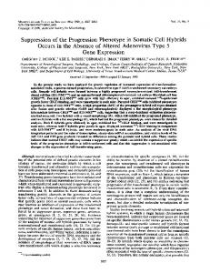
Figure 2: Chromatin fractionation on Sepharose CL-2B3 and analysis for DNA, total protein and LQ.a/p39. a: 0D260 profile. b: DNA agarose gel electrophoresis. c: PAGE of total proteins. d: Fluorogram of immunoprecipitated LQ.Wp39. N: unfractionated chromatin samples; M: size marker. The upper band of p39 comigrates with a protein that is unspecifically precipitated and not related toia or p39, as shown by precipitation of denatured lysates, peptide mapping and use of preimmune serum (data not shown). Likewise, some proteins of higher Mr are unspecifically precipitated. Pellets were then suspended in electrophoresis sample buffer, heated to 1 OOOC for 3 min, centrifuged and supernatants were run on SDSpolyacrylamide slab gels (16). Gels were prepared for fluorography (17) by fixation with a solution containing 10% trichloroacetic acid, 10% acetic acid, 30% methanol followed by treatment with EN3HANCE (NEN). Dried gels were exposed to Kodak XAR-5 or X-Omat 5 X-ray films. RESULTS AND DISCUSSION_ Sucrose gradient centrifugation analysis of the c-fos/p39 complex Sedimentation experiments were carried out to estimate the size of the c-LQ.j/p39 complex. A nuclear protein mixture, enriched in cLQ.ap39 by DEAE-cellulose chromatography, was used for sucrose gradient centrifugation and immunoprecipitation analyses. Fig. 1 shows the result of this experiment. More than 95% of the c-Lai_/p39 283
Nucleic Acids Research
sediments as a rather sharp and symmetrical band, indicating that the complex consists largely of monodisperse aggregates. To estimate the molecular weight of the complex 14C-labeled protein markers were run in a separate tube. Comparison with the sedimentation pattern of the protein markers (delineated as horizontal lines at top of Fig. 1) shows that the c-Lfo/p39 complex sediments at a slightly lower rate than bovine serum albumin (BSA). If BSA and c-Laa/p39 were similar in conformational shape the molecular weight of the c-f.Q.a/p39 complex would be similar to that of BSA, namely 69 000. The rigidity (e.g. shape) of c-fLos/p39, however, is at least for two reasons unlikely to be comparable to that of BSA: (i) c-LQ/p39 is a tight but non-covalently linked protein complex, and (ii) both c-fos and p39 are exceptionally highly modified proteins (1-7). Both conformation and modifications contribute most likely to the observed low apparent molecular weight which is less than the sum of the molecular weights of the components (c-LQ plus p39) of the complex. The main finding of the sedimentation analysis is that the complex does not seem to be composed of more than one molecule c-Lu and one molecule p39. Association of c-fos/p39 with chromatin To investigate if c-fos/p39 is a chromosomal protein chromatin was isolated from nuclei and fractionated by gel chromatography (Fig. 2a). Fractions were analyzed for DNA size by agarose gel electrophoresis (Fig. 2b), for proteins by PAGE (Fig. 2c) and for cLQ_o/p39 by immunoprecipitation analysis (Fig. 2d). About 20% of the isolated chromatin consists of rather large chromosomal fibers (Fig. 2a, fraction 3 and Fig. 2b, fraction 3) with the usual histone composition (Fig. 2c, fraction 3). (The nature of the additional, abundant protein of somewhat larger molecular weight than histone H3 is unknown). Interestingly, there is only very little, if any c-foQs/p39 associated with this chromatin fraction (Fig. 2d, fraction 3). In contrast, almost all c-fLQ./p39 is bound to the smaller chromatin fragments (compare Fig. 2d, fractions 6/7 with Fig. 2b, fractions 6/7). The experiments described below show that c-fos/p39 is not dissociated from chromatin at the ionic conditions (40 mM NaCI) used. When the experiment displayed in Figure 2 was performed at high salt concentrations (0.5 M NaCI) leading to the complete dissociation of c-fos/p39 from chromatin (see below) the unbound c-Los/p39 complex appeared in fractions 8/9 (data not shown). 284
Oi)
M.N. DNasel 400 1200 40 120 S P S P S P S P
(©
~~
Nucleic Acids Research
0
M.N. ONasel s sP
DNaseI Chr.CM.N. S P S P M
__H DNasel ....B
p39
[~~
H4
Figure 3: Release of c-LQ./p39 by nucleases. Nuclei were digested with 400 or 1200 units micrococcus nuclease (M.N.) or with 40 or 120 units DNasel, separated into nuclear pellets (P) and supernatants (S). a: Fluorogram of immunoprecipitated fQ.s/p39. b: PAGE of total proteins. Lanes 1,2: 1200 units M.N.; lanes 3,4: 120 units DNasel. c: DNA agarose gel electrophoresis. Lanes 2 through 5 as lanes 1 through 4 in panel b. Chr.: nucleosome ladder; M: size marker, X DNA digested with fcRI and
tiLadd I. c-fos/p39 is released from nuclei by nucleases To confirm the observed chromatin binding property of c-Lfo/p39 nuclei from stimulated NIH3T3 cells were treated with two unspecific nucleases, namely micrococcal nuclease and DNase I respectively. After extensive digestion the nuclear suspensions were centrifuged without prior lysis of the nuclei. Supernatants and pellets were analyzed for cfgV/p39, total proteins and DNA as described above. The majority of the c-fos/p39 complex is released from nuclei by nuclease treatment (Fig. 3a, lanes 3 and 7) along with a small amount of other proteins (Fig. 3b, lanes 1 and 3) and very little DNA (Fig. 3c, lanes 2 and 4), whereas the bulk of proteins and DNA remains in the nucleus (Fig. 3b, lanes 2 and 4; Fig. 3c, lanes 3 and 5). The amount of c-LQa/p39 appearing in the supernatant is clearly dependent on the concentration of nuclease used showing that the complex is indeed released by the nuclease activity 285
Nucleic Acids Research
ji~
~~~~~~~~.
. ...
Figure 4: Dissociation of c-LQ.a/p39 from nuclei by NaCI. Fluorogram of immunoprecipitated nuclear sediments and supernatants after incubation of nuclei in buffers containing the indicated concentrations of NaCI (in mM). C: RIPA-extracted nuclei. rather than by another aspect of the treatment (Fig. 3a; compare lanes 1,2 with lanes 3,4 and lanes 5,6 with lanes 7,8). In contrast, no cLQ.i1p39 is released when nuclei are treated with RNase A (2 mg/mI for 15 min at OC; data not shown). Ionic strength dependence of the c-fos/p39-chromatin interaction To assess the importance of electrostatic interactions of cLQ_$i/p39 with chromatin nuclei were extracted with solutions containing increasing concentrations of NaCI. After centrifugation nuclear pellets and supernatants were analyzed by immunoprecipitation. Fig. 4 shows the result of such an experiment. No c-Lo-a/p39 is released from nuclei in buffers containing up to 100 mM NaCI. At 200 mM NaCI, however, about 50% of c-LQ.a/p39 dissociates and at 300 mM NaCI only very little remains bound to chromatin; at 400 mM NaCI >90% of LgW.p39 dissociates. c-InQ./p39 thus belongs to a subset of nonhistone 286
Nucleic Acids Research
0
nuclei 10 30 90
supernatant 0
~~~~~-
10 30 90
4.
_-
_
3Exw- w tJP~~~]55
Figure 5: Extraction of c-LQ.s/p39 with tRNA. Nuclei were incubated with the indicated amounts of tRNA (in pg) and, after separation into nuclear pellets and supernatants, immunoprecipitated.
chromosomal proteins which are released from chromatin at moderate ionic strength. Addition of tRNA to nuclei releases c-fos/p39 Some of the nonhistone proteins as well as histone Hi are transferred from chromatin to exogenous nucleic acids added to isolated nuclei (16,17), presumably due to competition with DNA binding sites. It was therefore of interest to investigate whether cfQ.s/p39 belongs to this subclass of nonhistone proteins. To address this question increasing amounts of tRNA were added to nuclear suspensions. After centrifugation nuclear pellets and supernatants were analyzed by immunoprecipitation. Fig. 5 shows that indeed a substantial part of cLfo/p39 is transferred to exogenous tRNA when a 4-fold excess of tRNA (90g) over chromosomal DNA was used. DNA-binding properties of c-fos/p39 Most of the experiments described so far suggest that c-1Qa/p39 might be a DNA binding protein complex. DNA cellulose column chromatography was used to verify this supposition. A sample enriched 287
Nucleic Acids Research *
, 0) c'
0)
r
CL
Is
zn .CC o Cs a)r D
a
X
_ 1s
s:
I. .
..
0 00 Co a0)
7CO C')
±s
C
,2A, ,_
I 0
co
C.)
I
';
T
E
~.
_0 E CL:=
n as
L'.,
0Z Q0
'I i
C Ge
GA
LI)I U 01 .
o 0as o
'I
CE E c 00 jt,
cm
I I
0'
n0
I--L
o Z C_ (D
.2 ;1~~~~~~~~~~~~ iDL. C&\1 0) * Z Co
Z)
-_1 as (LT
0 0
f3 0J .-
0
z!S5i
0
E=
.f
HO.
U-
288
COO c LLo..CZ 0 o ' .1 '(0
0E. 3F-r
0aCs a fiC) c
+
Nucleic Acids Research in c-jfoj/p39 by NaCI extraction (see Materials and Methods) was applied to a single-stranded calf thymus DNA cellulose column at 50 mM NaCI and, after washing with a buffer containing 50 mM NaCI, the column was stepwise eluted with solutions of increasing ionic strength. About 25% of c-Lfo/p39 does not bind to single-stranded DNA at 50 mM NaCI (Fig. 6b, flow through and wash). At 0.15 M NaCI about 20% of the complex is released whereas the majority is eluted with a buffer containing 0.3 M NaCI (Fig. 6b, lanes 0.15 and 0.3). Since no binding of cfos/p39 to plain cellulose was observed (Fig. 6a), these findings show that the c-fQosp39 complex has an affinity for single-stranded DNA. Comparison of the protein pattern in the c-L region of the 3 column fractions, namely the run through-wash, the 0.15 M and the 0.3 M NaCI eluate, reveals small but recognizable differences in electrophoretic mobility of c-La proteins, in that the faster migrating protein elutes at higher salt concentrations. In contrast, the relative intensities of the three p39 bands as well as the ratio of c-fa:p39 seems to be very similar in all three column fractions. These findings suggest that different modified forms of c-LQ/p39 may exhibit differential binding to single-stranded DNA. In all likelyhood, the c-L protein rather than p39 is responsible for these different interactions. To study the binding of c-fos/p39 to double stranded DNA a similar experiment was performed using a double-stranded DNA cellulose column. A qualitatively similar picture emerges. Some of the c-fos/p39 does not interact with double-stranded DNA at 50 mM NaCI (Fig. 6c, flow through and wash), but most of the complex remains bound to the column as was the case with the single-stranded DNA column. Again, the slower migrating c-LQ molecules are enriched in the 0.15 M NaCI fraction (compare Fig. 6b, lane 0.15 with Fig. 6c, lane 0.15). The binding strength, however, is different for double- and single-stranded DNA: while all c-Lfsp39 is eluted at 0.3 M NaCI from the single-stranded DNA column most of it still binds to the double-stranded DNA column and is released only at higher ionic strength (compare Fig. 6b and 6c).
CONCLUSIONS The data presented in this paper strongly suggest an association of the c-Los1/p39 complex with chromatin. First, c-Lfo/p39 is released from nuclei by micrococcus nuclease and DNasel. Second, the c-fL.a/p39 complex cofractionates with chromatin in gel filtration experiments. 289
Nucleic Acids Research
Third, c-f.o/p39 binds to DNA in vitro and can be released from nuclei with tRNA, presumably by competition with DNA binding sites. Two lines of evidence suggest that c-LQa/p39 is not randomly distributed in chromatin. First, DNasel treatment releases >90% of the c-LQzi/p39 complex from intact nuclei under conditions where >90% of the DNA and other nuclear proteins remain in the nucleus. The fragments released from the nucleus together with c-Lfa/p39 were found to be very short. Second, after separation of chromatin by gel filtration, c-LQf./p39 was found preferentially in those fractions containing small chromatin pieces. These observations indicate that c-LgQa/p39 may be associated with chromatin regions showing nuclease hypersensitivity, e.g., transcriptionally active regions (18). The subnuclear location of cLfa/p39 thus seems to be similar to that of the v-myb and c-myb gene products (19), but different from SV40 large T antigen (20) which is predominantly found in the nucleoplasm and nuclear matrix. The subnuclear location of myc proteins does not seem to be resolved at this point. The previously reported association of myc proteins with the nuclear matrix (21) has recently been shown to be due to the particular experimental conditions, i.e., due to irreversible precipitation of the protein during incubation of the nuclei at 370C (22). Other investigators have found association of myc protein with chromatin (23 and quoted as unpublished data in ref. 19). It is thus difficult to judge whether the subnuclear location of myc protein is similar to or different from that of the c-LQfop39 complex. Interestingly, c-foQ.s/p39 does not show the thermolability described for myc and myb, in that it remains soluble after incubation of nuclei at 370C (our unpublished observations). Of particular interest are the DNA binding properties of c-fo/p39, although at this point no sequence-specific interaction with DNA is evident. However, sequence-specific binding has not been described to date for any retroviral or cellular oncogene product, although it is well documented for DNA tumor virus-encoded proteins, such as SV40 large T antigen, where the transforming protein interacts with defined viral sequences (24). The unequivocal identification of cellular target sequences for SV40 large T protein, however, remains the subject of future investigations. The relevance of the in vitro DNA binding properties of c-Lfo/p39 (and other oncogene-encoded proteins) for its (their) normal or transforming functions remains unclear. The fact, however, that c-ffo/p39 seems to be associated with chromatin and 290
Nucleic Acids Research that this association can apparently be disrupted by competition with tRNA suggests that interaction with DNA may be crucial to the function of cThree important questions need to be addressed now: (i) Does cLQ.f/p39 interact with specific DNA sequences? This question can be studied, for instance, with end-labeled restriction fragments of phage X DNA, which may contain degenerate versions of recognition sites for DNA binding proteins (25), or by molecular cloning of DNA sequences associated with LQ-/p39. (ii) What is the role in DNA binding of nuclear cofactors, such as p39? This can be addressed, for instance, by analyzing the DNA-binding properties of purified 1a protein in the presence or absence of nuclear extract. (iii) What is the role of posttranslational modifications? The data shown in Fig. 6b,c could suggest that highly modified protein has a lower affinity for DNA. This possibility, which needs to be investigated in detail, is intriguing in view of the previously presented hypothesis that posttranslational modifications may regulate the biological (i.e., transforming) activity of the protein (6,7).
ACKNOWLEDGEMENT We are grateful to Wendy Moses for preparation of the manuscript.
REFERENCES 1. Muller, R. (1986) Biochim. Biophys. Acta 823:207-225. 2. Verma, l.M. (1986) Trends Genet. 2:93-96. 3. Curran, T., Miller, D.A., Zokas, L. and Verma, I.M. (1984) Cell 36:259-268. 4. Curran, T. and Teich, N.M. (1982) Virology 116:221-235 5. Curran, T., Van Beveren, C., Ling, N. and Verma, l.M. (1985) Mol. Cell. Biol. 5:167-172. 6. Kruijer, W., Cooper, J.A., Hunter, T. and Verma, l.M. (1984) Nature 312:711-716. 7. Muller, R., Bravo, R., Burckhardt, J. and Curran, T. (1984) Nature 312:716-720. 8. Bishop, J.M. (1985) Cell 42:23-38. 9. Kreipe, H., Radzun, H.J., Heidorn, K., Parwaresch, M.R., Verrier, B and Muller, R. (1986) Differentiation (in press). 10. Muller, R., Verma, I.M. and Adamson, E.D. (1983) EMBO J. 2.:679684. 11. MUller, R. and Wagner, E.F. (1984) Nature 311:438-442. 12. ROther, U., Wagner, E.F. and MUller, R. (1985) EMBO J. 4:1775-1781. 13. Miller, A.D., Curran, T. and Verma, l.M. (1984) Cell 36:61-60. 14. MUller, R., MUller, D., Verrier, B., Bravo, R. and Herbst, H. (1986) EMBO J. 5:311-316. 15. Verrier, B., MUller, D., Bravo, R. and MUller, R. (1986) EMBO J. 5:913-917
291
Nucleic Acids Research 16. 17. 18. 19. 20. 21. 22. 23. 24. 25.
292
Jensen, R.H. and Chalkley, R. (1968) Biochem. 7:4388-4395. Ilyin, Y.V., Varshavsky, A.Y., Mickelsaar, U.N. and Georgiev, G.P. (1971) Eur. J. Biochem. 22:235-245. Elgin, S.C.R. (1984) Nature 309:213-214. Klempnauer, K.H. and Sippel, A.E. (1986) Mol. Cell. Biol. 6:62-69. Staufenbiel, M. and Deppert, W. (1983) Cell 33:173-181. Eisenman, R.N., Tachibana, C.Y., A rams, H.D. and Hann, S.R. (1985) Mol. Cell. Biol. 5:114-126. Evans, G.I. and Hancock, D.C. (1985) Cell 43:253-261. Bunte, T., Greiser-Wilke, I., Donner, P. and Moelling, K. (1982) EMBO J. 1:919-927. Lane, D.P., Simanis, V., Bartsch, R., Yewdell, J., Gannon, J. and Mole, S. (1985) Proc. Roy. Soc. Lond. B226:25-42. Desplan, C., Theis, J. and O'Farrell, P.H. (1985) Nature 318:630635.