0034. Expression, Isolation, and Characterization of a Signal. Sequence-Appended Chimeric Precursor Protein. Naheed Kaderbhai and Mustak A. Kaderbhai1.
PROTEIN EXPRESSION AND PURIFICATION ARTICLE NO.
7, 237–246 (1996)
0034
Expression, Isolation, and Characterization of a Signal Sequence-Appended Chimeric Precursor Protein Naheed Kaderbhai and Mustak A. Kaderbhai1 Institute of Biological Sciences, The University of Wales, Aberystwyth, Dyfed SY23 3DD, United Kingdom
Received August 8, 1995, and in revised form November 17, 1995
This report describes the properties and the functional utility of an unprocessed precursor protein overproduced in Escherichia coli. The precursor protein is from a fusion between DNA sequences coding for the alkaline phosphatase signal sequence and the full-length of rat liver cytochrome b5 . The intact precursor protein accumulated in the membranes represented to over 5% of the total bacterial protein. A procedure involving disruption of the bacterial cells by sonication, isolation of the membranes by differential centrifugation, solubilization with a polar solvent, and ion-exchange chromatography provided milligram quantities of the undegraded precursor in a homogeneous and soluble form. The chimeric precursor protein displayed a characteristic b-type hemoprotein spectrum, identical to that of the native cytochrome b5 . The properties of the precursor protein have been examined by a range of biophysical and biochemical methods. Molecular modeling suggests an amphipathic structure in which a fully preserved soluble core of cytochrome b5 is terminally bonded by hydrophobic interactions between the amino-terminal signal sequence and the carboxy-terminal membrane anchoring hemoprotein sequence. The precursor substrate was recognized and efficiently cleaved by signal peptidase. q 1996 Academic Press, Inc.
Over the last decade there have been significant advances in our understanding of the protein secretory pathways of both the prokaryotes and eukaryotes (1). As protein secretion is an essential bioactivity of all living cells, the prokaryotic secretory pathway has been proposed as a viable target for intervention with novel therapeutic or biocidal agents (2). An essential component of the prokaryotic secretory pathway (3) identified as a potential biocidal target is the signal peptidase 1
(4). This endopeptidase processes the N-terminal signal sequences in the secretory as well as the cytoplasmic and outer membrane precursor proteins (5,6). Sequence comparisons of signal peptidases of Gram-positive and Gram-negative organisms indicate that the enzyme is highly conserved across the prokaryotic kingdom (7). The Escherichia coli enzyme is a monomeric polypeptide of about 37 kDa, coded for by the lepB gene and found N-terminally anchored in the cytoplasmic membrane exposing the catalytically active core toward the periplasmic space (8). A variety of in vivo and in vitro studies has shown that signal peptidase accomplishes the processing of structurally diverse signal sequences in precursor proteins with high fidelity (9). Site-directed mutagenesis studies suggest that the E. coli signal peptidase is a serine protease type with the catalytic mechanism involving an unusual serine–lysine dyad (10,11). However, the enzyme is insensitive to inhibition by all the classic inhibitors of proteases (12) and the reaction mechanism of signal peptidase remains obscure because of lack of detailed structure. Moreover, determinations of kinetically meaningful activities of signal peptidase are presently hampered by the lack of substrate-level quantities of suitable precursor protein. Natural precursor substrates from normally growing cells have proved inappropriate for this purpose because they are short-lived (T1/2 õ 1 min) and are unstable (13). Although recombinant approaches have been successfully applied for overproduction of full-length precursor proteins (14– 16), the accumulation of the latter in the form of inclusion bodies, despite efforts to renature them (17), has precluded their uses in studying the processing events and for assaying the signal peptidase activity. We have been using a mammalian cytochrome b5 (b5T)2 as a model protein to investigate protein topogen2 Abbreviations used: b5 , globular domain of cytochrome b5 ; b5-T, full-length cytochrome b5 ; SS, signal sequence of E. coli alkaline phosphatase; SS-b5-T, signal sequence-appended full-length cyto-
To whom correspondence should be addressed.
237
1046-5928/96 $18.00 Copyright q 1996 by Academic Press, Inc. All rights of reproduction in any form reserved.
/ 6q05$$0565
03-20-96 17:17:55
pepa
AP-PEP
238
KADERBHAI AND KADERBHAI
esis in E. coli. The b5-T of liver endoplasmic reticulum is a well-characterised, small hemoprotein of 15.5 kDa which plays a central role in a variety of electron transfer reactions related to fatty acid desaturation, redox cycling of estrogen, cholesterol synthesis, and detoxification of xenobiotics (18,19). The hepatic b5-T is an amphipathic protein made up of two domains: a soluble, enzymically active, heme-containing globular core (b5) domain comprosed of the first 99 amino acid residues and a smaller membrane-anchoring domain at the carboxy terminus composed of 35 amino acid residues (T). B5-T belongs to a class of wide-spread, integral proteins with a monotopic membrane arrangement in which the globular b5 domain is laterally disposed facing the cytoplasm (20). Anchorage of b5-T in the endoplasmic reticular membrane is provided by a hydrophobic stretch of 22 uncharged amino acid residues in the T domain (21). The mechanism by which the T domain targets b5-T specifically to the endoplasmic is presently unknown. Our analyses of the membrane insertion sequence of b5-T suggested that the T portion contains a 25 amino acid residue helical motif which has comparable characteristics to stop-transfer sequences found in the transmembrane segments of biotopic proteins (22). Many proteins utilize an N-terminal, cleavable signal sequence in combination with a stop-transfer sequence to gain their characteristic bitopic membrane disposition (23). Therefore, to test the functionality of the potential stop-transfer sequence in b5-T, we have constructed a gene coding for a fusion form of b5-T carrying a secretory signal at its N terminus. Previously, we have reported that the soluble b5 core, although lacking a secretable homologue in nature, can be efficiently exported from E. coli by appending a signal sequence (SS) at its N terminus (24,25). In the present study we reinstated the T to the C terminus of SS-b5 to investigate the outcome of targeting of SS-b5-T in E. coli. Surprisingly, the expressed SS-b5-T was overproduced in an unprocessed form and selectively targeted to the inner membrane of E. coli. Here we describe the isolation and characterization of the unprocessed SS-b5-T precursor. Substrate-level quantities of the SS-b5-T precursor protein can be readily purified and it has proved to be an effective macromolecular substrate for E. coli signal peptidase. MATERIALS AND METHODS
DNA Manipulations New England Biolabs (U.K.) and NBL Gene Sciences (U.K.) supplied restriction endonucleases and DNA modifying enzymes; the reaction conditions used were chrome b5 ; T, C-terminal tail or membrane anchoring portion of cytochrome b5 .
/ 6q05$$0565
03-20-96 17:17:55
pepa
as recommended by the manufacturer. The standard operations involving DNA manipulations, i.e., purification, digestion, agarose electrophoresis, and bacterial transformation, were performed as described previously (26). The plasmid pA-sct harboring the precursor SS-b5-T gene and bearing the ampicillin resistance gene was constructed as follows: plasmid pAA-cyt (24) which expresses the secretory form of SS-b5 under the control of phoA promoter was doubly cleaved with XhoI and HindIII. Four staggered segments of double-stranded oligonucleotides coding for the extended portion of b5T were synthesised on glass beads using diisopropyl phosphoramidite intermediates on an automated Pharmacia-LKB Gene Assembler. The deprotected oligonucleotides were purified by filtration through NAP-10 columns, phosphorylated by the action of polynucleotide kinase and repurified using the Wizard DNA purification system (Promega, U.K.). Equimolar portions of the synthetic oligonucleotides were annealed with the larger fragment of XhoI–HindIII-cleaved pAA-cyt and the duplexes ligated by the action of T4 DNA ligase. Following their transformation and amplification in E. coli TB1, the recombinant plasmids were mapped with XhoI and HindIII restriction endonucleases. The sequence of SS-b5-T in the plasmid DNA isolated from one cell line was confirmed by dideoxy DNA sequencing (27). This E. coli TB1 strain/pA-sct was used in the present study. A 1200-bp DNA fragment coding the full-length of E. coli signal peptidase I gene (including the native ribosomal binding site) (8) was amplified by PCR. E. coli chromosomal DNA and the following pair of PstIadapted oligonucleotides served as the template and the primers, respectively. PstI primer 1: 5*-TTA CTGCAG AAATAACCCT TAGGAGTTGG-3* PstI primer 2: 5*-GTA CTGCAG A TCTCATAAAT AATTCACGTT-3*
The PCR reaction was performed in a final 50 ml volume containing 10 mM Tris–HCl (pH 8.8 @ 257C), 1.5 mM MgCl2 , 50 mM KCl, 0.1% (v/v) Triton X-100, 0.2 mM each dNTP, 10 mg E. coli chromosomal DNA, 25 pmol of each primer, and 2 units Thermus brockianus DNA polymerase. The mixture, overlaid with 50 ml paraffin oil, was subjected to 10 rounds of thermocycling involving denaturation at 947C for 1 min, annealing at 557C for 2 min, and extension at 727C using a programmed Hybaid thermocycler. DNA amplification was continued for 20 additional cyclings with the annealing temperature raised to 627C. The PstI-digested, agarose-purified, 1200-bp amplified DNA was ligated
AP-PEP
PRECURSOR SUBSTRATE FOR SIGNAL PEPTIDASE
into PstI-cleaved plasmid pMCY-ntp (University of Wales Plasmid Bank, Aberystwyth) in an orientation placing the signal peptidase gene downstream of the pho promoter. The resultant plasmid pMC-spase bearing the ampicillin resistance gene was contained in a E. coli TB1 host. Cultures The E. coli TB1 strains were maintained and propagated in LB liquid or solid media without or with ampicillin (75 mg/ml). For production of the recombinant proteins, E. coli strains carrying the relevant plasmid were batch cultivated for 6 h in a phosphate-limited (0.1 mM) MOPS medium (24) containing ampicillin (75 mg/ml); the incubation was at 357C with shaking at 125 rev/min. Purification of Signal Peptidase We purified the signal peptidase enzyme from the membranes derived from 1 liter of the low phosphateinduced E. coli/pMC-spase cells employing the procedure described by Wolfe et al. (5). Purification of SS-b5-T Protein The following procedures were performed at 47C. E. coli/pA-sct cells harvested (5000g 1 5 min) from 1 liter of culture were resuspended in 10 ml of 10 mM Tris– HCl (pH 8), 1 mM EDTA (TE) containing 10 mM bovine heme. The cells were treated with 0.1 mg lysozyme/ml and left to stand on ice for 30 min. Lysis was promoted by subjecting the suspension to 10 1 15-s sonications (Soniprep 150, MSE) with a 20-s interval between each pulse. The membranes were prepared from a post-cellular fraction (1000g for 20 min) by centrifugation at 105,000g for 1 h, resuspended in 2.5 ml TE buffer, and gradually added to 50 ml of 40% (v/v) acetonitrile while stirring in a beaker. A low-speed (8000g for 30 min) supernatant fraction representing the recombinant hemoprotein extract was diluted with an equal volume of 25 mM Tris–acetate (pH 8) (TA) and applied onto a TA-preequilibrated DEAE Sepharose CL-6B column (1 cm i.d. 1 10 cm high). The column was washed with 100 ml TA and the bound hemoprotein was then eluted as a broad peak during the application of a linear NaCl gradient (0 to 0.5 M) in TA containing 1% (w/v) NP-40. Hemoprotein-enriched fractions were pooled, extensively dialyzed against TA, and rechromatographed on an HR5/5 Mono Q column (Pharmacia-LKB, U.K.) using an automated Pharmacia-LKB FPLC system, following the procedure essentially as described above. Passage of the final preparation through an Amberlite XAD-2 resin column depleted residual NP-40.
/ 6q05$$0565
03-20-96 17:17:55
pepa
239
Assays The amounts of SS-b5-T protein present in the biological samples were estimated spectrally from the heights of the Soret absorption peak at 413 nm for the oxidized form and/or 423 nm for the reduced form using the millimolar absorption coefficients of 115 mM01rcm01 and 185 mM01rcm01, respectively (28). Protein content was estimated by the Bradford method (29) using bovine serum albumin as the standard. Precursor processing assays were conducted in a final reaction volume of 35 ml (or multiple volumes thereof) containing 0.4% (w/v) Triton X-100, 50 mM Tris–HCl (pH 7.5), 7.15 mM purified SS-b5-T protein and E. coli signal peptidase (amounts specified in the text) with incubation at 377C for varying durations. Reactions were terminated by mixing with 0.25 vol of SDS sample buffer (20% SDS, 75% (w/v) glycerol, 0.05% bromophenol blue) and immediately placing the mixtures, contained in tightly capped Eppendorf tubes, over a boiling water bath for 5 min. The proteins present were separated using a standard 15% polyacrylamide gel containing SDS (30). The Coomassie bluestained gels were densitometrically scanned using a Sharp JX325 scanner and the protein profiles quantified using Phoretix 1D software (Phoretix, U.K.) operating under MS Windows 3.1. Since the known amount of the precursor protein used at the start of the reaction was known, product formation at a given time point was determined from comparison of the integrated SS-b5 (corrected for the cleaved signal peptide) and SS-b5-T band areas. Laser Desorption Mass Spectroscopy The mass spectra of SS-b5-T and b5-T were generated using a Perspective Biosystems laser desorption mass spectrometer (40). After extensive dialysis against 0.1% Na deoxycholate, the protein samples were mixed with an equal volume of a matrix solution composed of sinapinic acid saturated in CH3CN:water (1:3 (v/v)). The dried mixture was irradiated with 337 nm laser light in order to destroy the matrix and the sample accelerated in a voltage field of 28 kV. The mass was evaluated by timing the flight of the released sample to the detector. The sizes of proteins were derived from the deconvoluted mass spectra. Protein Sequencing This was carried out according to the manufacturers instructions on an Applied Biosystems Model 470A gas phase sequencer. RESULTS
Expression of SS-b5-T in E. coli Figure 1A shows the schematic map of the gene construct coding for the tripartite SS-b5-T protein. The
AP-PEP
240
KADERBHAI AND KADERBHAI
tion of a hemoprotein within the bacterial cells. This was substantiated by SDS–PAGE analyses of total protein fractions from the induced bacteria which showed a time-dependent, massive accumulation of a 21-kDa polypeptide (Fig. 2B) that strongly and specifically cross-reacted with sheep anti-b5 polyclonal antibodies (data not presented). Thus, amounts of the 21-kDa polypeptide throughout bacterial growth corresponded closely with the changes in the induced hemoprotein detected spectroscopically. Purification of 21-kDa Hemoprotein
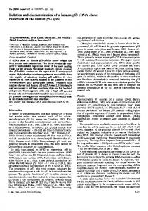
FIG. 1. (A) Schematic map of the gene coding SS-b5-T. phoA, promoter; S/D, Shine–Dalgarno sequence; SS, alkaline phosphate signal sequence; b5 and T, globular and tail portions of cytochrome b5 , respectively. (B) Amino acid sequence of SS-b5-T. The italicized and bold residues denote, respectively, the SS and the T portions of the fusion protein.
fusion protein (Fig. 1B) sequencially composed of the 21 residues of alkaline phosphatase SS, one remnant N-terminal residue of alkaline phosphatase, 99 residues of b5 , and 35 residues of T. The b5-T portion of the chimera represents the full-length endoplasmic reticulum cytochrome b5 of rat liver. The fusion was created by an in-phase linkage of the synthetic T DNA with SS-b5 in the plasmid pAA-cyt which was previously reported (24) to direct high-level secretion of the b5 protein into the periplasm of E. coli. The placement of the SS-b5-T sequence downstream under the control of the E. coli phoA promoter assures the gene is tightly repressed during growth of the bacteria in a standard rich medium. However, the gene can be optimally expressed in E. coli cultured in the Mops medium containing low (0.1 mM) phosphate. When E. coli TB1 cells harboring the plasmid pA-sct were cultured in the phosphate-limited Mops medium, they displayed an absorption spectrum characteristic of a b-type hemoprotein in the reduced state. The observation that the magnitude of the Soret absorption peak at 423 nm (Fig. 2A) increased concomitantly with culture growth indicated de novo synthesis and accumula-
/ 6q05$$0565
03-20-96 17:17:55
pepa
Electrophoretic analysis of the bacterial subcellular fractions showed that the 21-kDa protein was almost entirely present in the (cytosolic) membrane fraction with insignificant amounts detectable in the remaining soluble fraction (Fig. 3). The hemoprotein content in the isolated membranes was enriched threefold over that estimated in the total cell lysates (Table 1). In their physical association with the lipid bilayer, both the 21 kDa and hemoprotein appeared to be integral components since neither was extractable by treatment of the isolated membranes with 0.1 to 1 M NaCl or 0.1 M Na2CO3 (data not shown). Treatment of the membranes with 1% (w/v) Nonidet P-40 efficiently extracted the hemoprotein but the solubilization initiated a time-dependent loss of the 21-kDa protein with concomitant appearance of an 18-kDa component (data not presented) which was later identified as a derivative lacking the SS portion. An alternative approach of treatment of membranes with 40% (v/v) acetonitrile resulted in extraction of the 21-kDa protein in an intact form with solubilization of about 50% of the membrane hemoprotein (Fig. 3, lane 5). After loading onto the DEAESepharose CL-6B column and development with a salt gradient, the hemoprotein eluted at 300 mM NaCl as a well-defined peak, representing a 78% hemoprotein purity (Table 1). Further purification by Mono Q chromatography on an FPLC system separated a single major peak representing a hemoprotein at a specific content of 94% (Fig. 3, lane 7) from some minor impurities of lower molecular mass. Typical yields of the purified 21-kDa protein were around 5 mg protein/liter of cell culture at a cell density of Ç2 O.D. at 600 nm. Characterization of the 21-kDa Protein as SS-b5-T The isolated 21-kDa protein was subjected to spectral analyses in order to investigate its relatedness to native b5-T (Fig. 4). The 21-kDa protein had Soret band at 413 nm in the oxidized state, and on reduction, shift in absorbance to 423 nm was observed with emergence of peaks at 555 and 527 nm. The absorption spectrum of the oxidized versus reduced forms of the recombinant hemoprotein was identical to the trypsin-digested b5 purified from mammalian tissue, in having a character-
AP-PEP
PRECURSOR SUBSTRATE FOR SIGNAL PEPTIDASE
241
FIG. 2. Production of the 21-kDa protein in E. coli. (A) Spectral detection of the hemoprotein in intact bacteria. (B) Electrophoretic detection of the 21-kDa fusion protein in E. coli lysates by staining of the gel with Coomassie blue; the arrow marks the position of the induced band.
istic maximum at 423 nm and a minimum at 409 nm. The absorption coefficients determined for the oxidized and reduced forms of the 21-kDa hemoprotein at the various absorption peaks were close to the values previously reported for the mammalian liver and erythrocyte b5 (28). A preparation of the purified 21-kDa protein was subjected to 10 rounds of automated peptide degradation. This yielded an N-terminal amino acid sequence identical to that deduced from the nucleotide sequence of SS except that the GTG-encoded initiator methio-
/ 6q05$$0565
03-20-96 17:17:55
pepa
nine was found to be unprocessed in the synthesised product. The presence of T sequence in the 21-kDa protein was indicated by comparison of its electrophoretic mobility with the related derivatives, i.e., SS-b5 , b5-T, and b5 . In accord with their relative sizes the mobilities were SS-b5-T ú b5-T ú SS-b5 ú b5 . Processing of the 21kDa protein by signal peptidase (see below) converted it to a form which electrophoretically migrated with a mobility identical to that of b5-T. The mass of SS-b5-T determined by laser desorption spectroscopy yielded a
AP-PEP
242
KADERBHAI AND KADERBHAI
FIG. 3. Purification of the 21-kDa protein. Protein loadings: marker proteins, 25 mg; total cellular fraction, 75 mg; cytosolic fraction, 75 mg; membranes, 60 mg; acetonitrile extract, 60 mg; Sepharose CL-6B eluate, 12 mg; Mono Q chromatographic peak, 4 mg.
molecular weight of 17,665 that agreed well with the calculated molecular mass 17,669. These findings established the identity of the 21-kDa protein as being the SS-b5-T protein. Processing of SS-b5-T by Signal Peptidase Incubation of a 50-fold molar excess SS-b5-T with the purified signal peptidase resulted in a time-dependent loss of the 21-kDa band with concomitant formation of an 18-kDa band that was almost quantitatively recovered at the end of the reaction (Fig. 5). The processed protein was immunoelectrophoretically identical with native b5-T from rat liver (data not shown). The mass spectroscopically determined molecular mass of 15,516 of the signal peptidase-processed product matched well with the calculated size (15,514) of (arg)-b5-T. Moreover, the N-terminal sequence of the signal peptidaseprocessed product of SS-b5-T yielded the predominant sequence as (Arg)–Met–Ala–Glu–Gln, indicating that
FIG. 4. Spectral characteristics of SS-b5-T. Oxidised (O), dithionitereduced (R) and reduced versus oxidised (R-O) spectra of a 20 mg purified preparation of SS-b5-T.
precursor processing complied with the reaction specificity of signal peptidase (6,9). Processing was virtually unaffected by any of the following commercially available protease inhibitors of serine-, metallo-, thiol-, and aspartyl-proteases: 2.5 mM 4-(aminophenyl)-methanesulphonyl fluoride, 0.15 mM pyridyldisulfide, 1 mM benzamidine, 50 mM aprotinin, 0.5 mM phenylmethanesulfonyl fluoride, 1 mM 1,10 o-phenanthroline, 1 mM e-amino-n-caproic acid, 10 mM dithiothreitol, 2 mM Na-tosyl-L-lysine-chloromethylketone, 2.5 mM leupeptin, 2.5 mM iodoacetamide, 10 mM b-mercaptoethanol, 2.5 mM Na2 EDTA, 100 mM antipain dihydrochloride, 150 mM chymostatin, 250 mM bestatin, 5 mM pepstatin, 2.5 mM n-carboxybenzoxy-L-phenylalanine chloromethyl ketone. The catalysis was unaffected when the enzyme was chemically modified or cross-linked by ethyl-3-(3dimethyl-) aminopropyl-carbodiimide prior to assaying. However, the Group II heavy metal ions Pb2/, Cu2/, Zn2/, Mg 2/, Fe2/, Mn2/, and Co2/ proved particularly inhibitory at concentrations ranging from 5 to 10 mM. HgCl2 was particularly potent inhibitor with 50%
TABLE 1
Purification of SS-b5-T
Fraction/step
Volume (ml)
Protein (mg)
Hemoproteina (mg)
Hemoprotein yield (%)
Hemoprotein specific content (%)
Purification (fold)
Homogenateb Membranes Acetonitrile extract DEAE-Sepharose FPLC Mono-Q
10 3 52 20 3
935 148 54 13 6.4
49 26 13 10 6
100 52 26 20 12
5 17 24 78 94
1 3 4.5 15 18
a b
Spectrally estimated. Cell lysate from 1 liter culture.
/ 6q05$$0565
03-20-96 17:17:55
pepa
AP-PEP
PRECURSOR SUBSTRATE FOR SIGNAL PEPTIDASE
FIG. 5. Processing of SS-b5-T by signal peptidase. The assays were performed for varying periods as described under Materials and Methods. The concentrations of the substrate and enzyme were 7.15 and 0.14 mM, respectively. Extent of cleavage (top) monitored electrophoretically and (bottom) quantitated densitometrically from (top).
inhibition at 0.5 mM. The effect of varying the amounts of signal peptidase on the rate of SS-b5-T processing at the fixed concentration of 7.2 mM is shown in Fig. 6. Cleavage was sufficiently slowed down at the substrate to enzyme ratio of 800 to yield about 50% cleavage after 1 h. At substrate:enzyme ratios of 1800 or above the rates of SS-b5-T cleavage were linear within the first 30 min. Structure of SS-b5-T A hydropathy plot of the SS-b5-T primary structure based on the algorithms described by Kyte and Doolittle (31), with a moving average window size of five residues, displayed the distinctive tripartite amphipathic structure. The plot showed that the soluble hydrophilic b5 core was flanked on both sides by the membrane-seeking, hydrophobic amino- and carboxy- terminal-linked SS and T segments. This amphipathic character of the SS-b5-T protein was evidenced by its complete recovery with Triton X-114 following temper-
/ 6q05$$0565
03-20-96 17:17:55
pepa
243
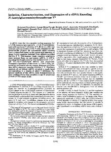
ature-induced phase separation (32) of the membranes or the isolated SS-b5-T protein (data not presented). The crystal structure of the 86 residue globular bo˚ resolution (33). vine b5 has been reported at a 2.8A In common with all of the sequenced mammalian b5 homologues, the bovine b5 is highly conserved in its primary structure (21). A large proportion of the b5 molecule (residues 26 to 83) is devoted to the formation of a crevice to accommodate the heme which fits tightly into the hydrophobic lining of the pocket. The two histidines at positions 45 and 68, located in the nonhelical loops at the top of the crevice, extend into the center of the pit to bind the heme iron. The heme-linked histidine residues are held rigidly by a variety of intermolecular forces and cannot be displaced from the iron atom by other ligands without disrupting the protein structure. Since the spectral properties of SS-b5-T protein are indistinguishable from either b5 or b5-T, it is reasonable to deduce that the b5 domain must be fully preserved in the fusion protein. In this respect it is possible to gain an insight into the likely, higher ordered structure of the SS-b5-T chimera by modeling on the known crystal structure backbone of b5 . The b5 is a compacted domain the interior of which is distinctly apolar and the exterior is extensively combed with side chains of charged and hydrophilic amino acids. The surface-exposed residues near the amino and carboxy ends are thought to have no apparent structural role and form flexible linkages. With these insights, we extended the surface-exposed termini of b5 with the predicted structures (34) of the (i) amino-terminal SS and (ii) carboxy-terminal T portions (35). In this structure (Fig. 7) the b5-T linkage has a flexible, randomly coiled segment which demarks the zone between the larger compact b5 domain and the lipophilic tail; this is the trypsin-susceptible region of the membrane-bound b5T that enables proteolytic generation of a soluble b5 form (36). Likewise, the protrusion of the amino-terminal SS linkage in the model is based on the premise that the SS is clearly accessible for both the recognition and the precise cleavage by the bulkier signal peptidase molecule. Both the lipophilic termini extensions have a high propensity to form a-helices which would thermodynamically favor their positioning away from the hydrophilic b5 core. Three-dimensional modeling of the SS-b5-T protein on Hyperchem Molecular Modeller V3 indicated that the terminally exposed hydrophobic segments in the tripartite protein possess sufficient flexibility and dimensions to establish interactions via their central hydrophobic cores and the terminal ionic groups. Moreover, the adjoining hydrophobic a – a helical cylinders are of a length sufficient to traverse the lipid bilayer. DISCUSSION
In this study we have shown that SS-b5-T expression in E. coli resulted in substrate level production of the
AP-PEP
244
KADERBHAI AND KADERBHAI
FIG. 6. The effect of varying signal peptidase concentration on the cleavage of SS-b5-T. The reactions were performed for 60 min at the enzyme concentrations ranging from 2 to 20 nM with the concentration of substrate fixed at 7.15 mM. Extents of processing (top) monitored electrophoretically and (bottom) quantitated from densitometric traces.
unprocessed SS-b5-T protein. Following 6 h of growth as much as 50 mg of SS-b5-T protein was produced per liter of batch-cultivated E. coli, without the formation of inclusion bodies. The SS-b5-T protein was specifically integrated with the cytoplasmic membranes where it represented about 5% of the total bacterial protein. Only a proportion (35%) of the bacterially produced SSb5-T existed in its holoform. However, supplementation of the lysed cells with exogenous bovine heme converted the apo-protein pool to holocytochrome. The presence of a significantly large pool of the apo-SS-b5T within the cells could be attributed to the limited capacity of the E. coli host to synthesize the prosthetic heme group (37) during overproduction of the recombinant protein. A procedure has been developed for the isolation of the intact SS-b5-T protein. This procedure involves extraction of the membranes with acetonitrile which solubilises SS-b5-T in an undegraded form. In turn the extracted SS-b5-T can be isolated in a homogeneous state
/ 6q05$$0565
03-20-96 17:17:55
pepa
by the conventional anion-exchange chromatography. The authenticity of the isolated SS-b5-T has been verified by a variety of criteria including N-terminal sequencing, laser desorption mass spectroscopy, immunoelectrophoretic analyses, phase separations, spectrophotometric analyses, and its processing by purified signal peptidase. The fact that SS-b5-T was spectrally indistinguishable from the native hemoprotein is indicative of the structural retention of the tightly folded heme-binding globular domain which must, therefore, have been correctly folded and preserved against likely perturbations by the neighboring SS and the native T peptide extensions. Molecular modeling of SS-b5-T on Hyperchem 3, based on combined data derived from prediction methods and the crystal structure of b5 , supports this postulated amphipathic structure of the chimera in which a spatially distinct hydrophobic core is formed by apolar interactions between the T and the SS segments. Based on thermodynamic considerations, the
AP-PEP
PRECURSOR SUBSTRATE FOR SIGNAL PEPTIDASE
FIG. 7. Potential structure of SS-b5-T. The schematic backbone structure of the three domains is shown; charged residues are shown in the SS and T segments. The crystal structure-derived backbone of the globular b5 domain extends from the amino acid residues 8 to 86 (34). The rectangle encompasses the probable regions of the SS and T helices that traverse the lipid bilayer. Signal peptidase cleavage site is denoted by the arrow.
hydrophobic segments must protrude away from the charged surface of the globular b5 and function to doubly anchor the protein in the membrane. Like many rod-shaped proteins (26), the anomalous, slower electrophoretic mobility of the recombinant SS-b5-T resolved under the denaturing effect of SDS may arise from preservation of the head-tail interactions imparting a decreased level of detergent binding (38). The mode of SS-b5-T targeting to integration and eventual organization in the membrane remains intriguing as the SS cleavage site appears to be inaccessible to the signal peptidase unless the membrane integrity is disrupted by detergent treatment. In contrast, a comparable SS-b5 derivative (lacking the T portion) was efficiently exported to the periplasm in E. coli and the signal processed according to the 03, 01 rule, at the site between Ala01 and Arg/1 (25). Thus appendage of T to the carboxy-end of SS-b5 coverts the derivative into a nonexportable, membrane bound form. Of course, several models could explain the mechanism by which SS-b5-T becomes assembled into the cytoplasmic membrane of E. coli. A potential transmembrane assembly could arise by loop insertion of the SS, initiating translocation of the b5 domain toward the exterior of the cell membrane. Following extracellular discharge of the entire b5 domain the stop-transfer sequence, identifiable in residues 104–128 (Fig. 1b) within the T core could have terminated the polypeptide translocation. The 23 residue hydrophobic a-helix flanked by charged amino acids in the T portion displays the classic characteristics of stop-transfer sequences found in
/ 6q05$$0565
03-20-96 17:17:55
pepa
245
many transmembrane proteins (23). It is plausible that the hydrophobic interactions between the helical portions of the T and SS within the lipid phase, and possibly between the charged residues adjacent to the membrane phase (Fig. 7), may have displaced the processing site, rendering it to be inaccessible to signal peptidase. However, this apparent inhibition was overcome by detergent treatment of the membranes. This model raises a strong possibility that SS cleavage of precursors by signal peptidase is an event that could occur after the polypeptide is almost fully translocated. Our postulated topography of SS-b5-T in the inner membrane remains to be investigated, employing selective chemical modification and progressive proteolysis approaches. The isolated SS-b5-T is a macromolecular substrate that is faithfully and efficiently processed by the signal peptidase. The specificity of the cleavage reaction is verified by N-terminal sequence of the processed protein band which matches the expected maturation site of the precursor in vivo. The cleavage of SS-b5-T by signal peptidase being insensitive to all the classic protease inhibitors is yet another indicator of the unusual reaction catalyzed by this endopeptidase (10–12). As a precursor protein that is expressible and isolatable in abundant quantities in a unique ‘‘soluble’’ form, SS-b5T can be exploited as a nonlabeled reagent for deciphering the reaction mechanism of the signal peptidase and as a probe for exploring events in protein translocation. ACKNOWLEDGMENT This work was supported by a grant from the University of Wales Research Funds. We are most grateful to Prof. M. Akhtar and Peter Robichaud for the mass spectroscopic measurements.
REFERENCES 1. Pugsley, P. A. (1993) The complete general secretory pathway in Gram-negative bacteria. Microbiol. Rev. 57, 50–108. 2. Misra, R., and Silhavy, T. J. (1992) ‘‘Emerging Targets in Antibacterial and Antifungal Chemotherapy’’ (Sutcliffe, J., and Georgopapadakou, P., Eds.), pp. 163–169. Chapman and Hall, London/New York. 3. Date, T. (1983) Demonstration by a novel genetic technique that leader peptidase is an essential enzyme of Escherichia coli. J. Bateriol. 154, 76–83. 4. Allsop, A. E., Brooks, G., Bruton, G., Coulton, S., Edwards, P. D., Hatton, K. I., Kaura, A. C., McLeon, S. D, Pearson, N. D., Smale, T. C., and Southgate, R. (1995) PENEM inhibitors of bacterial signal peptidase. Bioorg. Med. Letts. 5, 443–448. 5. Wolfe, P. B., Silver, P., and Wickner, W. (1982) The isolation of homogeneous leader peptidase from a strain of Escherichia coli which overproduces the enzyme. J. Biol. Chem. 257, 7898–7902. 6. von Heijne, G. (1993) The signal peptide. J. Membrane Biol. 115, 195–201. 7. van Dijl, J. M., de Jong, J., Vehmaanpera, G., Venema, G., and Bron, S. (1993) Signal peptidase I of Bacillus subtilis: Patterns
AP-PEP
246
KADERBHAI AND KADERBHAI
of conserved amino acids in prokaryotic and eukaryotic type I signal peptidase. EMBO. J. 11, 2819–2828. 8. Wolfe, P. B., Wickner, W., and Goodman, J. M. (1983) Sequence of the leader peptidase gene of Escherichia coli and the orientation of leader peptidase in the bacterial envelope. J. Biol. Chem. 258, 12073–12080.
24.
25.
9. Perlman, D., and Halvorson, H. O. (1983) A putative signal peptidase recognition site and sequence in eukaryotic and prokaryotic signal peptides. J. Mol. Biol. 167, 391–409. 10. Black, M. T., Munn, J. G. R., and Allsop, A. E. (1992) On the catalytic mechanism of prokaryotic leader peptidase. Biochem. J. 282, 539–543. 11. Sung, M., and Dalbey, R. E. (1992) Identification of potential active-site residues in Escherichia coli leader peptidase. J. Biol. Chem. 267, 13154–13159.
26.
27.
12. Black, M. T. (1993) Evidence that the catalytic activity of prokaryote leader peptidase depends upon the operation of a serinelysine dyad. J. Bacteriol. 175, 4957–4961.
28.
13. Ito, K. (1982) Purification of the precursor form of maltose-binding protein, a periplasmic protein of Escherichia coli. J. Biol. Chem. 257, 9895–9897.
29.
14. Bowden, A. U., Paredes, A. M., and Georgiou, G. (1991) Structure and morphology of protein inclusion-bodies in Escherichia coli. Bio/Technology 9, 725–730.
30.
15. Anba, J., Pages, J-M., Bernadac, A., and Lazdunski, C. (1987) in ‘‘Phosphate Metabolism and Cellular Regulation in Microrganisms’’ (Torriani-Gorini, A., Rothman, F. G., Silver, S., Wright, A., and Yagil, E., Eds.), pp. 73–77. Am. Soc. Microbiol., Washington, DC. 16. Pages, J. M., Anba, J., Bernadac, A., Shinagawa, H., Nakata, A., and Lazdunski, C. (1984) Normal precursors of periplasmic proteins accumulated in the cytoplasm are not exported posttranslationally in Escherichia coli. Eur. J. Biochem. 143, 499– 505. 17. Laminet, A. A., and Pluckthun, A. (1989) The precursor of betalactamase—purification, properties and folding kinetics. EMBO J. 8, 1469–1477.
31.
32. 33.
34.
35.
18. Schenkmann, J. B., Jansson, I., and Robie-Suh, K-M. (1976) Many roles of cytochrome b5 in hepatic microsomes. Life Sci. 19, 611–624.
36.
19. Roy, D., Strobel, H. W., and Liehr, J. G. (1991). Cytochrome b5mediated redox cycling of estrogen. Arch. Biochem. Biophys. 285, 331–338.
37.
20. Borgese, N., D’Arrigo, A., Silvestris, M. D., and Pietrini, G. (1993) NADPH-cytochrome b5 reductase and cytochrome b5 isoforms as models for study of post-translational targeting to the endoplasmic reticulum. FEBS Letts. 325, 70–75. 21. Ozols, J. (1989) Structure of cytochrome b5 and its topology in the microsomal membrane. Biochim. Biophys. Acta 997, 121– 130. 22. Blobel, G. (1980) Intracellular protein topogenesis. Proc. Natl. Acad. Sci. USA 77, 1496–1500. 23. Kuroiwa, T., Sakaguchi, M., Mihara, K., and Omura, T. (1991)
/ 6q05$$0565
03-20-96 17:17:55
pepa
38.
39.
Systematic analysis of stop-transfer sequence for microsomal membrane. J. Biol. Chem. 266, 9251–9255. Karim, A., Harding, V., Evans, A., Kaderbhai, N. N., and Kaderbhai, M. A. (1993) Efficient bacterial export of a eukaryotic cytoplasmic cytochrome. Bio/Technology 11, 612–618. Harding, V., Karim, A., Kaderbhai, N., Jones, A., Evans, A., and Kaderbhai, M. A. (1993) Processing of chimeric mammalian cytochrome b5 precursors in Escherichia coli: Reaction specificity of signal peptidase and identification of an aminopeptidase in post-translational processing. Biochem. J. 293, 751–756. Maniatis, T., Fritsch, E. F., and Sambrook, J. (1989) ‘‘Molecular Cloning. A Laboratory Manual,’’ Cold Spring Harbor Laboratory, Cold Spring Harbor, New York. Sanger, F., Coulson, S. R., Barrell, B. G., Smith, A. J. H., and Roy, B. A. (1980) Cloning in single-stranded bacteriophage as an aid to rapid DNA sequencing. J. Mol. Biol. 143, 161–178. Estabrook, R. W., and Werringloer, J. (1978) The measurement of difference spectra: Application to cytochromes of microsomes. Methods Enzymol. 52, 212–220. Bradford, M. (1976) A rapid and sensitive method for the quantitation of protein utilising the principle of protein-dye binding. Anal. Biochem. 72, 248–254. Laemmli, U. K. (1970) Cleavage of structural proteins during assembly of the head of bacteriophage T4. Nature 227, 680–685. Kyte, J., and Doolittle, R. K. (1982) A simple method for displaying the hydropathic character of a protein. J. Mol. Biol. 157, 105–132. Bordier, C. (1981) Phase-separation of integral membrane-proteins in Triton X-114 solution. J. Biol. Chem. 25, 1604–1607. Matthews, F. S., and Czerwinski, E. W. (1986). Cytochrome b5 and cytochrome b5 reductase from a chemical and X-ray diffraction viewpoints, in ‘‘The Enzymes of the Biological Membranes’’ (Martonosi, A., Ed.), Vol. 4, pp. 143–197. Plenum, New York. Carmenes, R. S., Freije, J. P., Molina, M. M., and Martin, J. M. (1988) PREDICT 7, a program for protein structure prediction. Biochem. Biophys. Res. Commun. 159, 687–693. Vergeres, G., Ramsden, J., and Waskell, L. (1995) The carboxyl terminus of the membrane-binding domain of cytochrome b5 spans the bilayer of the endoplasmic reticulum. J. Biol. Chem. 270, 3414–3422. Spatz, L., and Strittmatter, P. (1971) A form of cytochrome b5 that contains an additional hydrophobic sequence of 40 amino acid residues. Proc. Natl. Acad. Sci. USA 68, 1042–1046. Gallagher, J., Kaderbhai, N., and Kaderbhai, M. A. (1992). Genedose dependent expression of soluble cytochrome b5 in Escherichia coli. Appl. Microbiol. Biotechnol. 38, 77–83. Takano, E., Maki, M., Mori, H., Hatanaka, N., Marti, T., Tikani, K., Kannagi, R., Ooi, T., and Murachi, T. (1988) Pig-heart calpastatin: Identification of repetitive domain-structures and anomalous behaviour in polyacrylamide-gel electrophoresis J. Biol. Chem. 27, 1964–1972. Egner, B. J., Langley, G. J., and Bradley, M. (1995) solid-phase chemistry—direct monitoring by matrix-assisted laser-desorption ionization time-of-flight mass-spectrometry—A tool for combinatorial chemistry. J. Org. Chem. 60, 2652-2653.
AP-PEP