Th is is an Open Access article distributed under the terms of the Creative Commons Attribution License (http://creativecommons.org/ licenses/by/2.0), which permits unrestricted use, distribution, and reproduction in any medium, provided the original work is properly cited.
84
ENDOCRINE REGULATIONS, Vol. 51, No. 2, 84–95, 2017 doi:10.1515/enr-2017-0008
Expression of genes encoding IGFBPs, SNARK, CD36, and PECAM1 in the liver of mice treated with chromium disilicide and titanium nitride nanoparticles 1,2
Minchenko DO, 1Tsymbal DO, 2Yavorovsky OP, 2Solokha NV, 1Minchenko OH
1
Department of Molecular Biology, Palladin Institute of Biochemistry, National Academy of Sciences of Ukraine, Kyiv, Ukraine; 2Department of Pediatric and Department of Hygiene and Occupational Pathology, National Bohomolets Medical University, Kyiv, Ukraine E-mail:
[email protected]
Objective. The aim of the present study was to examine the effect of chromium disilicide and titanium nitride nanoparticles on the expression level of genes encoding important regulatory factors (IGFBP1, IGFBP2, IGFBP3, IGFBP4, IGFBP5, SNARK/NUAK2, CD36, and PECAM1/CD31) in mouse liver for evaluation of possible toxic effects of these nanoparticles. Methods. Male mice received 20 mg chromium disilicide nanoparticles (45 nm) and titanium nitride nanoparticles (20 nm) with food every working day for 2 months. The expression of IGFBP1, IGFBP2, IGFBP3, IGFBP4, IGFBP5, SNARK, CD36, and PECAM1 genes in mouse liver was studied by quantitative polymerase chain reaction. Results. Treatment of mice with chromium disilicide nanoparticles led to down-regulation of the expression of IGFBP2, IGFBP5, PECAM1, and SNARK genes in the liver in comparison with control mice, with more prominent changes for SNARK gene. At the same time, the expression of IGFBP3 and CD36 genes was increased in mouse liver upon treatment with chromium disilicide nanoparticles. We have also shown that treatment with titanium nitride nanoparticles resulted in down-regulation of the expression of IGFBP2 and SNARK genes in the liver with more prominent changes for SNARK gene. At the same time, the expression of IGFBP3, IGFBP4, and CD36 genes was increased in the liver of mice treated with titanium nitride nanoparticles. Furthermore, the effect of chromium disilicide nanoparticles on IGFBP2 and CD36 genes expression was significantly stronger as compared to titanium nitride nanoparticles. Conclusions. The results of this study demonstrate that chromium disilicide and titanium nitride nanoparticles have variable effects on the expression of IGFBP2, IGFBP3, IGFBP4, IGFBP5, SNARK, CD36, and PECAM1 genes in mouse liver, which may reflect the genotoxic activities of the studied nanoparticles. Key words: mRNA expression, IGFBPs, SNARK, CD36, PECAM1, chromium disilicide nanoparticles, titanium nitride nanoparticles, mouse liver
A rapid development of nanoscience and nanotechnologies has given a rise to the wide range of applications of man-made nanomaterials in specific biomedical fields for treatment, diagnosing, controlling, and repairing of biological systems at the mo-
lecular level. Titanium nitride nanoparticles have favorable properties and are used for the coating of orthopedic implants, coronary stents, and syringe needles as well as for improving long-term implants in the dental medicine. However, very little is known
Corresponding author: Dmytro O. Minchenko, M.D., PhD., Department of Molecular Biology, Palladin Institute of Biochemistry, National Academy of Sciences of Ukraine, Leontovycha 9, Kyiv 01601, Ukraine; e-mail:
[email protected].
Unauthenticated Download Date | 7/4/17 10:54 PM
Minchenko, et al. about the toxicity, including genotoxicity, induced by these nanoparticles (van Hove et al. 2015; Chu et al. 2016; Karjalainen and Nammas 2016; Ritz et al. 2016). At the same time, for ultrafine titanium dioxide nanoparticles, which are widely used in the number of applications, including white pigment in paint, ceramics, food packaging materials, food additives, cosmetic creams, toothpastes, and components of surgical implants, it has been shown that the longterm exposure to titanium dioxide nanoparticles led to their accumulation in brain, oxidative stress, overproliferation of all glial cells, tissue necrosis as well as hippocampal cell apoptosis, which resulted in neurogenic disease states in mice (Ze et al. 2014; Rollerova et al. 2015). Significant changes in brain gene expressions were also found: increases in collagen A1 (COL1A1), serine/threonine-protein kinase 1 (AKT1), catenin beta 1 (CTNNB1), cysteine and serine rich nuclear protein 1 (CSRNP1), DDIT4 (DNA damage inducible transcript 4), cytochrome P450 family 2 subfamily E member 1 (CYP2E1), and Krev interaction trapped protein 1 (KRIT1) expressions and great decreases in dopamine receptor D2 (DRD2), neuraminidase 1 (NEU1), Fc receptor-like A (FCRLA), and 7-dehydrocholesterol reductase (DHCR7) expressions (Ze et al. 2014). Furthermore, Yang et al. (2016) have shown that oral administration of titanium dioxide nanoparticles disrupts hepatic metabolic functions in a mouse model. Recently, it has been shown that titanium dioxide nanoparticles have possible impact on health due to their specific properties supported by their size and geometry (Simon et al. 2017). These nanoparticles, which were synthesized as nanoneedles, titanate scrolled nanosheets, and gelsol-based isotropic nanoparticle, were internalized at various degrees and their toxicity depended on both the titanium content and nanoparticles shape. Diverse titanium dioxide nanoparticles affect intracellular calcium homeostasis, thereby leading to endoplasmic reticulum stress-dependent toxicity (Simon et al. 2017). There are data that chromium disilicide nanowires are attractive choices for future applications in magnetic storage, photovoltaic, and field emitters (Hsu et al. 2015), but the toxicity of chromium disilicide nanoparticles has not been studied yet. Recently, Alarifi et al. (2016) have shown that chromium oxide nanoparticles had significant cytotoxic effects on murine fibrosarcoma cells. At the same time, there are data that chromium is well-established human carcinogen causing various cancers upon long term exposure (Humphries et al. 2016) and that quercetin inhibits chromium-induced malignant cell transformation (Pratheeshkumar et al. 2016).
85
The insulin-like growth factor binding proteins (IGFBPs) as extracellular matrix proteins bind and regulate the availability of both IGFs and inhibit or stimulate the growth promoting effects of the IGFs through IGF/INS receptors and through other signaling pathways. Although the primary site of action of ECM proteins is extracellular, evidence is emerging for non-canonical intracellular roles of IGFBPs, which regulate cell proliferation and survival as well as tumor angiogenesis and cell migration (Zhu et al. 2014; Bach 2015; Hellewell and Adams 2016; Kim et al. 2016). IGFBPs are now understood to have many actions beyond their endocrine role in the transport of IGF (Zhu et al. 2014; Bach 2015). Additionally, IGFBPs bind non-IGF ligands in the extracellular space, cell membrane, cytoplasm and nucleus, thereby modulating cell proliferation, survival and migration in an IGF-independent manner. Both IGFBP1 and IGFBP2 preferentially stimulate tumor growth through variable mechanisms. Thus, Kim et al. (2016) have shown that IGFBP1 and ALDH1A1 are differentially overexpressed in the colorectal cancer liver metastasis and may play a dual role, functioning as both tumor suppressors and metastasis promoters in colorectal cancer. Furthermore, there are data that glioblastoma-derived MCSF (macrophage colony-stimulating factor) induces microglial release of IGFBP1 to promote angiogenesis (Nijaguna et al. 2015). IGFBP2 is highly up-regulated in glioblastoma tissues. It has been also shown that exogenous IGFBP2 promotes proliferation and invasion in glioma cells via the integrin β1-ERK pathway and that this IGFBP with MDA9/syntenin promotes angiogenesis in human melanoma (Han et al. 2014). Moreover, IGFBP2 enhances VEGF gene promoter activity and consequent promotion of angiogenesis by neuroblastoma cells (Azar et al. 2011). Recently, Phillips et al. (2016) revealed that IGFBP2 not only is a driver of glioma progression and a prognostic factor but also is also required for tumor maintenance and can be blocked using anti-IGFBP2 strategies. Furthermore, cell viability during glucose deprivation is enhances by TRIB3 (pseudokinase Tribbles homolog 3) through upregulation of IGFBP2, which is recognized as a novel nutrient deficiency survival factor (Ord et al. 2015). IGFBP3 is an N-linked glycosylated, phosphorylated protein, which has been reported to regulate cancer growth because it has proapoptotic and growth-inhibitory actions as well as cytoprotective and growth-potentiating properties (Johnson and Firth 2014). This protein participates in transcription, cell-stress responses, autophagy and cancer (Hellewell and Adams 2016). Antiprolif-
Unauthenticated Download Date | 7/4/17 10:54 PM
86
TiN and CrSi2 nanoparticles affect the expression of regulatory genes
erative and apoptotic effects of IGFBP3 are IGF-independent and mediated by its receptor TMEM219 and within the nucleus IGFBP3 appears to have a role in transcriptional regulation (Ingermann et al. 2010; Baxter 2015; Ranke 2015). IGFBP-3 is TP53-inducible and in response to DNA damage, forms a complex with the epidermal growth factor receptor (EGFR), translocating to the nucleus to interact with DNAdependent protein kinase (Baxter 2015). It is interesting to note that IGFBP1, IGFBP2, and IGFBP3 are linked to insulin resistance, obesity, and the type 2 diabetes mellitus, and metabolic syndrome, because they are at the interface of growth and metabolism (Sabin et al. 2011; Gokulakrishnan et al. 2012; Kim and Lee 2015). Thus, IGFBP4 preferentially binds to IGF2 and regulates growth and development of tissues and organs by negatively regulating IGF signaling. It has also IGF-independent effects including inhibition of angiogenesis and promotion of cancer cell migration (Praveen Kumar et al. 2014). Among most cancers, IGFBP4 has growth inhibitory role and is reported as a down-regulated gene, except for renal cell carcinoma and some gliomas, wherein it promotes tumor progression (Praveen Kumar et al. 2014). Thus, IGFBP4 modulates ligand-dependent estrogen receptor-alpha activation in breast cancer cells in an IGF-independent manner through activation of the Akt/PKB signaling pathway (Hermani et al. 2013). Recently, it has been shown that inhibition of tumor-associated αvβ3 integrin regulates the angiogenic switch by enhancing the expression of IGFBP4, an important negative regulator of IGF-1 signaling, leading to reduced melanoma growth and angiogenesis in vivo (Contois et al. 2015). The IGFBP5, which is often dysregulated in human cancers, plays a crucial role in the carcinogenesis and cancer development, preferentially acts as an important tumor suppressor, and the proliferation of IGFBP5-mutated cancer cells is selectively blocked by IGF-1R inhibitors (Wang et al. 2015; Ding et al. 2016). Overexpression of IGFBP5 suppresses epithelial-mesenchymal transition and decreases the expression of E-cadherin, exerts its inhibitory activities by reducing the phosphorylation of IGF1R, ERK1/2, and p38-MAPK kinases and abating the expression of HIF1α and its target genes, VEGF and MMP9 (Wang et al. 2015; Hwang et al. 2016). SNARK (SNF1/AMP kinase-related kinase), also known as NUAK2 (NUAK family SNF1-like kinase 2), is a stress-activated kinase involved in tolerance to glucose starvation, induces cell-cell detachment by increasing F-actin conversion to G-actin, partici-
pates in the control of cell proliferation and apoptosis (Namiki et al. 2011; Liu et al. 2012; Sun et al. 2013). Furthermore, miR-143 inhibits oncogenic traits by degrading NUAK2 in glioblastoma because this miRNA as well as miR-1 directly regulate NUAK2 oncogene (Fu et al. 2016; Miller et al. 2016). CD36 (cluster determinant 36), also known as leukocyte differentiation antigen CD36, thrombospondin receptor, collagen type I receptor, and FAT (fatty acid translocase), seems to have numerous potential physiological functions (Pascual et al. 2017). CD36 functions as a cell adhesion molecule and its inhibition impairs metastasis. Recently, it has been shown that exogenous FABP4 increases breast cancer cell proliferation and activates the expression of fatty acid transport proteins CD36 and FABP5 in MCF-7 breast cancer cells (Guaita-Esteruelas et al. 2017). CD36 gene may also play some role in the pathogenesis of impaired fasting glucose/impaired glucose tolerance and type 2 diabetes (Wang et al. 2012). PECAM1 (platelet and endothelial cell adhesion molecule 1), also known as CD31, is involved in angiogenesis, leukocyte migration, and integrin activation and its enhanced expression correlates with hypoxia inducible factor-1α in human glioblastoma multiforme (Avci et al. 2015; Musumeci et al. 2015; Ilhan-Mutlu et al. 2016). Previously, we have shown that C 60 fullerene and cerium dioxide nanoparticles affect the expression of some endoplasmic reticulum stress response genes, which are linked to cell proliferation, cell survival and death processes (Minchenko et al. 2013a,b). Multiple studies have clarified the link between endoplasmic reticulum stress, which controls numerous processes including cell proliferation and survival and various diseases including cancer and metabolic diseases (Bravo et al. 2013; Manie et al. 2014; Chevet et al. 2015; Dejeans et al. 2015). It is possible that long-term treatment of mice with chromium disilicide and titanium dioxide nanoparticles also led to endoplasmic reticulum stress and altered the expression of stress related genes, which are integrated into the unfolded protein response signaling pathways and regulate cell proliferation and apoptosis. The main goal of this work was to study the effect of chromium disilicide and titanium nitride nanoparticles on the expression level of genes encoding important regulatory factors and enzymes (IGFBP1, IGFBP2, IGFBP3, IGFBP4, IGFBP5, PECAM1, CD36, and SNARK) in mouse liver for evaluation of possible genotoxic effects of these nanoparticles.
Unauthenticated Download Date | 7/4/17 10:54 PM
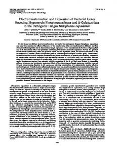
Minchenko, et al. Materials and Methods Nanoparticles and treatment conditions. Titanium nitride and chromium disilicide nanoparticles were produced by prof. A.V. Ragulya at I.M. Frantsevich Institute for Problems of Materials Science of the National Academy of Sciences of Ukraine. Titanium nitride nanoparticles have medium size of 20 nm and can create conglomerates (Figures 1, 2). Chromium disilicide nanoparticles have medium size of 45 nm and also can create conglomerates (Figures 1, 2). For the study, we used 60 age-matched male mice, which were kept in the animal facilities of Bohomolets National Medical University and housed in a quiet, temperature controlled room, and were provided with water and dry food pellets ad libitum. Before removing the liver, mice were sacrificed by cervical dislocation. All procedures conformed to the guidelines of the Bohomolets National Medical University. For treatment by nanoparticles, animals received 20 mg titanium nitride or chromium disilicide nanoparticles with food every working day for 2 months. Before usage, both titanium nitride and chromium disilicide nanoparticles were treated by special procedure for disruption of conglomerates. RNA isolation. Frozen liver tissue (100 mg) was homogenized into 0.4 ml of Trizol reagent (Invitro-
87
gen, Carlsbad, CA, U.S.A.) and total RNA was extracted according to manufacturer’s protocol. RNA pellets were washed with 75% ethanol and dissolved in nuclease-free water. For additional purification, RNA samples were re-precipitated with 95% ethanol and re-dissolved again in nuclease-free water. RNA concentration and spectral characteristics were measured using NanoDrop Spectrophotometer ND1000 (PEQLAB, Biotechnologie GmbH). Reverse transcription and quantitative PCR analysis. The expression levels of IGFBP1, IGFBP2, IGFBP3, IGFBP4, IGFBP5, SNARK/NUAK2, CD31/ PECAM1, and CD36 mRNAs as well as ACTB mRNA were measured in liver tissue of control mice and animals treated by chromium disilicide and titanium nitride nanoparticles (4 mice in each group) using realtime quantitative polymerase chain reaction. Thermo Scientific Verso cDNA Synthesis Kit (Lithuania) was used for cDNA synthesis according to manufacturer’s protocol. Maxima SYBR Green/ROX qPCR Master Mix (Thermo Fisher Scientific, U.S.A.) and “7500 HT Fast Real-Time PCR System” (Applied Biosystems, U.S.A.) were used for amplification. Polymerase chain reaction was performed in triplicates. The amplification of IGFBP1 cDNA for real time RCR analysis was performed using two oligonucleotides primers: forward – 5’-agatcgccgacctcaagaaa-3’
Figure 1. The scanning electron microscopy images of titanium nitride (TiN) and chromium disilicide (CrSi2) nanoparticles.
Unauthenticated Download Date | 7/4/17 10:54 PM
and reverse – 5’-ccagggatgtctcacactgt-3’. The nucleotide sequences of these primers correspond to sequences 705–724 and 875–856 of mouse IGFBP1 cDNA (GenBank accession number NM_008341). For amplification of IGFBP2 cDNA, we used forward (5’-catccccaactgtgacaagc-3’ and reverse (5’-tcctgctgctcgttgtagaa-3’) primers. The nucleotide sequences of these primers correspond to sequences 811–830 and 984–965 of mouse IGFBP2 cDNA (GenBank accession number NM_008342). The amplification of IGFBP3 cDNA for real time RCR analysis was performed using two oligonucleotides primers: forward – 5’-cgtccacatcccaaactgtg-3’ and reverse – 5’-tgaggcaatgtacgtcgtct-3’. The nucleotide sequences of these primers correspond to sequences 838–857 and 945–926 of mouse IGFBP3 cDNA (GenBank accession number NM_008343). For amplification of IGFBP4 cDNA, we used forward (5’-gagcgaacatcccaacaaca-3’ and reverse (5’-tgcggtcacagtttggaatg-3’) primers. The nucleotide sequences of these primers correspond to sequences 597–616 and 844–825 of mouse IGFBP4 cDNA (GenBank accession number NM_010517). For amplification of IGFBP5 cDNA, we used forward (5’-gacaggaatccgaacaaggc-3’ and reverse (5’-aagtccccatccacgtactc-3’) primers. The nucleotide sequences of these primers correspond to sequences 1274–1293 and 1505–1486 of mouse IGFBP5 cDNA (GenBank accession number NM_010518). For amplification of SNARK (SNF1/AMP-activated protein kinase 2 NUAK family; NUAK2) cDNA, we used forward (5’-ccaggcatttcttccgacag-3’ and reverse (5’-tcccgttgactatctcaggc-3’) primers. The nucleotide sequences of these primers correspond to sequences 572–591 and 787–768 of mouse SNARK cDNA (GenBank accession number NM_028778). The amplification of PECAM1 (platelet/endothelial cell adhesion molecule 1), also known as CD31 antigen, cDNA for real time RCR analysis was performed using two oligonucleotides primers: forward – 5’-agtcagagtcttccttgccc-3’ and reverse – 5’-agttcagaagtggagcagct-3’. The nucleotide sequences of these primers correspond to sequences 1969–1988 and 2135–2116 of mouse PECAM1 cDNA (GenBank accession number NM_001032378). For amplification of CD36 (cluster determinant 36), also known as thrombospondin receptor PAS-4 protein, and fatty acid translocase (FAT), cDNA we used forward (5’-gaatgggctgtgatcggaac-3’ and reverse (5’-acgtcatctgggttttgcac-3’) primers. The nucleotide sequences of these primers correspond to sequences 459–478 and 687–668 of mouse CD36 cDNA (GenBank accession number NM_001159555). The amplification of ACTB (beta-actin) cDNA for real time RCR analysis was
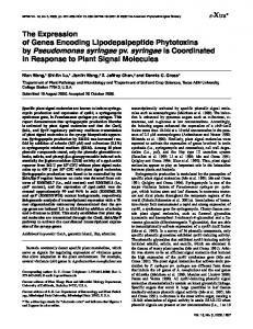
Number (u.a.)
TiN and CrSi2 nanoparticles affect the expression of regulatory genes
Size (nm)
Number (u.a.)
88
Size (nm)
Figure 2. The size dispersion by number of titanium nitride (TiN) and chromium disilicide (CrSi2) nanoparticles.
performed using forward – 5’-cctctatgccaacacagtgc-3’ and reverse – 5’-cctgcttgctgatccacatc-3’ primers. These primers nucleotide sequences correspond to 985–1004 and 1190–1171 of mouse ACTB cDNA (NM_007393). The expression of beta-actin mRNA was used as control of analyzed RNA quantity. The primers were received from Sigma-Aldrich (St. Louis, MO, U.S.A.). Quantitative PCR analysis was performed using a special computer program “Differential expression calculator”. The values of IGFBP1, IGFBP2, IGFBP3, IGFBP4, IGFBP5, SNARK/NUAK2, CD31/PECAM1, and CD36 gene expressions were normalized to the expression of beta-actin (ACTB) mRNA and represented as percent of control (100%). All values are expressed as mean ± SEM from triplicate measurements performed in four independent experiments. Statistical analysis was performed as described previously (Bochkov et al. 2006). The amplified DNA fragments were also analyzed on a 2% agarose gel and that visualized by SYBR* Safe DNA Gel Stain (Life Technologies, Carlsbad, CA, U.S.A.).
Results To determine whether a long-term treatment of mice with chromium disilicide or titanium nitride nanoparticles have genotoxic effects, we studied the expression level of a subset of genes encoding im-
Unauthenticated Download Date | 7/4/17 10:54 PM
Minchenko, et al. portant regulatory factors, such as IGFBP1, IGFBP2, IGFBP3, IGFBP4, IGFBP5, CD31/PECAM1, and CD36, and enzyme SNARK/NUAK2 in the liver. We showed that long-term treatment of mice by titanium nitride nanoparticles did not significantly change the body weight: before – 20.6±0.51 g and after 2 months – 19.3±0.90 g (n=18), but in the group of control mice, the body weight is significantly increased: before – 21.0±0.72 g and after 2 months – 26.9±1.02 g (+28%; n=20; p