Keywords: angiogenesis inhibitors; transgenic mice; thymidine kinase; Lewis lung carcinoma; ... angiogenesis by ablating endothelial cells using a suicide gene.
Gene Therapy (2003) 10, 1170–1178 & 2003 Nature Publishing Group All rights reserved 0969-7128/03 $25.00 www.nature.com/gt
RESEARCH ARTICLE
Expression of thymidine kinase driven by an endothelial-specific promoter inhibits tumor growth of Lewis lung carcinoma cells in transgenic mice A Dancer1, S Julien1, S Bouillot1, H Pointu2, M Vernet2 and P Huber1 CEA, Laboratoire de De´velopement et Vieillissement de l’Endothe´lium, INSERM EMI 02-19, France; and 2Atelier de Transgene`se, DRDC, 17, rue des Martyrs, Grenoble, France 1
The possibility of inhibiting tumor growth by limiting angiogenesis has raised considerable interest. In this study, we examined the feasibility of inhibiting tumor growth by targeting a suicide gene in the endothelium. Toxicity must be directed solely to angiogenic cells. Therefore, we used the herpes simplex virus-thymidine kinase (TK) gene, in combination with the prodrug ganciclovir (GCV), which affects replicative cells. To test this strategy, we produced transgenic mice carrying the TK gene driven by the vascular endothelial (VE)-cadherin promoter. Lewis lung carcinoma cells were injected subcutaneously to establish tumors and to test the effect of GCV on tumor growth. In two independent transgenic lines, GCV treatment (75 mg/kg/day) resulted in a
66–71% reduction of tumor volume at day 20 postimplantation compared to wild-type mice (650 and 550 versus 1930 mm3, Po0.02 and 0.01, respectively), whereas no significant difference was observed when vehicle alone was injected. Tumor growth inhibition was accompanied by a marked reduction in tumor vascular density (151 versus 276 vessels/mm2, Po0.05) and an increase in tumor cell death, suggesting that tumor growth inhibition was caused by a reduction in tumor angiogenesis. Our data support the potential utility of endothelial targeting of suicide genes in cancer therapy. Gene Therapy (2003) 10, 1170–1178. doi:10.1038/ sj.gt.3301981
Keywords: angiogenesis inhibitors; transgenic mice; thymidine kinase; Lewis lung carcinoma; endothelium; ganciclovir
Introduction There is a lot of evidence establishing that tumor growth is dependent upon neovessel formation.1–3 Angiogenesis is essential because it provides oxygen and nutrients to tumor cells.4 Moreover, tumor vessels provide a path for metastatic cell dissemination.4 Angiostatic molecules such as endostatin, angiostatin and many others have been shown to slow or even eradicate tumors in experimental systems.3,5–8 Therefore, angiostatic approaches – alone or in combination with radiotherapy, surgery or regular chemotherapy – are considered as potential therapeutic routes to cure cancer. However, high doses of angiostatic molecules are necessary to combat tumor growth, and variations in preparation quality have led to dramatic differences in their inhibition activity.6,9–11 Endothelial cells are attractive targets for gene therapy as they are easily accessible through the systemic injection of DNA transfer vectors,12–15 and thus angiostatic gene transfer may represent a potent alternative to drug delivery. One major advantage of angiostatic gene therapy is that the strategies for endothelial targeting and gene delivery may be similar for most cancer types. It is therefore important to evaluate all possible systems leading to tumor angiogenCorrespondence: Dr P Huber, DRDC-DVE, CEA-Grenoble, 17, rue des Martyrs, 38054 Grenoble cedex 9, France Received 26 June 2002; accepted 26 November 2002
esis inhibition. Delivery of angiostatic genes in mice resulted in efficient tumor growth inhibition.16–26 However, ectopic expression of therapeutic proteins in healthy tissues may eventually lead to side effects such as organ dysfunction. Therefore, it is desirable to restrict therapeutic gene expression to specific tissues. In this paper, we examined a way to prevent tumor angiogenesis by ablating endothelial cells using a suicide gene. The herpes simplex 1 thymidine kinase (TK) gene has been widely used in gene therapy of animal tumors, providing a conditional and efficient suicide system.27,28 TK enzyme becomes cytotoxic in the presence of ganciclovir (prodrug ganciclovir, GCV), a nucleoside analogue, commonly administered in antiherpetic therapies. TK phosphorylates GCV, and phospho-GCV is thought to act as a DNA replication inhibitor by preventing DNA chain elongation after its incorporation by DNA polymerase.28 In that respect, only proliferative cells are concerned by the suicide mechanism. However, it has been reported that in some situations, the TK/GCV system may also affect quiescent cells by an unknown mechanism.29 Owing to its toxicity, TK gene expression must be limited exclusively to the targeted cells. Gene delivery to the neovasculature could be accomplished using an integrin avb3-targeting ligand.30 However, tissue-specific suicide gene expression can also be achieved by transcriptional control of conventional vectors, using tissue-specific promoters. Accordingly, TK gene expres-
Targeting of TK to tumor vasculature A Dancer et al
sion could be targeted in vivo at various cell types, namely hepatocytes,31,32 megakaryocytes,33 astrocytes,34 as well as pituitary,29,35 thyroid36 and ovarian37 cells. In this study, we produced transgenic mice using an endothelial-specific promoter, the vascular endothelial (VE)-cadherin gene promoter, to drive TK gene expression in the endothelium. VE-cadherin is an adhesive transmembrane protein exclusively located at intercellular junctions of all endothelial cell types.38–40 In particular, VE-cadherin has been localized in endothelial cells within tumors.41 VE-cadherin promoter has been characterized by our group and we have shown that it specifically drives reporter gene expression in the endothelium of transgenic mice.42–45 Here, the antitumor activity of the TK/GCV system was tested in a primary tumor model implanted in VEcadherin/TK transgenic mice in order to substantiate the principle of an angiotoxic gene therapy.
Individual hygromycin-resistant clones were examined for TK gene insertion into genomic DNA by PCR (not shown) and for TK activity by testing their GCV sensitivity. Cells were expanded in GCV containing medium (40 mM) and observed after 15 days of treatment. Cells transfected by TK gene without promoter were not sensitive to GCV treatment whereas three out of four clones transfected by TK gene with phosphoglycerokinase promoter died shortly after GCV addition. On 11 clones transfected with VE-cad-TK construct, six were highly sensitive to GCV, leading to total cell death at day 15. For all clones except one, GCV sensitivity was correlated to TK expression levels as determined by RT-PCR (not shown). These results indicate that VE-cadTK expressing construct is functional in endothelial cells. The fact that some clones did not express TK suggests that VE-cadherin promoter may be sensitive to positional effect of genome insertion.
Results
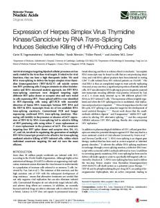
Establishment and characterization of transgenic mice with VE-cad-TK construct A suicide gene strategy, based on endothelial expression of TK, was evaluated by producing transgenic mice with the VE-cad-TK construct. These mice were subsequently analyzed in a tumor angiogenesis model produced by subcutaneous injection of Lewis lung carcinoma (LLC) cells. In this model, tumor growth has previously been shown to be angiogenesis dependent.6 In order to have mice syngenic with LLC cells, transgenesis was performed in C57Bl/6 genetic background. Two transgenic founders were obtained, females 23 and 28, for which the number of transgene copies was estimated to be 4 and 4100, respectively (Figure 1b). The progeny of the founders were used after two generations to allow possible copy segregation. However, no variation in the electrophoretic profiles could be noted in F2 mice (not shown).
A DNA construct containing the TK gene under the control of the VE-cadherin promoter (VE-cad-TK) is active in murine endothelial cells The herpes simplex virus TK gene was inserted downstream of the murine VE-cadherin promoter (positions �2486/+24) (Figure 1a), which was previously shown to promote chloramphenicol acetyl-transferase gene expression in the endothelium of transgenic mice.44 Expression of TK from this construct (VE-cad-TK) as well as control vectors (ie, TK without promoter (pJL104) or with the ubiquitous phosphoglycerokinase promoter (pPNT)) was first evaluated by stable transfection into a murine endothelial cell line, H5V, expressing VE-cadherin (Garlanda et al46 and our own observations). Cotransfection with vector pcDNA3/Hygro allowed selection of transfected cells.
1171
Figure 1 Transgenic mice and transgene expression. (a) The injected DNA consisted of the VE-cadherin promoter (�2486/+24) upstream of the TK gene and polyadenylation signal. (b) Genomic DNA from F0 newborns was digested by SspI and hybridized with a probe in the VE-cadherin promoter. Two transgenic lines (23 and 28) contained tandem repeats of the transgene (4.15 kb) and inverse repeats (line 28; 7.4 kb). The endogenous promoter band (6.9 kb) allowed estimation of transgene copy number by radioactive counting. (c) Expression of TK gene was assessed by RT-PCR on total lung RNA. Bands are present only when RT is added, indicating that RNA are not contaminated by genomic DNA. HPRT controls are shown below. Gene Therapy
Targeting of TK to tumor vasculature A Dancer et al
1172
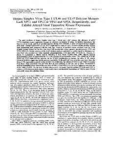
Expression of TK gene was first assessed by RT-PCR starting from lung RNA as it is the organ where VEcadherin promoter harbored the strongest activity in previous transgenic mice.44 Figure 1c shows amplified products at the correct size demonstrating transgene expression in both lines. Interestingly, the signal for line 23 appeared to be only slightly lower than that for line 28, notwithstanding the dramatic difference in transgene copy number. TK tissue distribution was further analyzed by immunostaining studies on various tissue sections using a mouse monoclonal anti-TK antibody and a polyclonal anti-von Willebrand factor (VWF) antibody, which labels endothelial cells. TK (in red) and VWF (in green) were codistributed in the vasculature of LLC implanted tumors (see Materials and methods) in both mouse lines (Figure 2a–l). Anti-TK antibody labelled all VWF positive structures (although not in the same subcellular localization), with very few exceptions, estimated to be less than 5% (arrowheads in Figure 2d–f). This result indicates that TK is expressed in pathological vessels. In contrast, TK immunoreactivity was negative or weak in normal vasculature. For instance, TK could not be found in the brain, the thymus and the spleen, and only faint signals could be detected in the lung, the kidney and the liver (data not shown). In the heart, TK was localized in the endocardium (Figure 2m, n, p, q), but was absent or in very low amounts in myocardial vessels (Figure 2o, r). As for RT-PCR, differences in TK expression levels between these two lines were not obvious in immunofluorescent studies. In conclusion, although TK expression is low and variable in normal vasculature, its expression in tumor vessels indicates that the VETK transgenic mouse model is adequate for testing the efficacy of an antitumoral suicide gene strategy based on endothelial expression of TK.
VE-cad-TK transgenic mice showed growth retardation of subcutaneously growing tumors compared to wildtype mice We first examined whether GCV administration would be toxic for the transgenic mice. Even though the vascular system is known to be essentially quiescent in adults, (i) toxicity of TK/GCV in nonproliferative cells has been formally proved in one situation,29 (ii) TK may be efficient at very low levels and (iii) the effect on the few replicative endothelial cells may be sufficient to damage the normal vasculature. All the mice used in the following studies were aged between 3 and 5 months. Intraperitoneal injections of 75 mg/kg/day of GCV did not alter viability as well as body weight of transgenic or wild-type animals, whereas injections of 150 mg/kg/day led to general edema in some mice from line 28 (not shown). The fact that the toxic effect was observed only in transgenic mice strongly suggests that vascular TK expression combined with high GCV doses may alter the barrier function of the endothelium. Therefore, in all following studies, GCV was administered at a dose of 75 mg/kg/day. To examine the effect of GCV on pathological angiogenesis and tumor growth, 2 � 105 LLC cells were implanted subcutaneously in the back of anesthetized mice (wild type, lines 23 and 28; n¼5 in each group) at day 1. GCV diluted in PBS, or PBS alone, was daily Gene Therapy
Figure 2 Tissue distribution of TK in transgenic mice. LLC tumors (a–l) from line 23 (a–f) and line 28 (g–l) were immunolabelled with VWF antibody (green, left panels) and TK antibody (red, middle panels). The framed regions in (a) and (g) are shown at higher magnification in (d–f) and (j–l), respectively. Merged images (right panels) show a codistribution of both proteins, with few exceptions (arrowheads). In the heart (m–r), TK expression is located in the endocardium of line 23 (p) and line 28 (q). TK labelling was observed in some myocardial vessels from line 28 (r). (m–o) The phase contrast images corresponding to (p–r), respectively.
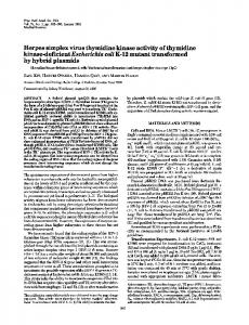
injected from day 5, which is the time at which tumors began to be visible, and tumor size was measured from day 12. Under these conditions, tumor volume in wild type reached about 2000 mm3 at day 20 and mice were subsequently euthanized. Representative tumor growth curves are shown in Figure 3. When PBS alone was injected, tumor growth was similar in all three mouse types, whereas GCV administration led to tumor growth retardation in both transgenic lines compared to wildtype mice. Differences were significant from day 17 onwards and, at day 20, the average tumor volumes in lines 23 and 28 were 34 and 29%, respectively, of that of the wild type. Autopsy of mice killed at day 20 did not
Targeting of TK to tumor vasculature A Dancer et al
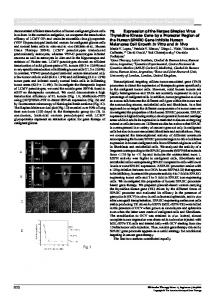
than beyond, and the histology is thus more homogeneous in small tumors than in the large ones. For each tumor, eight vascular ‘hot spot’ areas were chosen and vessel density was counted by visual observation. Figure 4a,b shows two representative immunostainings of tumors implanted in line 28 and wild-type mice, respectively. Quantification of vessel density of four independent tumors in each case revealed a significant decrease (Po0.05) in tumor volumes when implanted in transgenic versus wild-type mice (Figure 4c). These results strongly suggest that the tumor growth retardation observed in transgenic mice is because of a limitation of vessel development within the tumor.
Figure 3 Specific efficacy of GCV treatment on tumor growth. LLC tumor cells (2 � 105) were implanted subcutaneously in the back of wild-type (WT) and transgenic (VETK23, VETK28) mice on day 1. From day 5, mice were treated by daily intraperitoneal injections of GCV (75 mg/kg/ day in PBS; upper panel) or PBS alone (lower panel). Tumor size was measured and the volume was calculated using the formula length � width2 � 0.52. Each point represents the mean value of five animals (error bars: SEM). Significant differences of transgenic versus wild-type mice could be determined from day 17. Maximal probability is indicated, according to Student’s t-test.
1173
Tumors implanted in transgenic mice contained higher numbers of apoptotic cells The tumors described above were also analyzed for their apoptotic cell content. Sections were immunolabelled with a polyclonal antibody detecting only the active form of caspase 3, a specific apoptosis marker.47 Immunolabelling was correlated with nucleus fragmentation, as determined by Hoechst staining. One such caspase 3positive cell is shown in Figure 5a. The average cell count was 49% higher in tumors from transgenic mice compared to those from wild types (Figure 5b). We noticed a very small number of caspase 3-positive endothelial cells (data not shown), suggesting that the reduction in vessel density may be brought about through a mechanism different from endothelial cell apoptosis. Alternatively, it is possible that apoptosis of very few endothelial cells may lead to a significant decrease in vessel density. Apoptosis was also assessed by terminal dUTP nick-end labelling (TUNEL), a marker of DNA fragmentation.48 The number of TUNEL-positive cells (Figure 5c) was significantly higher in tumors
reveal abnormalities like inflammations or hemorrhages in nonpathological organs, such as skin, liver, brain, lung and heart. Histological data of lung, heart and skin did not show signs of edema (not shown). These results indicate that TK expression in the vasculature leads to a specific and pronounced GCV effect on LLC tumor growth.
Tumor growth retardation is correlated with a reduction in vessel density in transgenic mice To know whether growth retardation in transgenic mice was because of a lack of blood vessels in the tumor, histological sections of tumors implanted on wild type or line 28 mice were stained with a monoclonal anti-CD31 antibody to specifically label endothelial cells. For this purpose, tumors were removed when they reached a volume of B400 mm3, because (i) this corresponds to the divergent point of wild-type versus transgenic tumor growth curves (Figure 3, upper panel) and therefore the origin of this relative difference may be viewed at this stage and (ii) there are fewer necrotic areas at this stage
Figure 4 TK expression induces a decrease in tumor vascular density. Tumor induction and GCV treatment were as in Figure 2. Tumors were harvested when they reached B400 mm3 and were snap-frozen for preparation of histological sections. Vessels were visualized by CD31 immunolabelling (brown) and nuclei were counterstained with hematoxylin (blue). Eight highly vascularized areas (‘hot spots’) were identified for each tumor and vessels were counted on numerized images. (a, b) Two representative images of LLC tumor sections implanted on VETK28 and wild-type mice, respectively (scale bars: 200 mm). (c) Histograms of mean vascular densities (n¼4 in each case). Error bars represent SEM. Data show a significant decrease (Po0.05) in tumor vascularity when implanted in transgenic mice compared to wild types. Gene Therapy
Targeting of TK to tumor vasculature A Dancer et al
1174
Figure 5 Tumors in transgenic mice contain a higher number of apoptotic cells. Apoptosis was evaluated in 400 mm3 tumors by caspase 3 active immunolocalization (a, b) and TUNEL (c, d). One caspase 3-positive cell is represented in (a). Nuclei were stained with Hoechst. The average numbers of caspase 3 active cells in tumors of wild-type (WT) and transgenic (VETK28) mice are shown in (b) (n¼4 in each case). Similarly, TUNELpositive cells, as shown in (c), were counted and mean values are reported in (d). Nuclei were stained with hematoxylin. In both assays, the number of positive cells was significantly higher in transgenic versus wild-type mice (***Po0.01; *Po0.05). Scale bars: 10 mm. Error bars in (b, d) represent the SEM.
implanted in transgenic mice (Figure 5d), further suggesting that tumor growth reduction was caused by an increase of tumor cell apoptosis. Interestingly, the number of positive cells/mm2 were not in the same range in caspase 3 labelling and in TUNEL assays (Figure 5b, d). This may be because of distinct assay sensitivities or to differential kinetics in caspase 3 activation and DNA cleavage during the apoptotic process, as previously reported.49 Altogether, the data show a correlation between vessel density and LLC cell apoptosis, suggesting that vasculature reduction may cause an increase in tumor cell death.
Discussion Our study demonstrates that endothelial targeting of TK, in combination with GCV administration, is a relevant and promising strategy to reduce tumor growth. Three previous studies reported the effectiveness of TK targeting in tumor endothelium as antitumoral strategy.50–52 In two cases,50,51 TK was also expressed in tumor cells as Gene Therapy
nonspecific promoters were used. In the experimental model established by Mavria and Porter,52 the ecotropic retrovirus expressing TK could only infect the vasculature and not the human tumor cells. This latter study illustrates the relevance of vascular TK expression in tumor growth reduction, compared to tumor cell transduction alone. To our knowledge, this is the first time that TK is specifically targeted to the endothelium in a mouse tumor model. The data reported here support the general concept that angiogenesis sustains tumor growth.1,3 Partial inhibition of vessel development dramatically reduced tumor size in this model, indicating a fine adjustment of tumor growth by the vascular system. Reduction of vessel density in size-matched tumors implanted in transgenic mice was accompanied with an increase in tumor cell apoptosis. LLC cell death is likely to be caused by hypoxia generated by the lack of vessels, as hypoxia induces apoptosis;53 however, we cannot exclude a bystander effect possibly because of contact with dying endothelial cells, diffusion of phosphorylated GCV or through an immune system response, as previously reported for TK/GCV strategies.54–59 Interestingly, very few endothelial cells were activated-caspase 3-positive indicating that reduction in vessel density did not involve a caspase-dependent apoptosis mechanism but possibly a necrosis process, as previously reported.57,60,61 Accordingly, GCV-sensitive VE-cad-TK transfected H5V cells subjected to GCV were not labelled by anti-caspase 3 antibody and did not harbor signs of apoptosis, such as nuclei fragmentation (data not shown). Toxicity of TK/GCV system precludes the use of ubiquitous promoters such as viral transcriptional regulatory sequences. Specific targeting of TK can be achieved by the use of tissue-specific promoters. However, a major concern about tissue-specific versus viral promoters is their low gene expression activation capability. Indeed, VE-cadherin promoter did not yield high TK protein expression in normal vasculature, with the exception of the endocardium; nevertheless TK could be detected in endothelial cells within tumors. This feature suggests that VE-cadherin promoter is more active in angiogenic cells than in resting vessels. If this finding is correct, VE-cadherin promoter may be useful to specifically target other types of therapeutic proteins to the angiogenic endothelium. In a previous transgenic study using chloramphenicol acetyl-transferase gene as reporter, we showed that VEcadherin promoter was active in adult vasculature by monitoring enzyme activity in tissue extracts or by in situ hybridization.44 Both assays are much more sensitive than immunological methods, which may explain why TK was barely detected in VE-cad-TK transgenic mice. In the previous model, chloramphenicol acetyl-transferase gene expression was not tested in tumors, which does not allow a comparison with VE-cad-TK mice. Altogether, the data herein confirm the endothelial-specific activity of VE-cadherin promoter as shown by colocalization with VWF and demonstrate that the 2.5 kb VEcadherin promoter fragment used to drive TK expression is active in tumor vasculature. Interestingly, no obvious difference could be observed between lines 23 and 28, in terms of TK expression pattern and TK activity on tumor growth. These features suggest either a genome insertion
Targeting of TK to tumor vasculature A Dancer et al
position effect of the transgene or the existence of other mechanisms limiting TK gene expression. From the standpoint of a somatic transfer of VE-cadherin/TK construct, this may not be a relevant issue, as each transduced cell may integrate the DNA in a different genome site. Ablation of endothelial cells within tumors was not complete, although sufficient to induce a significant tumor growth reduction. In other studies, elimination of TK-targeted cells was also found to be partial,33,62 possibly indicating that cells may acquire resistance to TK/GCV action. Improvement of TK/GCV efficacy may be obtained (i) by promoter engineering, that is, adding enhancer elements that will not compromise tissuespecificity, (ii) by coexpression with a herpes virus structural protein, which enhances intercellular trafficking of TK,63 or (iii) through the administration of ponicidin, a plant diterpenoid, which increases TK activity.64 Alternatively, TK/GCV therapy may be combined with conventional anticancer treatments.27 For instance, several publications reported synergistic effects achieved with a combination of TK/GCV therapies and ionizing radiation.64–66 As discussed by Jain,67 in case of combination with standard chemotherapy, ‘normalization’ of vessel growth is preferable to angiogenic cell ablation in order to achieve drug delivery. In conclusion, our study provides a proof-of-principle for the use of TK/GCV targeted to tumor vessels. A rational explanation of the observed effect is that reduced vascularity induced by GCV in transgenic mice limits blood supply, which in turn leads to tumor hypoxia, cell death and tumor growth reduction. The next step is to use gene therapy vectors to transfer VEcadherin/TK in adult mice, to further evaluate the feasibility of a gene therapy based on this approach. In a preliminary study, the VE-cadherin promoter was able to drive LacZ expression in mouse endothelium, when targeted with adenoviral vectors (T. Moll, I. Dreher and A. Muhs, personal communication), indicating that the promoter is still active using this approach. However, alternative means to target TK in tumor endothelium of adult mice exist: (i) although adenovirus vectors have been extensively used,12,14,51,68 other gene therapy vectors were shown to efficiently transduce endothelial cells in vivo, such as liposomes30,69,70 or retroviruses,50,52 and (ii) promoters showing endothelial-specific transcriptional activity have been described including those for tie-271 and flk-172 genes. Therefore, several routes should be tested and compared to achieve optimal TK delivery and tumor growth inhibition.
Materials and methods Plasmid construction The TK gene was inserted downstream of the murine VE-cadherin promoter. To this end, the NotI-BglII fragment of pJL104 (kindly provided by Dr Fong), containing the herpes simplex virus TK gene as well as the SV40 polyadenylation signal, was inserted by bluntend ligation into the XhoI and SmaI sites of �2486CAT plasmid,44 thereby replacing the CAT gene. For transfection, the recombinant plasmid, VE-cad-TK, was linearized by SalI, pJL104 by BglII, and both pPNT73 and pcDNA3/Hygro (Invitrogen) by XhoI. For transgenesis,
the DNA fragment containing the VE-cadherin promoter, the TK gene and polyadenylation sequence was liberated from plasmidic sequences by SalI digestion, isolated from agarose gel and further purified on Elutip-d columns (Schleicher & Schuell).
1175
Cell culture, transfection and selection H5V cells46 were grown in Dulbecco’s modified essential medium (DMEM glutamax I) supplemented with 10% fetal calf serum, 50 U/ml penicillin, 50 mg/ml streptomycin (all from Gibco-BRL). For transfection, 2 � 105 cells/well were seeded in six-well dishes and transfected with 1 mg of either VE-cad-TK, pJL104 or pPNT using the Fugene transfection reagent (Roche Applied Science). In each condition, 100 ng of pcDNA3/Hygro was cotransfected for selection of transfectants. After 1 day, 200 mg/ml of hygromycin (Roche Applied Science) was added to the culture medium until colonies were picked at day 10 and grown independently. Each colony was tested for vector integration by PCR with TK primers (not shown). To test cell sensitivity to GCV, 2 � 105 cells/well were seeded in six-well dishes and grown in the presence of 40 mM of GCV (Cymevan, Roche). Viable cells were observed at day 15 after trypan blue staining. Transgenic mice DNA microinjection into C57Bl/6 fertilized eggs was performed using established procedures.74 Until F2 generation, the animals were screened by Southern blot, using standard protocols.75 Briefly, 15 mg of tail genomic DNA was digested with SspI, electrophoresed in 0.7% agarose gel, transferred onto Hybond-N+ membrane (Amersham Pharmacia Biotech) and hybridized with a 32 P-labelled VE-cadherin promoter probe (�289/+24). Transgene copy number was estimated by comparative analysis with endogenous promoter on a PhosphorImager (Molecular Dynamics). Afterwards, mice were genotyped by PCR using two primers in the TK gene (Genbank AF303108): 50 -CCCCTGCCATCAACACGCG TCTGC and 50 -CGGCGTCGGTCACGGCATAAGGC (positions 12–35 and 412–390, respectively), and the following conditions: DNA (500 ng), in standard reaction mixture, was denatured for 5 min at 941C, then subjected to 30 cycles of amplification (1 min at 941C, 1 min at 551C, 1 min at 721C) in a Techne PHC-3 thermocycler. Mice were maintained heterozygotes for the transgene by mating transgenic females with C57Bl/6 males. As previously documented,76,77 TK gene induced male infertility. Analysis of TK gene expression Total lung RNA was extracted with Trizol reagent (Gibco-BRL) and RT-PCR was performed using standard procedures. Briefly, 500 ng RNA was treated with 1 U of RQ1 DNase (Promega) for 15 min at 371C. After enzyme inactivation (5 min at 901C), RNA was reverse-transcribed with 100 U of Moloney murine leukemia virus enzyme (Gibco-BRL) for 30 min at 371C and PCR was performed with above primers for 28 cycles using the same amplification conditions, which allows exponential amplification of TK gene. Hypoxanthine phosphoribosyltransferase (HPRT) cDNAs were amplified using primers described in Vittet et al78 under the same conditions. Gene Therapy
Targeting of TK to tumor vasculature A Dancer et al
1176
Immunohistofluorescence studies were done on 7 mm frozen sections, fixed for 30 min in 4% paraformaldehyde in phosphate-bufferd saline (PBS), saturated for 30 min with the Histo-mouse kit (Zymed) and incubated for 1 h with a mouse monoclonal anti-TK antibody (clone 18G32C4; a generous gift from Aventis) at 10 mg/ml in 2% bovine serum albumin/PBS together with a rabbit polyclonal anti-VWF antibody (Dako Corporation) diluted 1:500 in the same solution. Secondary antibodies were a cyanine 3-conjugated anti-mouse immunoglobulin antibody (Jackson Laboratories) and an alexa 488conjugated anti-rabbit immunoglobulin antibody (Molecular Probes). Sections were observed under a confocal fluorescence microscope (Leica TCS SP2). Images were acquired sequentially for alexa 488 (excitation 488 nm, collection 500–530 nm), cyanine 3 (excitation 543 nm, collection 560–650 nm) and bright field.
Tumor implantation and drug administration LLC cells were seeded on gelatinized dishes and cultured in supplemented DMEM, as above. On the day of implantation (day 1), a suspension of 2 � 105 cells in 100 ml PBS was injected subcutaneously in the back of anesthetized mice. GCV diluted in 100 ml PBS, or PBS alone, was administered daily from day 5 by intraperitoneal injection, at a dose of 75 mg/kg. At day 12, the tumor area was shaved and the tumor size was measured daily using a caliper. Mice were killed at day 20 by CO2 inhalation. Analysis of tumor vascular density and cell apoptosis LLC cells were implanted and mice were GCV treated as described above. Mice were euthanized when tumors reached B400 mm3 and tumors were harvested and snap-frozen. Vessel density was calculated as previously described.79,80 Histological sections (7 mm) were prepared, fixed for 20 min with 4% paraformaldehyde/ PBS, saturated for 30 min with 2% bovine serum albumin/PBS and incubated for 1 h at room temperature with a rat anti-mouse CD31 antibody (MEC13, Pharmingen). Secondarily, sections were incubated with a horseradish peroxidase-conjugated anti-rat immunoglobulin antibody (Jackson Laboratories) under the same conditions. Peroxidase activity was revealed in the presence of the substrate diaminobenzidine (Dako Corporation) for 5 min. Nuclei were stained with Harris hematoxylin (Sigma Diagnostic). Several sections were examined under a microscope (Axioplan, Zeiss, objective � 16) to define eight nonoverlapping vessel hot spots per tumor. CD31-positive signals were counted by a blinded observer on numerized pictures and a mean vessel density was calculated for each tumor. For caspase 3 active labelling, tumors were sectioned, acetone-fixed for 10 min at �201C and incubated for 1 h at room temperature with an anti-caspase 3 active antibody (R&D systems) diluted at 1:2500, followed by a cyanine 3-conjugated anti-rabbit immunoglobulin antibody (Jackson Laboratories). Nuclei were stained with Hoechst 33258 (Sigma Diagnostic) diluted 1:1000 for 3 min. Positive cells were counted in eight random areas per tumor with objective � 20. TUNEL was performed using the in situ Cell Death Detection kit from Roche Applied Science. Briefly, sections were fixed for 20 min in 4% paraformaldehyde/PBS, permeabilized for 2 min with 0.1% Triton XGene Therapy
100 in 50 mM sodium citrate and saturated for 30 min with 20% normal bovine serum (Dako Corporation) and 3% bovine serum albumin in PBS. The TdT/fluoresceindUTP mix was incubated with sections for 1 h at 371C. A second saturation step was performed for 30 min with 20% sheep serum (Dako Corporation) and 1% digoxygenin blocking reagent (Roche Applied Science) in PBS. The alkaline phosphatase-conjugated antifluorescein antibody was incubated for 30 min at 371C and labelling was revealed with the Fast-Red reagent (Sigma Diagnostic). Nuclei were stained with Harris hematoxylin. No positive cells were observed when TdT/fluoresceindUTP mix was omitted. Apoptotic cells were counted as for caspase 3 labelling, excluding section sides and necrotic foci.
Acknowledgements We thank Drs Tim Fong and Marielle Chiron at Aventis for providing protocols for the tumor model and the antiTK antibodies. We are grateful to Dr Didier Grunwald from DRDC-CIS lab for image acquisition on confocal microscope. This project was supported by grants from the CEA and Aventis.
References 1 Folkman J. Angiogenesis and breast cancer. J Clin Oncol 1994; 12: 441–443. 2 Fidler IJ, Ellis LM. The implications of angiogenesis for the biology and therapy of cancer metastasis. Cell 1994; 79: 185–188. 3 Carmeliet P, Jain RK. Angiogenesis in cancer and other diseases. Nature 2000; 407: 249–257. 4 Folkman J. Seminars in Medicine of the Beth Israel Hospital, Boston. Clinical applications of research on angiogenesis [see comments]. N Engl J Med 1995; 333: 1757–1763. 5 O’Reilly MS et al. Angiostatin: a novel angiogenesis inhibitor that mediates the suppression of metastases by a Lewis lung carcinoma. Cell 1994; 79: 315–328. 6 O’Reilly MS et al. Endostatin: an endogenous inhibitor of angiogenesis and tumor growth. Cell 1997; 88: 277–285. 7 Gasparini G. The rationale and future potential of angiogenesis inhibitors in neoplasia. Drugs 1999; 58: 17–38. 8 Bouma-ter Steege JC, Mayo KH, Griffioen AW. Angiostatic proteins and peptides. Crit Rev Eukaryot Gene Exp 2001; 11: 319– 334. 9 Hohenester E, Sasaki T, Mann K, Timpl R. Variable zinc coordination in endostatin. J Mol Biol 2000; 297: 1–6. 10 Dhanabal M et al. Endostatin: yeast production, mutants, and antitumor effect in renal cell carcinoma. Cancer Res 1999; 59: 189– 197. 11 Jouanneau E et al. Lack of antitumor activity of recombinant endostatin in a human neuroblastoma xenograft model. J Neurooncol 2001; 51: 11–18. 12 Lynch CM et al. Adeno-associated virus vectors for vascular gene delivery. Circ Res 1997; 80: 497–505. 13 Mavria G, Jager U, Porter CD. Generation of a high titre retroviral vector for endothelial cell-specific gene expression in vivo. Gene Ther 2000; 7: 368–376. 14 Qian HS et al. Improved adenoviral vector for vascular gene therapy: beneficial effects on vascular function and inflammation. Circ Res 2001; 88: 911–917. 15 Ma Z et al. Redirecting adenovirus to pulmonary endothelium by cationic liposomes. Gene Ther 2002; 9: 176–182.
Targeting of TK to tumor vasculature A Dancer et al
1177 16 Lin P et al. Antiangiogenic gene therapy targeting the endothelium-specific receptor tyrosine kinase Tie2. Proc Natl Acad Sci USA 1998; 95: 8829–8834. 17 Blezinger P et al. Systemic inhibition of tumor growth and tumor metastases by intramuscular administration of the endostatin gene. Nat Biotechnol 1999; 17: 343–348. 18 Compagni A et al. Fibroblast growth factors are required for efficient tumor angiogenesis. Cancer Res 2000; 60: 7163–7169. 19 Feldman AL et al. Antiangiogenic gene therapy of cancer utilizing a recombinant adenovirus to elevate systemic endostatin levels in mice. Cancer Res 2000; 60: 1503–1506. 20 Jin RJ et al. The application of an anti-angiogenic gene (thrombospondin-1) in the treatment of human prostate cancer xenografts. Cancer Gene Ther 2000; 7: 1537–1542. 21 Sacco MG et al. Systemic gene therapy with anti-angiogenic factors inhibits spontaneous breast tumor growth and metastasis in MMTVneu transgenic mice. Gene Ther 2001; 8: 67–70. 22 Ding I et al. Intratumoral administration of endostatin plasmid inhibits vascular growth and perfusion in MCa-4 murine mammary carcinomas. Cancer Res 2001; 61: 526–531. 23 Kuo CJ et al. Comparative evaluation of the antitumor activity of antiangiogenic proteins delivered by gene transfer. Proc Natl Acad Sci USA 2001; 98: 4605–4610. 24 Gyorffy S, Palmer K, Gauldie J. Adenoviral vector expressing murine angiostatin inhibits a model of breast cancer metastatic growth in the lungs of mice. Am J Pathol 2001; 159: 1137–1147. 25 Hampl M et al. Therapeutic effects of viral vector-mediated antiangiogenic gene transfer in malignant ascites. Hum Gene Ther 2001; 12: 1713–1729. 26 Jin X et al. Evaluation of endostatin antiangiogenesis gene therapy in vitro and in vivo. Cancer Gene Ther 2001; 8: 982–989. 27 Spencer DM. Developments in suicide genes for preclinical and clinical applications. Curr Opin Mol Ther 2000; 2: 433–440. 28 Greco O, Dachs GU. Gene directed enzyme/prodrug therapy of cancer: historical appraisal and future prospectives. J Cell Physiol 2001; 187: 22–36. 29 Allen RG et al. Targeted ablation of pituitary pre-proopiomelanocortin cells by herpes simplex virus-1 thymidine kinase differentially regulates mRNAs encoding the adrenocorticotropin receptor and aldosterone synthase in the mouse adrenal gland. Mol Endocrinol 1995; 9: 1005–1016. 30 Hood JD et al. Tumor regression by targeted gene delivery to the neovasculature. Science 2002; 296: 2404–2407. 31 Hirano T et al. HVJ-liposome-mediated transfection of HSVtk gene driven by AFP promoter inhibits hepatic tumor growth of hepatocellular carcinoma in SCID mice. Gene Ther 2001; 8: 80–83. 32 Ido A et al. Gene therapy targeting for hepatocellular carcinoma: selective and enhanced suicide gene expression regulated by a hypoxia-inducible enhancer linked to a human alpha-fetoprotein promoter. Cancer Res 2001; 61: 3016–3021. 33 Tronik-Le Roux D et al. Suppression of erythro-megakaryocytopoiesis and the induction of reversible thrombocytopenia in mice transgenic for the thymidine kinase gene targeted by the platelet glycoprotein alpha IIb promoter. J Exp Med 1995; 181: 2141–2151. 34 Vandier D et al. Inhibition of glioma cells in vitro and in vivo using a recombinant adenoviral vector containing an astrocytespecific promoter. Cancer Gene Ther 2000; 7: 1120–1126. 35 Lee EJ et al. Adenovirus-mediated targeted expression of toxic genes to adrenocorticotropin-producing pituitary tumors using the proopiomelanocortin promoter. J Clin Endocrinol Metab 2001; 86: 3400–3409. 36 Zhang R, Straus FH, DeGroot LJ. Adenoviral-mediated gene therapy for thyroid carcinoma using thymidine kinase controlled by thyroglobulin promoter demonstrates high specificity and low toxicity. Thyroid 2001; 11: 115–123.
37 Selvakumaran M et al. Ovarian epithelial cell lineage-specific gene expression using the promoter of a retrovirus-like element. Cancer Res 2001; 61: 1291–1295. 38 Lampugnani MG et al. A novel endothelial-specific membrane protein is a marker of cell-cell contacts. J Cell Biol 1992; 118: 1511– 1522. 39 Breviario F et al. Functional properties of human vascular endothelial cadherin (7B4/cadherin-5), an endothelium-specific cadherin. Arterioscler Thromb Vasc Biol 1995; 15: 1229–1239. 40 Breier G et al. Molecular cloning and expression of murine vascular endothelial-cadherin in early stage development of cardiovascular system. Blood 1996; 87: 630–641. 41 Liao F et al. Selective targeting of angiogenic tumor vasculature by vascular endothelial-cadherin antibody inhibits tumor growth without affecting vascular permeability. Cancer Res 2002; 62: 2567–2575. 42 Huber P et al. Genomic structure and chromosomal mapping of the mouse VE-cadherin gene (Cdh5). Genomics 1996; 32: 21–28. 43 Gory S et al. Requirement of a GT box (Sp1 site) and two Ets binding sites for vascular endothelial cadherin gene transcription. J Biol Chem 1998; 273: 6750–6755. 44 Gory S et al. The vascular endothelial-cadherin promoter directs endothelial-specific expression in transgenic mice. Blood 1999; 93: 184–192. 45 Lelievre E et al. ETS1 lowers capillary endothelial cell density at confluence and induces the expression of VE-cadherin. Oncogene 2000; 19: 2438–2446. 46 Garlanda C et al. Progressive growth in immunodeficient mice and host cell recruitment by mouse endothelial cells transformed by polyoma middle-sized T antigen: implications for the pathogenesis of opportunistic vascular tumors. Proc Natl Acad Sci USA 1994; 91: 7291–7295. 47 Thornberry NA, Lazebnik Y. Caspases: enemies within. Science 1998; 281: 1312–1316. 48 Gavrieli Y, Sherman Y, Ben-Sasson SA. Identification of programmed cell death in situ via specific labeling of nuclear DNA fragmentation. J Cell Biol 1992; 119: 493–501. 49 Liu X, Zou H, Slaughter C, Wang X. DFF, a heterodimeric protein that functions downstream of caspase-3 to trigger DNA fragmentation during apoptosis. Cell 1997; 89: 175–184. 50 Ram Z et al. The effect of thymidine kinase transduction and ganciclovir therapy on tumor vasculature and growth of 9L gliomas in rats. J Neurosurg 1994; 81: 256–260. 51 Pramudji C et al. In situ prostate cancer gene therapy using a novel adenoviral vector regulated by the caveolin-1 promoter. Clin Cancer Res 2001; 7: 4272–4279. 52 Mavria G, Porter CD. Reduced growth in response to ganciclovir treatment of subcutaneous xenografts expressing HSV-tk in the vascular compartment. Gene Ther 2001; 8: 913–920. 53 Brunelle JK, Chandel NS. Oxygen deprivation induced cell death: An update. Apoptosis 2002; 7: 475–482. 54 Freeman SM et al. The ‘bystander effect’: tumor regression when a fraction of the tumor mass is genetically modified. Cancer Res 1993; 53: 5274–5283. 55 Freeman SM, Ramesh R, Marrogi AJ. Immune system in suicidegene therapy. Lancet 1997; 349: 2–3. 56 Colombo BM et al. The ‘bystander effect’: association of U-87 cell death with ganciclovir- mediated apoptosis of nearby cells and lack of effect in athymic mice. Hum Gene Ther 1995; 6: 763–772. 57 Vile RG et al. Generation of an anti-tumour immune response in a non-immunogenic tumour: HSVtk killing in vivo stimulates a mononuclear cell infiltrate and a Th1-like profile of intratumoural cytokine expression. Int J Cancer 1997; 71: 267–274. 58 Frank DK, Frederick MJ, Liu TJ, Clayman GL. Bystander effect in the adenovirus-mediated wild-type p53 gene therapy model of human squamous cell carcinoma of the head and neck. Clin Cancer Res 1998; 4: 2521–2528. Gene Therapy
Targeting of TK to tumor vasculature A Dancer et al
1178 59 Gough MJ et al. Macrophages orchestrate the immune response to tumor cell death. Cancer Res 2001; 61: 7240–7247. 60 Kaneko Y, Tsukamoto A. Gene therapy of hepatoma: bystander effects and non-apoptotic cell death induced by thymidine kinase and ganciclovir. Cancer Lett 1995; 96: 105–110. 61 Melcher A et al. Tumor immunogenicity is determined by the mechanism of cell death via induction of heat shock protein expression. Nat Med 1998; 4: 581–587. 62 Canfield V, West AB, Goldenring JR, Levenson R. Genetic ablation of parietal cells in transgenic mice: a new model for analyzing cell lineage relationships in the gastric mucosa. Proc Natl Acad Sci USA 1996; 93: 2431–2435. 63 Liu CS et al. VP22 enhanced intercellular trafficking of HSV thymidine kinase reduced the level of ganciclovir needed to cause suicide cell death. J Gene Med 2001; 3: 145–152. 64 Hayashi K, Hayashi T, Sun HD, Takeda Y. Potentiation of ganciclovir toxicity in the herpes simplex virus thymidine kinase/ganciclovir administration system by ponicidin. Cancer Gene Ther 2000; 7: 45–52. 65 Valerie K et al. Substantially improved in vivo radiosensitization of rat glioma with mutant HSV-TK and acyclovir. Cancer Gene Ther 2001; 8: 3–8. 66 Kanazawa T et al. Gamma-rays enhance rAAV-mediated transgene expression and cytocidal effect of AAV-HSVtk/ ganciclovir on cancer cells. Cancer Gene Ther 2001; 8: 99–106. 67 Jain RK. Normalizing tumor vasculature with anti-angiogenic therapy: a new paradigm for combination therapy. Nat Med 2001; 7: 987–989. 68 Bainbridge JW et al. Inhibition of retinal neovascularisation by gene transfer of soluble VEGF receptor sFlt-1. Gene Ther 2002; 9: 320–326. 69 Liu Y et al. Factors influencing the efficiency of cationic liposome-mediated intravenous gene delivery. Nat Biotechnol 1997; 15: 167–173.
Gene Therapy
70 Uyechi LS, Gagne L, Thurston G, Szoka Jr FC. Mechanism of lipoplex gene delivery in mouse lung: binding and internalization of fluorescent lipid and DNA components. Gene Ther 2001; 8: 828–836. 71 Schlaeger TM et al. Uniform vascular-endothelial-cell-specific gene expression in both embryonic and adult transgenic mice. Proc Natl Acad Sci USA 1997; 94: 3058–3063. 72 Kappel A et al. Identification of vascular endothelial growth factor (VEGF) receptor-2 (Flk-1) promoter/enhancer sequences sufficient for angioblast and endothelial cell-specific transcription in transgenic mice. Blood 1999; 93: 4284–4292. 73 Tybulewicz VL et al. Neonatal lethality and lymphopenia in mice with a homozygous disruption of the c-abl proto-oncogene. Cell 1991; 65: 1153–1163. 74 Hogan B, Beddington R, Costantini F, Lacy E. Manipulating the Mouse Embryo. Cold Spring Harbor Laboratory Press: New York, NY, 1994. 75 Sambrook J, Fritsch EF, Maniatis T. Molecular Cloning: A Laboratory Manual. Cold Spring Harbor Laboratory Press: Cold Spring Harbor, New York, 1989. 76 Al-Shawi R et al. A Mup promoter-thymidine kinase reporter gene shows relaxed tissue- specific expression and confers male sterility upon transgenic mice. Mol Cell Biol 1988; 8: 4821–4828. 77 al-Shawi R et al. The herpes simplex virus type 1 thymidine kinase is expressed in the testes of transgenic mice under the control of a cryptic promoter. Mol Cell Biol 1991; 11: 4207–4216. 78 Vittet D et al. Embryonic stem cells differentiate in vitro to endothelial cells through successive maturation steps. Blood 1996; 88: 3424–3431. 79 Vermeulen PB et al. Quantification of angiogenesis in solid human tumours: an international consensus on the methodology and criteria of evaluation. Eur J Cancer 1996; 32A: 2474–2484. 80 Filleur S et al. In vivo mechanisms by which tumors producing thrombospondin 1 bypass its inhibitory effects. Genes Dev 2001; 15: 1373–1382.