ORIGINAL ARTICLE
Print ISSN 1738-5520 / On-line ISSN 1738-5555 Copyright © 2010 The Korean Society of Cardiology
DOI 10.4070/kcj.2010.40.12.651
Open Access
Expression Pattern of the Thioredoxin System in Human Endothelial Progenitor Cells and Endothelial Cells Under Hypoxic Injury Keon-Jae Park, BS, Yeon-Jeong Kim, BS, Eun Ju Choi, MS, No Kwan Park, PhD, Gi Hyun Kim, MD, Sang Min Kim, MD, Sang Yeub Lee, MD, Jang-Whan Bae, MD, Kyung-Kuk Hwang, MD, Dong-Woon Kim, MD and Myeong-Chan Cho, MD Division of Cardiology, Department of Internal Medicine, Chungbuk National University School of Medicine, Cheongju, Korea
ABSTRACT Background and Objectives: The thioredoxin (TRx) system is a ubiquitous thiol oxidoreductase pathway that regulates
cellular reduction/oxidation status. Although endothelial cell (EC) hypoxic damage is one of the important pathophysiologic mechanisms of ischemic heart disease, its relationship to the temporal expression pattern of the TRx system has not yet been elucidated well. The work presented here was performed to define the expression pattern of the TRx system and its correlation with cellular apoptosis in EC lines in hypoxic stress. These results should provide basic clues for applying aspects of the TRx system as a therapeutic molecule in cardiovascular diseases. Subjects and Methods: Hypoxia was induced with 1% O2, generated in a BBL GasPak Pouch (Becton Dickinson, Franklin Lakes, NJ, USA) in human endothelial progenitor cells (hEPC) and human umbilical vein endothelial cells (HUVEC). Apoptosis of these cells was confirmed by Annexin-V: Phycoerythrin flow cytometry. Expression patterns of TRx; TRx reductase; TRx interacting protein; and survival signals, such as Bcl-2 and Bax, in ECs under hypoxia were checked. Results: Apoptosis was evident after hypoxia in the two cell types. Higher TRx expression was observed at 12 hours after hypoxia in hEPCs and 12, 36, 72 hours of hypoxia in HUVECs. The expression patterns of the TRx system components showed correlation with EC apoptosis and cell survival markers. Conclusion: Hypoxia induced significant apoptosis and its related active changes of the TRx system were evident in human EC lines. If the cellular impact of TRx expression pattern in various cardiovascular tissues under hypoxia or oxidative stress was studied meticulously, the TRx system could be applied as a new therapeutic target in cardiovascular diseases, such as ischemic heart disease or atherosclerosis. (Korean Circ J 2010;40:651-658) KEY WORDS: Thioredoxins; Apoptosis; Endothelial cells; Cell hypoxia.
Introduction
tant roles in the development and advance of hypertension, atherosclerosis, myocardial ischemia, and restenosis after coronary intervention.3-5) There are various kinds of reactive oxygen species (ROS) that are correlated with some cardiovascular diseases, such as nitric oxide (NO·), superoxide (O2·-), hydrogen peroxide (H2O2), and peroxinitrite (ONOO·-).3) NO· is normally produced by endothelial nitric oxide synthase (NOS) in the vascular endothelium, and inducible NOS is expressed in macrophages and smooth muscle cells. Superoxide is generated by a one electron reduction of oxygen by various forms of oxidases such as xanthine oxidase, Nicotinamide Adenine Dinucleotide Phosphate (NADPH) oxidase, mitochondrial cytochrome P450 monooxygenase, heme oxygenase, and lipoxygenase.3) Peroxinitrite is rapidly generated by the reaction of NO· with O2·-.3) Because ROS have various harmful effects on subcellular organelles, a counterpart system to limit ROS action, known as
Oxidative stress is associated with various pathophysological states, such as chronic inflammatory diseases, malignancies, and neurodegenerative disorders.1-3) A number of studies have recently reported that oxidative stress also has imporReceived: May 9, 2010 Revision Received: June 21, 2010 Accepted: August 9, 2010 Correspondence: Myeong-Chan Cho, MD, Division of Cardiology, Department of Internal Medicine, Chungbuk National University School of Medicine, 62 Gaesin-dong, Heungdeok-gu, Cheongju 361-763, Korea Tel: 82-43-269-6356, Fax: 82-43-273-3252 E-mail:
[email protected] cc This is an Open Access article distributed under the terms of the Creative Commons Attribution Non-Commercial License (http://creativecommons.org/licenses/by-nc/3.0) which permits unrestricted non-commercial use, distribution, and reproduction in any medium, provided the original work is properly cited.
651
652
Thioredoxin System Expression in EPC and Endothelial Cells
the antioxidative system, is essential for cellular survival. The thioredoxin (TRx) system is one of the crucial antioxidative pathways and is a thiol-reducing system which is composed of TRx, TRx reductase (TRxR), and TRx interacting protein (TxNip). It reduces oxidized cystein groups on proteins through an interaction with the redox-active center of TRx.6) TRx reduces the oxidized form of TRx peroxidase, and the reduced TRx eliminates ROS. Endothelial cells (ECs) of the arterial intima constitute the crucial contact surface with blood. ECs and possess many highly regulated mechanisms to maintain vascular homeostasis that often go awry during the pathogenesis of vascular diseases. For example, ECs show antithrombotic action via expression of prostacyclin, heparan sulfate proteoglycan, and plasminogen activator inhibitor-1.9) ECs also secrete NO to control vascular tone and contractility. During EC desquamation, mobilized human endothelial progenitor cells (hEPC) from bone marrow populate the injured intima area.9) This function is closely related with the, severity of atherosclerosis, diabetes mellitus, smoking and age.10) TRx is expressed ubiquitously in ECs and protects ECs from various kinds of oxidative stresses.11) H2O2 increases TRx expression in ECs and low concentrations of H2O2 protect ECs from apoptosis by induction of TRx overexpression; however, high concentrations of H2O2 will exhaust the TRx reservoir and eventually induce EC apoptosis.12) Up to now, there has been little data available on the temporal expression pattern of the TRx system and its correlation with apoptosis in ECs under hypoxia. In this report, we describe the hypoxia-induced temporal expression of the TRx system, its correlation with apoptosis, and survival signal changes in hEPCs and human umbilical vein endothelial cells (HUVECs). We expected that this work will provide us the optimal time condition of TRx therapy such as gene delivery of TRx to overcome various cardiovascular diseases related with EC pathology in animal models.
Subjects and Methods Cell culture hEPCs were isolated from human peripheral blood with minor modification of the protocol of Kalka et al.13) Briefly, the mononuclear cells were fractionated from other components of peripheral blood by centrifugation at 1,200 rpm for 10 minutes on Histopaque-1077 (Sigma-Aldrich Co., St. Louis, MO, USA) gradients according to manufacturer’s instructions. Isolated mononuclear cells were resuspended using an EGM-2 BulletKit system (Lonza Ltd., Basel, Switzerland) consisting of endothelial basal medium, 5% fetal bovine serum (FBS), human endothelial growth factor, vascular endothelial growth factor, human fibroblast growth factor-B, insulin-like growth factor-1, ascorbic acid and heparin. Mononuclear cells were seeded at 1×10⁷ cells per well on 2% gelatin-
coated six-well plates (Sigma-Aldrich Co.). The media was changed on the 6th day after plating and every 3 days thereafter. Each cluster or colony was observed microscopically daily. Cells were passaged at confluence following dissociation with 0.05% trypsin and grown for 3 days in fresh medium which was consisted of M199 (Sigma-Aldrich Co.) supplemented with 10% FBS and antibiotics or the EGM-2 BulletKit system. HUVECs were isolated from human umbilical veins, as previously described.14) Umbilical cords were immersed into a culture dish and carried to the cell culture room. Both ends of the cord were bound and the ends of the umbilical vein were cannulated. The vein was washed through the cannula by sterile phosphate buffered saline (PBS). Then, the vein was filled with 0.02% collagenase in PBS, and placed in a CO2 incubator for 10 minutes at 37°C. HUVECs were isolated in centrifuge tube from the vessel by two times of washing with 10 mL M199. Cell suspension was centrifuged at 1,200 rpm for 10 minutes, and the supernatant was discarded and the cells were resuspended with EGM-2 BulletKit system on 2% gelatin-coated six-well culturing plates. Cells were passaged at confluence following dissociation with 0.05% trypsin and grown for 3 days in fresh medium which was consisted of M199 supplemented with 10% FBS and antibiotics or EGM2 BulletKit system.
Generation of hypoxic conditions To induce the hypoxic condition in the two EC lines, cultured hEPC and HUVEC were placed in the GasPakTM pouch system (BD, Franklin Lakes, NJ, USA) according to the manufacturer’s instructions. The GasPak pouch system was maintained in a CO2 incubator 37°C until cell harvesting at 6, 12, 24, 36, 48, 60 and 72 hours. Quantitative measurement of apoptosis by fluorescence-activated cell sorting Cultured hEPC and HUVEC were processed with the Annexin V: Phycoerythrin (PE) Apoptosis Detection Kit I (BD). The Annexin V: PE Kit was used to assess cell membrane integrity; 7-Aminoacetinomycin D (AAD, BD) was used to assess cell membrane permeability, and thus cell apoptosis. In brief, collected hEPC and HUVEC were washed twice with cold PBS and then resuspended in the binding buffer (10 mM HEPES, 140 mM NaCl, 2.5 mM CaCl2, pH 7.4) at a concentration 5×10⁶ cell/mL. After these mixtures were transferred to a 5 mL culture tube, 5 μL of Annexin-V and 7-AAD were added. After gentle vortexing, cells were incubated for 10 minutes at room temperature in the dark and analyzed by fluorescence-activated cell sorting (FACS). The FACSCalibur-S (BD) was equipped with an argon ion laser system which was adjusted to 488 nm excitation and 578 nm maximum emission. FACS analysis was performed 4 times on each
Keon-Jae Park, et al.
cell line and time point. Results were depicted as mean±standard deviation (SD). The proportion of apoptosis in the control (baseline time) was described as 100% in each cell line, the percent change in degree of apoptosis at each observation time was calculated and depicted by comparing it to apoptosis in the control.
TRx, TRxR, TxNip, Bcl-2, Bax-α, Akt, Survivin (Santa Cruz Biotechnology, Santa Cruz, CA, USA). The antibodies were diluted 1 : 2,000 in TBST, incubated, shaking, at room temperature for 1 hour, washed in TBST, and incubated with horseradish peroxidase-conjugated secondary antibody (1 : 2,0001 : 5,000) for 1 hour. After washing, blots were developed using the West-Zol kit (iNtRON Biotechnology) for 1 minute and chemiluminescence was detected using the LAS-3000 (Fujifilm, Minato, Tokyo, Japan) for 10-300 seconds. Optical density (OD) was evaluated by the MultiGauge Ver 3.1 (Fujifilm) program. ODs were calculated 5 times for each result and expressed as mean±SD. OD in the control (baseline time) was described as 100% in each cell line, and the percent change in OD at each observation time was calculated and depicted by comparing it to the OD of the control.
Western blot analysis To examine the expression pattern of the TRx system in hEPCs and HUVECs which were stressed by hypoxia, we performed a Western blot analysis. Harvested hEPCs and HUVECs were immersed in PRO-PREP Protein Extraction Solution (iNtRON Biotechnology, Seongnam, Korea) and incubated at 4°C for 2 hours. The extracts were centrifuged with 12,000 rpm at 4°C for 15 minutes to remove insoluble material. Protein concentrations were determined using the Bio-Rad protein assay kit (Bio-Rad Laboratories, Hercules, CA, USA). Equal amounts of total protein (20 μg) were diluted with sample buffer containing 100 mM dithiothreitol and heated to 98°C for 5 minutes and subjected to 10% sodium dodecyl sulfate-polyacrylamide gel electrophoresis in a gel apparatus (Hoefer, Holliston, MA, USA), subsequently transferred to polyvinylidene fluoride membranes according to the method of Gershoni and Palade15) Blots were blocked with 5% skim milk in Tris-Buffered Saline Tween-20 (TBST) for 2 hours (at RT or 4°C). The membrane was incubated with antibodies against 6 hours
Con
12 hours
653
Reverse transcription-polymerase chain reaction for thioredoxin messenger ribonucleic acid expression Total ribonucleic acid (RNA) was extracted from cultured cells using TRIzol® Reagent (Invitrogen, San Diego, CA, USA) according to the manufacturer’s instructions. Gene-specific primers for the human TRx gene (forward, 5’-TTCTTTCAT TCCCTCTGTG-3’; reverse, 5’-TCCGTAATAGTGGCTT CG-3’) and human glyceraldehyde-3-phosphate dehydrogenase (GAPDH) (forward, 5’-ATGGGGAAGGTGAAGGTCG
24 hours
36 hours
48 hours
60 hours
72 hours
HUVEC on hypoxia
*
hEPC
HUVEC
hEPC on hypoxia
500
600
*
*
400
*
300
*
200 100 0
Con
6
12
24
36
48
60
72
Apoptosis (% of control)
Apoptosis (% of control)
600
500
*
400
*
300
*
*
200 100 0
Con
6
12
24
36
48
60
72
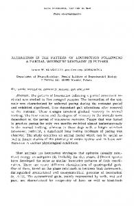
Hours Hours Fig. 1. Apoptosis in hEPC and HUVEC during hypoxia via FACS analysis. Hypoxia generated by the GasPak system, hEPC showed bimodal peaks of apoptosis at 12 and 24 hours (175.9±35.8%, 398.3±75.8%), 60 and 72 hours (220.5±68.1%, 368.8±20.3%) of hypoxia. HUVECs showed increased apoptosis at 12 hours (256.3±42.2%) of hypoxia initially and the highest degree of apoptosis was observed at 72 hours (500.6±45.6%) of hypoxia. *Significantly different from the control time (p