Oct 7, 1977 - by applying image enhancement techniques to electron mi- crographs of specimens that have been specifically labeled with. Fab fragments.
Proc. Nati. Acad. Sci. USA
Vol. 74, No. 12, pp. 5514-5518 December 1977 Biochemistry
New method for localizing proteins in periodic structures: Fab fragment labeling combined with image processing of electron micrographs (antigenic sites/electron microscopy/protein conformation/bacteriophage T-even)
U. AEBItt, B. TEN HEGGELER, L. ONORATO, J. KISTLER, AND M. K. SHOWEO Department of Microbiology, Biozentrum of the University of Basel, 4056 Basel, Switzerland
Communicated by A. H. Doermann, October 7, 1977
ABSTRACr Fab fragments prepared from antisera directed against purified bacteriophage T4 structural proteins- and head-related structures were used to label proteins on the surface of T-even giant phage capsids. Optically filtered electron micrographs of the Fablabeled capsids reveal both the location of specific proteins within the capsomeres and differing conformational states of the protein subunits. We describe parameters affecting the utility of this technique for the study of molecular organization and protein conformation in periodic biological structures.
phage T4 capsids we have been able to confirm the localization (2, 4) of these proteins within the capsomere. Furthermore, this technique has allowed us to demonstrate an induced conformational change on the binding of one of these proteins to the basic capsid matrix. MATERIALS AND METHODS Antigens and Antisera. The T4 outer capsid proteins hoc and soc were purified as described by Ishii and Yanagida (2). T4 coarse polyheads (13) composed only of the product of gene 23 (P23) obtained from a mutant in gene 20 were purified by differential centrifugation. Gene 23ts aberrant preheads containing most of the T4 head proteins including hoc and soc (L. Onorato, unpublished data) were purified by two successive sucrose gradients (14). A 200- to 500-,gg sample of each antigen was injected in Freund's complete adjuvant into the hind footpads of rabbits. Animals were bled twice weekly, starting 4 weeks after injection, and serum from several bleedings was pooled for the preparation of IgG and Fab fragments. The production of high-titer soc antiserum required bimonthly intravenous boosting with 50 jig of the purified antigen. The sera were~characterized on Ouchterlony plates and by immunoreplicate electrophoresis (15). The anti-hoc serum gave no reaction with T2 phage proteins, but reacted strongly with hoc and with another T4 protein, possibly derived from hoc. Antisoc serum reacted only with soc protein. Antiserum to 23ts aberrant preheads reacted strongly with hoc, weakly with P23 and P24, and gave no detectable precipitin reaction with soc or the proteolytically processed form of P23 found in the mature phage head, P23* (16). Anti-polyhead serum reacted with both P23 and P23*. IgG and Fab Fragments. IgG was purified from crude serum by precipitation three times with a saturated ammonium sulfate solution [33% (wt/vol) final concentration] followed by DEAE-cellulose chromatography (17). Monovalent Fab fragments were prepared from purified IgG by cleavage with mercuri-papain as described by Porter (12). Where the fragmentation was incomplete, Fab fragments were separated from undigested IgG by chromatography on Sephadex G-100. Immune Electron Microscopy. Samples containing 1 to 5 X 10⁷ T2L (18) or T4 (19) giant phage particles were incubated with Fab fragments (0.5-1000 ,g) in 20-200 Ail of 10 mM sodium phosphate buffer, pH 7.0, for 1 hr at 370, 4 hr at 20°, or
Many biological structures, including virus shells, contractile filaments, microtubules, and parts of bacterial cell walls, are built up as ordered arrays of one or several species of protein subunits. Electron microscopy of negatively stained specimens followed by image processing of the electron micrographs has proved to be a useful tool for establishing the supramolecular organization of these asetblies (1). The most common wayol localizing individual protwein subunits within their repeating unit has been to compare th: filiered images of related structures that differ in the*otein cporsition. These are usually obtained either from mutants that cannot synthesize one or more of the constituents (2, 3), by differential dissociationof the structures (4), or by in vitro complementation of the defrcient structures with the proteins they are lacking (2, 4-6). However, this approach cannot unambiguously identify the various stain-excluding regions of the repeating unit with particular constituent proteins, because binding of a protein to a periodic assembly may alter the unit cell morphology indirectly by inducing a change in tertiary or quaternary structure. This change may mask the direct change caused by the added protein. One way to localize the individual proteins or protein domains within a supramolecular structure is to label them with specific markers such as antibodies (7-10). However, the utility of this approach is limited both by the size of the antibody molecules [molecular weight (Mr) 150,000] and the dimensions and surface topography of the structure in which the protein is to be localized. To improve the precision of localizing antigenic sites on a structure, Craig and Offer (I1) have used monovalent Fab fragments (12) (Mr 50,000) rather than whole antibodies. We have extended this method for analyzing the molecular organization and conformation of periodic structures by applying image enhancement techniques to electron micrographs of specimens that have been specifically labeled with Fab fragments. By using Fab fragments prepared from specific antisera directed against the constituent proteins of bacterio-
Abbreviations: Mr, molecular weight; P23, product of bacteriophage T4 gene 23 (Mr 58,000); P23*, proteolytically processed form of P23 found in the mature phage head (Mr 47,700). t Molecular Biology Institute, University of California, Los Angeles,
The costs of publication of this article were defrayed in part by the payment of page charges This article must therefore be hereby marked "advertisement" in accordance with 18 U. S. C. §1734 solely to indicate this fact.
CA 90024.
t To whom reprint requests should be addressed.
5514
Proc. Natl. Acad. Sci. USA 74 (1977)
Biochemistry: Aebi et al.' T2L or T4hocsoc7
P236
T2L+hoc or T4soc
P236hoc1
T2L+soc or T4hoc
P236soc6
5515
T2L+hoc+soc or T4
P236soc6hoc,
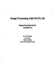
*:hoc, O:SoC * :P23FIG. 1. Schematic representation of bacteriophage T-even capsomere morphologies. The top line indicates the bacteriophage capsid and purified proteins which produce the capsomere types shown below. Each capsomere type is also labeled with its morphological type and protein composition.
12 hr at 4°. The phage particles were freed of excess label by two cycles of centrifugation and resuspended in 50 mM TrisCi, pH 7.2, at 5 X 10⁹ particles per ml. The conditions for antibody excess were established by showing that the supernatant remaining after the first centrifugation of the giant phage was able to label a second aliquot of phages to the same extent as the first. Both labeled and unlabeled samples were prepared for electron microscopy by either negatively staining them with 1% uranyl acetate, pH 4.5 (19), or by freeze-drying and shadowing them (20). Electron micrographs were recorded at a magnification of approximately 35,000 times as described previously (19). Image Processing of Electron Micrographs. Optical diffraction and subsequent filtering of electron micrograph areas containing approximately 200 well-ordered unit cells of the tubular part of giant phage capsids were carried out as described previously (19).
RESULTS The capsomere morphologies of bacteriophages T2 and T4 are shown schematically at the extreme left and right of Fig. 1. They were obtained from optically filtered electron micrographs of negatively stained giant phage capsids (2, 4, 21). These giant phages are viable polymorphic variants of normal T-even phage that are characterized by an abnormally elongated tubular part of the wild-type capsid (22). They can be induced by either genetic (19, 23) or chemical (18) means. The hexagonal T2 lattice is built exclusively from P23* molecules, which are clustered into 6-type capsomeres (21). Bacteriophage T4 is built from the same P23* lattice, but in addition contains the two outer capsid proteins hoc and soc in the ratio P23*6soc6hoc, (2). The localization of hoc and soc in the (6 + 6 + 1)-type T4 capsomeres was inferred from lattices whose capsomeres are shown between those of T2 and T4 in Fig. 1. These intermediate forms were obtained (as indicated in Fig. 1) from T4 mutants defective in production of hoc, soc, or both (2), by chemically extracting these proteins from the T4 lattice (4) and by the addition of hoc and soc in vitro to a T2 6-type lattice (2, 4, 24). The experiments we describe below were designed to test whether the new stain-excluding regions that appear in the capsomeres of (6 + 1)-, (6 + 6)- and (6 + 6 + 1)-types actually
localize hoc and soc, or result from the rearrangement of portions of the P23* molecules upon binding of hoc and soc. Localization of hoc within the P23* lattice Fig. 2A illustrates the change in surface morphology that is obtained when (6 + 1)-type giant phage capsids (top) are reacted-with an excess of anti-hoc Fab fragments (bottom). Fig. 2B shows areas of optically, filtered micrographs of the unlaibeled (top) and labeled (middle: noiMsaturated; bottom: saturated) capsids. Although the labeling does not look very regular on unprocessed rnicrographs, it is sufficiently so to enhance significantly the central afin-exludung region in the filtered images. When (6 + 6 + 1)-type giant phage capsids are reacted ywith the same Fab fragments, a similar enhancement of the central stain-excluding region is obtained. Preincubation of the Fab fragments with purified hoc protein completely inhibits this reaction. Therefore, the central stain-excluding region of (6 + 1)- and (6 + 6 + 1)-type capsomeres actually localizes the hoc molecule in the capsomere, as previously suggested (4, 24). Localization of soc within the P23* lattice Reaction of (6 + 6 + 1)-type giant phage capsids with Fab fragments prepared from a serum directed against purified soc protein did not give specific and regular labeling of the hexagonal surface lattice, even when the Fab fragments were added in great excess. However, when these capsids (Fig. 2C top) were reacted with an excess of Fab fragments prepared from a serum raised against soc-containing 23ts aberrant preheads (14) absorbed with both T2L phage and purified hoc protein prior to the reaction, specific and regular labeling of the hexagonal surface lattice was obtained (Fig. 2C bottom). As can be seen on optically filtered electron micrographs (Fig. 2D), saturation of the (6 + 6 + 1)-type lattice (top) with Fab fragments results in a unit.cell morphology that is characterized by six radially elongated stain-excluding regions surrounding a central one (Fig. 2D bottom). From superposition of the two stain-excluding patterns (shown schematically in the middle of Fig. 2D), we conclude that each of the radially elongated stain-excluding regions localizes a specifically bound Fab fragment. The only feature of the (6 + 6 + 1)-type capsomere that can still be visualized in the labeled pattern is the hoc molecule, which gives rise to a reduced central stain-excluding
5516
Biochemistry:
Aebi et al. A
:. ^_.S .-I _
Proc. Natl. Acad. Sci. USA 74 (1977)
C
as,
..
',
An>> -.'.,'. o
roe
;>