Manuscript
Click here to download Manuscript Diaz-Blancat et al_r2_manuscript.docx
Click here to view linked References
1 2 3 4 5 6 7 8 9 10 11 12 13 14 15 16 17 18 19 20 21 22 23 24 25 26 27 28 29 30 31 32 33 34 35 36 37 38 39 40 41 42 43 44 45 46 47 48 49 50 51 52 53 54 55 56 57 58 59 60 61 62 63 64 65
Fast neural dynamics of proactive cognitive control in a task-switching analogue of the Wisconsin Card Sorting Test Gema Díaz-Blancat1, Juan García-Prieto1,2, Fernando Maestú1,3, Francisco Barceló4
1
Laboratory of Cognitive and Computational Neuroscience (UCM-UPM), Centre for Biomedical
Technology, Universidad Politécnica de Madrid, Madrid, Spain 2
Laboratory of Electrical Engineering and Bioengineering, Department of Industrial Engineering,
Universidad de La Laguna, Tenerife, Spain 3
Department of Basic Psychology II, Complutense University of Madrid, Spain
4
Laboratory of Neuropsychology, University of the Balearic Islands, Spain
Keywords: Cognitive control, context processing, magnetoencephalography, prefrontal cortex, task-switching, Wisconsin card sorting test.
Running title: Proactive control in the WCST
Corresponding author: Francisco Barceló, PhD. Laboratory of Neuropsychology University of the Balearic Islands 07122 Palma de Mallorca Spain E-mail:
[email protected] (+34) 971 172750
1
Abstract 1 2 3 4 5 6 7 8 9 10 11 12 13 14 15 16 17 18 19 20 21 22 23 24 25 26 27 28 29 30 31 32 33 34 35 36 37 38 39 40 41 42 43 44 45 46 47 48 49 50 51 52 53 54 55 56 57 58 59 60 61 62 63 64 65
One common assumption has been that prefrontal executive control is mostly required for target detection (Posner and Petersen, 1990). Alternatively, cognitive control has also been related to anticipatory updating of task-set (contextual) information, a view that highlights proactive control processes. Frontoparietal cortical networks contribute to both proactive control and reactive target detection, although their fast dynamics are still largely unexplored. To examine this, we analyzed rapid magnetoencephalographic (MEG) source activations elicited by task cues and target cards in a task-cueing analogue of the Wisconsin card sorting test (WCST). A single-task (color sorting) condition with equivalent perceptual and motor demands was used as a control. Our results revealed fast, transient and largely switch-specific MEG activations across frontoparietal and cingulo-opercular regions in anticipation of target cards, including (1) early (100-200 ms) cue-locked MEG signals at visual, temporo-parietal and prefrontal cortices of the right hemisphere (i.e., calcarine sulcus, precuneus, inferior frontal gyrus, anterior insula and supramarginal gyrus); and (2) later cue-locked MEG signals at the right anterior and posterior insula (200-300 ms) and the left temporo-parietal junction (300-500 ms). In all cases larger MEG signal intensity was observed in switch relative to repeat cueing conditions. Finally, behavioral restart costs and test scores of working memory capacity (Forward Digit Span) correlated with cue-locked MEG activations at key nodes of the frontoparietal network. Together, our findings suggest that proactive cognitive control of task rule updating can be fast and transiently implemented within less than a second and in anticipation of target detection.
Díaz-Blancat et al.
2
Introduction 1 2 3 4 5 6 7 8 9 10 11 12 13 14 15 16 17 18 19 20 21 22 23 24 25 26 27 28 29 30 31 32 33 34 35 36 37 38 39 40 41 42 43 44 45 46 47 48 49 50 51 52 53 54 55 56 57 58 59 60 61 62 63 64 65
Functional neuroanatomy of cognitive control is a major hot topic in human neuropsychology, with a special interest for the anterior executive control system (Miller and Cohen, 2001; Petersen and Posner, 2012), as well as its complex frontoparietal architecture linking key hubs in medial and lateral prefrontal cortex (PFC) with posterior parietal cortex and subcortical structures (Corbetta and Shulman, 2002; Dosenbach, et al., 2006, 2008). The prevailing view has been that the anterior executive control system is mostly engaged during target detection (Posner and Petersen, 1990). These authors argued that when a target is identified and becomes consciously attended, it generates a global workspace of widespread cortical activation causing interference and conflict across the system, which in turn triggers activity in medial frontal/anterior cingulate cortices (ACC) (Petersen and Posner, 2012). This is also a common assumption behind the interpretation of classic neuropsychological assessment tools such as the Wisconsin Card Sorting Test (WCST), where clinicians and researchers alike often assume maximal cognitive effort and control for processing the target cards (Lezak, et al., 2012; Monchi et al., 2001), over and above any other accessory or contextual signals in spatial and/or temporal proximity with the sorting cards. An alternative view emphasizes the key role of PFC in the online representation and updating of the spatiotemporal context for goal-directed actions (Braver, 2012; Braver and Barch, 2002). For instance, Braver (2012) distinguishes between anticipatory, proactive control processes that serve to prepare the system for upcoming goaldirected control of behavior, and stimulus-driven, reactive control processes that are recruited to deal with relevant target information. However, the relative importance from proactive and reactive control modes in classical tests of executive function remains largely unexplored. Moreover, the fast neural dynamics during proactive and reactive control modes remains poorly understood, partly due to the limited temporal resolution of metabolic brain imaging studies (cf., Braver, et al., 2003; Konishi, et al., 1998; Monchi, et al., 2001).
Díaz-Blancat et al.
3
Converging evidence from event-related potential (ERP) studies in healthy controls and frontal 1 2 3 4 5 6 7 8 9 10 11 12 13 14 15 16 17 18 19 20 21 22 23 24 25 26 27 28 29 30 31 32 33 34 35 36 37 38 39 40 41 42 43 44 45 46 47 48 49 50 51 52 53 54 55 56 57 58 59 60 61 62 63 64 65
lesion patients suggests that cognitive control can be partly implemented proactively, either when targets are temporarily predictable (Karayanidis et al., 2003), or in response to contextual cues forerunning target onset (Barceló, 2003; Karayanidis et al., 2009). Task-cueing ERP studies suggest that task- set reconfiguration (TSR; also “task rule updating”) can be fully completed with long cue-target intervals (>1000 ms) and well before target onset (Adrover-Roig and Barceló, 2010; Jost, et al., 2008). Likewise, focal lesions to lateral PFC are compatible with relatively preserved target P3 potentials (Barceló, et al., 2000), even though the same PFC lesions disrupt P3-like potentials to informative contextual cues forerunning target onset (Barceló and Knight, 2007). Together, these studies suggest that proactive cognitive control is critical for efficient goal-directed behavior (i.e., target detection), at least under the wellstructured task conditions of conventional neuropsychological testing. If this hypothesis holds true, then an analysis of the fast neural dynamics in a task-cueing version of the WCST could help us clarify the relative contribution of frontoparietal cortical regions to proactive and reactive control modes. Whereas proactive control of task-switching (also i.e., “task rule updating”) is expected to occur mostly during the anticipatory period in cued task-switching, reactive control is mostly required for target detection and categorization, involving processes such as target-driven rule execution (Braver, 2012; Corbetta et al., 2008). The dual model of cognitive control argues that proactive and reactive control modes are subserved by distinct regions within the anterior executive system, with key roles for lateral PFC and posterior temporo-parietal cortices (Braver, 2012), as delineated by functional magnetic resonance imaging (fMRI) studies. Thus, Dosenbach et al., (2006) described a frontoparietal network —including lateral PFC and the intraparietal sulcus— involved in initiating and adapting task control on a trial-by-trial basis. In contrast, a cingulate-opercular network –including dorsal ACC, medial frontal cortex, frontal operculum and anterior insula (aINS)– has been related to both transient ‘start-cue’ and sustained maintenance of task goals over trials. Taking advantage Díaz-Blancat et al.
4
of the excellent trade-off between anatomical and temporal resolution offered by 1 2 3 4 5 6 7 8 9 10 11 12 13 14 15 16 17 18 19 20 21 22 23 24 25 26 27 28 29 30 31 32 33 34 35 36 37 38 39 40 41 42 43 44 45 46 47 48 49 50 51 52 53 54 55 56 57 58 59 60 61 62 63 64 65
magnetoencephalography (MEG), Periáñez et al., (2004) explored the fast dynamics of proactive cognitive control using a simplified task-cueing version of the WCST. These authors found the earliest switch-specific MEG activations at the inferior frontal gyrus (IFG) 100-300 ms post-cue onset, followed by recurrent peaks of MEG activity at the ACC and the supramarginal gyrus (SMG) from 300 to 600 ms post-cue onset. However, these authors did not compare MEG source dynamics between proactive (cue-locked) and reactive (target-locked) control modes. Up to date, only a few MEG studies have examined the fast dynamics of cognitive control during task-switching (Bayless, et al., 2006; Henaff, et al., 2010; Oh, et al., 2014; Periáñez, et al., 2004; Wang, et al., 2001), although none of those studies contrasted MEG dynamics during proactive and reactive control modes. Also many fMRI studies using WCST analogues analyzed feedback signals rather than switch cues prompting for task rule updating, which hindered the analysis of pure task-switching, making it difficult to disentangle switch-specific from reward-related neural processes in WCST performance (Konishi, et al., 1998; Monchi, et al., 2001).
In order to circumvent these limitations, task-cueing paradigms are well-suited for examining the fast neural dynamics during proactive and reactive control modes (Braver, et al., 2003; Karayanidis, et al., 2009). Here we used a task-cueing version of the WCST adapted for measuring event-related neural responses while participants sorted target cards following one of two rules (color or form; cf., Adrover-Roig and Barceló, 2010). The correct task rule switched intermittently as announced by auditory tonal cues signaling either switches or repetitions in the ongoing stimulus-response (S-R) mapping. Visual feedback was delivered on a trial-by-trial basis, and the analyses focused on correct color trial runs only, in order to avoid contamination from negative feedback effects. A single-task “color only” condition with equivalent sensory and motor response demands served as a control. Here, the infrequent deviant sound (i.e., the “switch cue” in the task-switching condition) acted as a mere distractor against a background of Díaz-Blancat et al.
5
repetitive standard sounds (i.e., “repeat cues” in the task-switching condition). This procedure 1 2 3 4 5 6 7 8 9 10 11 12 13 14 15 16 17 18 19 20 21 22 23 24 25 26 27 28 29 30 31 32 33 34 35 36 37 38 39 40 41 42 43 44 45 46 47 48 49 50 51 52 53 54 55 56 57 58 59 60 61 62 63 64 65
has yielded reliable estimates of two indexes of behavioral (restart and mixing) switch costs (cf., Adrover-Roig and Barceló, 2010). On the one hand, restart costs are observed on first trials following any interrupt signal instructing to switch or repeat the task. Restart costs have been related to the suppression of conflicting S-R mappings from the previous task rule (Allport and Wylie, 2000; Poljac, et al., 2009), and are known to be large on first cued repetition trials (i.e., paradoxical “repetition cost”) in conditions where both the task rule and the sensory cue change regarding the previous trial (Forstmann et al., 2007; Periañez & Barceló, 2009). Such type of restart costs can be assumed to require conflict resolution at both “higher” (i.e., rule updating) and “lower” (S-R re-mapping) levels in the hierarchy of control (Miller & Cohen, 2001; Schneider & Logan, 2006). Therefore, restart costs were expected to correlate with ACC activity, since this structure is thought to play a pivotal role in conflict monitoring (Braver, 2012). In contrast, mixing costs measure sustained rule interference during task repetitions in mixed task blocks relative to homogeneous blocks (Monsell, 2003). For the sake of simplicity and cleanness of MEG signals, long cue-target intervals (>1000 ms) were employed here to allow enough preparation time for full task-set reconfiguration prior to target onset (Barceló et al., 2006). Finally, under the assumption that anticipatory task rule updating involves executive control, we expected cue-locked MEG activations to correlate with behavioral restart costs and with neuropsychological test scores of executive function.
In sum, the present study examined the hypothesis whether prefrontal executive control can be rapidly engaged during the processing of task cues for proactive rule updating (Braver, 2012; Braver and Barch, 2002), as distinct from the reactive processing of the target WCST cards (Petersen and Posner, 2012). In examining this hypothesis, we used a computerized task-cueing version of the WCST adapted for measuring event-related MEG activations associated with anticipatory task cues (prompting for proactive control) and target cards (demanding reactive Díaz-Blancat et al.
6
control) under both task-switching and single-task conditions (cf., Adrover-Roig and Barceló, 1 2 3 4 5 6 7 8 9 10 11 12 13 14 15 16 17 18 19 20 21 22 23 24 25 26 27 28 29 30 31 32 33 34 35 36 37 38 39 40 41 42 43 44 45 46 47 48 49 50 51 52 53 54 55 56 57 58 59 60 61 62 63 64 65
2010).
Methods Participants Eighteen young adults took part in the study (mean age 26.7 ± 4.3 years, range 21-36 years, 11 females). Three participants were excluded from the final analyses due to outlier behavioral data (n= 1) and corrupted MEG data (n= 2), thus leaving a final sample of fifteen participants (mean age 26.4 ± 4.7 years, range 21-36 years, 9 females). All participants were recruited at the Center for Biomedical Technology, and were graduate or postgraduate students at the Universidad Politécnica de Madrid. They all had normal or corrected to normal visual acuity. History for neurological disease, psychiatric illness, head injury, stroke, substance abuse (excluding nicotine), learning disabilities, or any other clinical conditions that could interfere with behavioral testing were criteria for exclusion. Experimental procedures and behavioral testing was performed in accordance with the Declaration of Helsinki, informed consent was obtained from all participants, and the study was approved by the ethics committee of the Center for Biomedical Technology.
Neuropsychological Assessment All participants completed a 45-min battery of neuropsychological tests before MEG scanning, including the MiniMental State Examination, forward and backward Digit Span, Trail Making Test -forms A and B, Stroop test, Boston naming test, semantic fluency (animals) and phonological fluency (COWA-FAS form, as described in Adrover-Roig and Barceló, 2010). The neuropsychological assessment confirmed that all participants showed normal scores compared to their age-matched normalized sample (Lezak, et al., 2012).
Díaz-Blancat et al.
7
Task design and Procedures 1 2 3 4 5 6 7 8 9 10 11 12 13 14 15 16 17 18 19 20 21 22 23 24 25 26 27 28 29 30 31 32 33 34 35 36 37 38 39 40 41 42 43 44 45 46 47 48 49 50 51 52 53 54 55 56 57 58 59 60 61 62 63 64 65
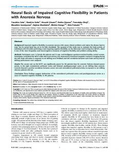
We used two versions of a computerized task-cueing paradigm inspired in the WCST (cf., Adrover-Roig and Barceló, 2010), each corresponding to the task-switching and single-task conditions, respectively. In the task-switching procedure participants were requested to start sorting cards following the color rule, and then to alternate between the color and shape rules. Tonal cues informed participants when to switch (low-pitched tone) or repeat (high-pitched tone) the previous rule. In the single-task condition, participants were to sort cards always by the color rule, and the tones were mere distracters to be ignored for efficient performance. Target cards in both task conditions were restricted to the 24 WCST choice cards that can be unambiguously sorted based on just one stimulus dimension (Fig. 1). The colored geometrical shapes were outlined in black against a white background to improve visual contrast. The same sequence of target cards was used both in single-task and task-switching conditions for all participants. Thus, both task conditions consisted of two blocks of 240 trials each, with a 5-min rest period between blocks. Each trial began with a tonal cue (either 500 Hz or 1000 Hz tones, 200 ms duration, 10 ms rise/fall times, 75 dB sound pressure level), followed by a visual target display with the four key cards on top of one choice card. The mean probabilities of tonal cues were p= 0.25 for the low-pitched tone, and p= 0.75 for the high-pitched tone, with a pseudorandom sequential arrangement to allow for a minimum of three repeat trials following any switch trial. The cue-target interval was jittered with a squared distribution with values ranging between 1000 and 1100 ms (Fig. 1). Participants used a 4-button panel with a horizontal arrangement to match the choice card with one of the key cards on top. The far left button designated the key card on the far left of the display; the far right button designated the key card on the far right, and so on. Participants used their left and right thumbs to press the leftand right-hand side buttons in a response pad, respectively. Immediately after responding, the Spanish word for ‘‘right’’ or ‘‘wrong’’ was visually displayed for 200 ms as feedback. Likewise, the Spanish words for “too fast” or “too slow” appeared whenever the button was pressed Díaz-Blancat et al.
8
before 300 ms or after 3000 ms from target onset, respectively. Finally, a jittered interval of 1 2 3 4 5 6 7 8 9 10 11 12 13 14 15 16 17 18 19 20 21 22 23 24 25 26 27 28 29 30 31 32 33 34 35 36 37 38 39 40 41 42 43 44 45 46 47 48 49 50 51 52 53 54 55 56 57 58 59 60 61 62 63 64 65
between 700-750 ms elapsed between the button press and the next tonal cue (Fig. 1). Visual stimuli were projected onto a screen 1.35 m away from the participant, and subtended a visual angle of 4.44˚ horizontally and 2.86˚ vertically (cf., Adrover-Roig and Barceló, 2010). Trial sequence and image display were controlled with Presentation® software (Neurobehavioral Systems Inc., Albany, CA).
As said before, the same trial structure was used both in single-task and task-switching conditions. Trial runs contained at least three target cards between any two consecutive lowpitched tones, with a variable number of runs containing either three (20%), four (45%), five (30%) or six (5%) target cards in between any two low-pitched tones. This variable length of trial runs made the next switch tone relatively unexpected, so that task-switching operations were effectively time-locked to the cueing events. This task structure favored that trials in the taskswitching condition could be classified into switch, first-, second- and third-repeat target trials. Correspondingly, in the single-task condition trials were classified as deviant, first-, second- and third-standard trials, depending on their sequential order following the infrequent deviant lowpitched tone. The order of the single-task and task-switching conditions was counterbalanced across subjects. Before the MEG testing session, participants were fully instructed and practiced each task until they reached a criterion of 100% correct trials during 5 min, so as to optimize their performance during the testing session (cf., Adrover-Roig and Barceló, 2010). ************************ Insert Figure 1 about here ************************ Behavioral switch costs and distraction costs were estimated from correct (mean reaction times, RTs) and failed trials (error rates) in task-switching and single-task conditions, respectively. In the single-task condition, failed trials were defined as those where subjects did Díaz-Blancat et al.
9
not sort the choice card by its color. In the task-switching condition, failed trials were defined as 1 2 3 4 5 6 7 8 9 10 11 12 13 14 15 16 17 18 19 20 21 22 23 24 25 26 27 28 29 30 31 32 33 34 35 36 37 38 39 40 41 42 43 44 45 46 47 48 49 50 51 52 53 54 55 56 57 58 59 60 61 62 63 64 65
those where subjects (a) did not follow the instruction cue to switch or repeat the previous rule, or (b) failed to select the correct response within the currently relevant task rule (Barceló, 2003). In both task conditions, any responses performed earlier than 300 ms (false alarms) or later than 3000 ms (omissions) were also computed as errors. To avoid contamination from negative feedback and post error slowing effects, the analyses of behavioral and MEG data considered correct trial runs only, that is, runs containing no errors (cf., Barceló, 2003).
Two behavioral indexes of residual switch costs were estimated for each participant. Restart costs were computed as the difference in mean RT between the first and third repeat targets in the task-switching condition (Rushworth, et al., 2002), as well as in the single-task condition. Mixing costs were computed as the difference in mean RT between third repeat targets and third standard targets in the task-switching and single-task conditions, respectively (cf., Monsell, 2003). Given the long cue-target intervals (>1000 ms), switch-specific costs indexing fast and transient rule updating were expected to be absent (Monsell, 2003), particularly for the subset of correct trials targeted in the MEG analyses. Only the color rule in the task-switching condition was used for comparison of behavioral and MEG results in the single-task (color) condition. Statistical analyses were conducted using SPSS v.20 software. All post-hoc tests of simple effects were performed using the Bonferroni correction with a significance level of p < .05.
MEG Data Acquisition and Analyses MEG data were acquired with a 306-channel Vector view system (Elekta-Neuromag) at the Center for Biomedical Technology (Madrid, Spain). The system comprises 102 magnetometers and 204 planar gradiometers on a sensor array, located inside a magnetically shielded room. Sampling frequency was 1kHz, and an online anti-alias filter (0.1 – 330 Hz) was applied. A 3D spatial digitizer was used to digitize the head shape (Polhemus Fastrak, Polhemus Inc., Díaz-Blancat et al. 10
Colchester, VT, USA). A head position indicator (HPI) determined the position of the head with 1 2 3 4 5 6 7 8 9 10 11 12 13 14 15 16 17 18 19 20 21 22 23 24 25 26 27 28 29 30 31 32 33 34 35 36 37 38 39 40 41 42 43 44 45 46 47 48 49 50 51 52 53 54 55 56 57 58 59 60 61 62 63 64 65
respect to the sensor array at the beginning of the recordings. Four HPI coils were attached to the subject (one on each mastoid, two on the forehead), and their position with respect to the 3 fiducials (nasion, left and right pre-auricular points) was determined. Vertical eye movements were recorded using two electrodes attached above and below the left eye in a bipolar montage.
External noise was removed from MEG data using the temporal extension of Signal-Space Separation (tSSS) (Taulu and Kajola, 2005) in MaxFilter (version 2.2, Elekta-Neuromag). All recordings were visually inspected to identify artifacted segments, including eye blinks, eye movements or muscular movement, which was removed from subsequent analyses. Raw artifact-free segments where cleaned with the aid of Brainstorm software toolbox (Tadel, et al., 2011), creating signal space projection vectors corresponding to each type of artifact. These SSP vectors were then factored out of the MEG recordings. The resulting clean single-trial MEG segments consisted of 310 ms pre-stimulus baseline periods and 1,000 ms post-cue and posttarget periods.
Source Reconstruction and Statistical Analyses Source reconstruction was performed using Brainstorm toolbox software (Tadel, et al., 2011). Since the participants’ MRI scans were not available, source reconstruction was based on the cortical surface of the standard MNI/Colin27 brain template (Collins, et al., 1998). A 3D grid with 15,003 sources was created in the template brain and later adapted onto each subject’s head using Colin27’s scalp and the subject’s head shape. A forward model was computed using an overlapping spheres approach (Huang, et al., 1999). Source time-series were computed using a minimum norm estimation algorithm (Tadel, et al., 2011), and all 306 channels, including both magnetometers and planar gradiometers, were considered in the reconstruction. Minimum Díaz-Blancat et al. 11
norm estimates (MNE) produced a measure of the current density flowing at the surface of the 1 2 3 4 5 6 7 8 9 10 11 12 13 14 15 16 17 18 19 20 21 22 23 24 25 26 27 28 29 30 31 32 33 34 35 36 37 38 39 40 41 42 43 44 45 46 47 48 49 50 51 52 53 54 55 56 57 58 59 60 61 62 63 64 65
cortex. To visualize these results and compare them between subjects, we normalized the MNE values using Z-scores to get a standardized level of activation with respect to noise in the baseline. Source orientations were constrained perpendicular to the mesh surface (Tadel, et al., 2011). Averaged trials were projected to the 15,003 sources, and the 1,000 ms window after each stimulus was segmented into 100 ms intervals (cf., Periáñez, et al., 2004). Average density was obtained in each interval for statistical comparisons. Only trials sorted by the color rule in both the single-task and task-switching conditions entered these MEG analyses.
Statistical analysis was performed with Brainstorm (Tadel et al. 2011). We realized paired t-test comparisons on the average source activation between the following three task conditions, for the cue and target periods: switch vs. repeat, switch vs. deviant, and repeat vs. standard. Only third repeat (repeat3) and third standard (standard3) target trials were considered in these contrasts to avoid contamination by carryover effects from the infrequent switch and deviant tones. The present task design allowed us to examine both switch specific and task-level MEG differential activations during both cue-locked and target-locked periods. Task-switch specific MEG activations were explored with the contrast between switch vs. repeat trials during the cue and target periods. Task-level differential MEG activations were explored with contrasts: switch vs. deviant, and repeat vs. standard trials, also during the cue and target periods.
To protect whole-brain analyses against false positive activations, we used the false discovery rate (FDR) correction. This procedure is designed to control the expected proportion of false positives (type I errors in null hypothesis testing) against all positive activations, following the Benjamini–Hochberg step-up procedure with a corrected p-value threshold of 0.01 as implemented in Brainstorm (Benjamini and Hocheberg, 1995). Pearson product–moment correlations were used to examine the association between MEG activations and behavioral Díaz-Blancat et al. 12
measures. Restart and mixing time costs, as well as five neuropsychological test scores (forward 1 2 3 4 5 6 7 8 9 10 11 12 13 14 15 16 17 18 19 20 21 22 23 24 25 26 27 28 29 30 31 32 33 34 35 36 37 38 39 40 41 42 43 44 45 46 47 48 49 50 51 52 53 54 55 56 57 58 59 60 61 62 63 64 65
and backward Digit Span scores, FAS total score, TMT B:A, and Stroop color-word; cf., AdroverRoig & Barceló, 2010) were then correlated with the absolute source MEG amplitude for each significant ROI and time window. Finally, correlations between behavior and MEG source activity were considered significant for p-values lower than 0.01 after a non-parametric permutation correction (N= 5,000).
Results Behavioral Results Across tasks and participants, accuracy was always better than 94% (mean= 98.4%, SD= 1.5% correct trials) in the single task, and better than 92% (mean= 96.1%, SD= 2.1% correct trials) in the task-switching condition. The analysis of error rates revealed a main Task effect (F 1,14 = 49.8, p< .0001, ηp2= 0.71) indicating more errors under task-switching compared to the single-task (3.9% vs. 1.5%). An interaction between Task and Trial sequence (F3,42 = 3.8, p< .04, ηp2= 0.21), indicated that errors were evenly distributed across trials in the single-task (all ps = 1.0), whereas switch and first-repeat trials were more error prone than later repeat trials in the taskswitching condition (switch vs repeat1, p= 1.0; repeat1 vs repeat2, p= .05; repeat1 vs repeat3, p= 0.03; repeat2 vs repeat3, p= 1.0). No other effects or interactions reached statistical significance for error rates.
For mean reaction times, there were main effects for Task (F1,14 = 43.8, p< .0001, ηp2= 0.76), Trial sequence (F3,42 = 8.5, p< .001, ηp2= 0.37), as well as their interaction (F3,42 = 6.9, p< .001, ηp2= 0.33), indicating similar response speed across all trials in the single-task condition (mean RTs difference between deviant with first, second and third standard trials, 1.1, 9.2 and 1.5 ms, respectively, all ps = 1.0). In contrast, first repeat trials were responded to slower than switch, second and third repeat trials in the task-switching condition (mean RTs differences 64, 62 and Díaz-Blancat et al. 13
73 ms, respectively, all p< .01, see Table 1). In turn, mean RTs to switch, second and third repeat 1 2 3 4 5 6 7 8 9 10 11 12 13 14 15 16 17 18 19 20 21 22 23 24 25 26 27 28 29 30 31 32 33 34 35 36 37 38 39 40 41 42 43 44 45 46 47 48 49 50 51 52 53 54 55 56 57 58 59 60 61 62 63 64 65
trials did not differ significantly (all ps= 1.0) No other effects or interactions reached significance for mean reaction times. A mean restart cost of 73 ms was found between first and third repeat trials under task-switching conditions (95% CI [38.2 – 106.6 ms]) , and an average mixing cost of 152 ms was measured in third repeat trials relative to third standard target trials in the singletask condition (95% CI [77.0 – 227.0 ms]; see Table 1).
************************ Insert Table 1 about here ************************ MEG Results Figure 2 presents the significant differences in averaged MEG signal intensity for the switch versus repeat comparison during the cue period under the task-switching condition only at four latency windows: 100-200 ms, 200-300 ms, 300-400 ms and 400-500 ms post-cue onset. No other planned contrasts in averaged MEG signal intensity during the cue or target periods reached significance levels (threshold p-value < 0.01; with FDR correction). Table 2 presents a summary of regions of interest (ROIs) showing significant differential MEG signal amplitude for the relevant contrast (switch > repeat) under different time windows in the cue period only. Of note, the group-averaged (N=15) MEG signal waveforms for switch cues, repeat cues, and switch-repeat difference waveforms revealed mostly phasic and transient MEG activations from 100 to 500 ms post-cue onset (see Supplementary Fig. 1).
Switch-specific differential MEG activations were observed from 100 to 500 ms post-cue onset. At an early 100 to 200 ms time window, significantly larger MEG signal intensities in response to switch compared to repeat cues were observed in the IFG, anterior and posterior insula, precentral and postcentral gyri, SMG, precuneus and calcarine sulcus, all of them in the right Díaz-Blancat et al. 14
hemisphere (Fig. 2; 100-200 ms). Further, the same comparison yielded significant differences 1 2 3 4 5 6 7 8 9 10 11 12 13 14 15 16 17 18 19 20 21 22 23 24 25 26 27 28 29 30 31 32 33 34 35 36 37 38 39 40 41 42 43 44 45 46 47 48 49 50 51 52 53 54 55 56 57 58 59 60 61 62 63 64 65
in MEG signal intensity in the right insula (200-300 ms post-cue), followed by the left superior temporal gyrus and left inferior parietal lobe (300-400 ms post-cue), and the right parietal lobe (400-500 ms post-cue). Without exception, the direction of the differences in all these significant contrasts revealed increased MEG power in the switch as compared to the repeat condition. No significant differences in MEG signal activity were observed between task cues and single-task distracters at later latency windows in the cue period. No planned contrasts in averaged MEG signal intensity reached significance levels during the target period (threshold pvalue < 0.01; with FDR correction). ************************ Insert Table 2 about here ************************ Insert Figure 2 about here ************************ Correlation Analyses Two measures of residual behavioral costs (restart and mixing), and five neuropsychological scores (forward and backward Digit Span scores, COWA-FAS total score, TMT B:A, and Stroop color-word) were correlated with 26 maxima of MEG source activation, one per condition, at those ROIs and time windows showing significant differential Switch > Repeat activation (Table 2), thus totaling 182 attempted correlations that were corrected for multiple comparisons using 5,000 permutations and a corrected p-value < 0.01. A significant positive correlation was observed between the Forward Digit Span score and cue-locked MEG signal intensity at the right IFG (repeat condition: R= 0.73, p< 0.01; Fig. 3A), and the right SMG (switch condition: R= 0.76, p< 0.01; Fig. 3B). Further, restart costs were negatively correlated with cue-locked MEG signal intensity at the right SMG (repeat condition: R= -0.73, p< 0.01; Fig. 3C). All significant correlations were observed in the early 100-200 ms time window only. Díaz-Blancat et al. 15
************************ 1 2 3 4 5 6 7 8 9 10 11 12 13 14 15 16 17 18 19 20 21 22 23 24 25 26 27 28 29 30 31 32 33 34 35 36 37 38 39 40 41 42 43 44 45 46 47 48 49 50 51 52 53 54 55 56 57 58 59 60 61 62 63 64 65
Insert Figure 3 about here ************************ Discussion This study examined the hypothesis that prefrontal executive control can be rapidly engaged during proactive processing of contextual information for efficient goal-directed behavior (Braver, 2012; Miller and Cohen, 2001). Towards this end, the temporal dynamics of MEG source activity were examined using a task-cueing WCST analogue adapted to assess both proactive and reactive control modes. As expected, reliable switch-specific differential MEG activations were found in several nodes of the frontoparietal and cingulo-opercular networks, such as anterior and posterior insula, IFG, SMG, superior temporal gyrus, inferior parietal lobe, and precuneus (Table 2). Importantly, these switch-specific effects occurred proactively in the cue period, evolved fast and transiently within half a second post-cue onset, and then subsided well before target onset. Task-level differential MEG activations among switch and single-task conditions (switch vs. deviant and repeat vs. standard) did not reach significance after correction for false discovery rate (FDR), neither during the cue nor the target periods, thus attesting for an adequate control of general non-specific attention and stimulus-response (S-R) selection effects in our task-cueing paradigm. Behavioral restart costs and neuropsychological test scores of working memory capacity (Forward Digit Span) showed a linear association with cue-locked MEG activations at key frontoparietal regions. Together, these findings reveal fast and transient switch-specific MEG source activity in key frontoparietal and cingulo-opercular regions during the proactive control of task rule updating and in anticipation to target onset.
Early proactive task rule updating (100-200 ms post-cue) The contrast between switch and repeat cues revealed early and transient switch-specific MEG activations in a distributed network of frontoparietal and cingulo-opercular regions, including Díaz-Blancat et al. 16
anterior insula, IFG, SMG, and precuneus, all of which showed a right hemisphere 1 2 3 4 5 6 7 8 9 10 11 12 13 14 15 16 17 18 19 20 21 22 23 24 25 26 27 28 29 30 31 32 33 34 35 36 37 38 39 40 41 42 43 44 45 46 47 48 49 50 51 52 53 54 55 56 57 58 59 60 61 62 63 64 65
predominance (Table 2). Of note, no task-level contrasts of differential MEG activation (switch vs. deviant, repeat vs. standard) reached significance within the cue period.
In line with past studies, we observed early (100-200 ms) cue-locked differential (switch > repeat) MEG activation in the IFG. Thus, Periáñez et al. (2004) observed a switch-specific increase in the number of MEG activity sources bilaterally in the IFG from 100 to 300 ms postcue onset. Similarly, Oh et al. (2014) reported transient MEG activity in IFG from 100 to 350 ms of target onset during extradimensional set-shifting, although their study did not segregate proactive from reactive stages of control. Instead, here we found evidence for an early and transient switch-specific involvement of the right IFG during anticipatory task rule updating, with increased MEG signals to switch compared to repeat cues, which is consistent with past fMRI studies of task-switching (Derrfuss, et al., 2005; Kim, et al., 2012), and with WCST studies that used negative feedback signals as switch cues (Konishi, et al., 1998; Monchi, et al., 2001). Hence, our results suggest an implication of the right IFG in anticipatory rule updating, without the confound with reactive target detection and reward-related feedback processing.
Early differential MEG activation in the right anterior and posterior insula (aINS, pINS) revealed increased MEG signal intensity to switch cues relative to repeat cues. Such early switch-specific effects might index phasic top-down modulation during proactive task rule updating in response to the behaviorally more relevant switch cues compared to repeat cues. This proposal concurs with the purported role of the aINS in task rule updating (Derrfuss, et al., 2005; Dosenbach et al., 2008). At this early time window, both the right aINS and pINS were concurrently activated, suggesting these two structures interact to modulate physiological reactivity to salient stimuli (see Menon and Uddin, 2010 for a review).
Díaz-Blancat et al. 17
The right precuneus was also more activated in response to switch cues (Table 2, Fig. 2). At least 1 2 3 4 5 6 7 8 9 10 11 12 13 14 15 16 17 18 19 20 21 22 23 24 25 26 27 28 29 30 31 32 33 34 35 36 37 38 39 40 41 42 43 44 45 46 47 48 49 50 51 52 53 54 55 56 57 58 59 60 61 62 63 64 65
two previous studies found similar early precuneus MEG activations during task-switching. Bayless et al. (2006) and Oh et al (2014) reported early 100-350 ms MEG activity at precuneus during extra-dimensional shifts in attention –the equivalent of our switch cues. Likewise, Barber and Carter (2005) reported increased precuneus activity during switch compared to repeat trials in the preparatory cue-target period, and suggested that this region contributes to the anticipatory component of task-switching, perhaps pre-activating cortical regions for the upcoming detection of stimulus features necessary for S-R associations.
The right SMG showed concurrently enhanced MEG signals in this early time window in response to switch cues compared to repeat cues (Fig. 2, Table 2). Together with the significant effects reported above, this result is consistent with the purported role of the right SMG as a key node within a ventral frontoparietal network for reorienting attention to new sources of information as part of a stimulus-driven “circuit breaker” mechanism (Corbetta et al., 2008). The early timing of these MEG activations, together with our improved task-cueing design, all suggest that key nodes of this ventral frontoparietal network may play a role in interrupting ongoing selection of relevant information not only for detection of specific targets (Corbetta & Shulman, 2002; Petersen and Posner, 2012), but also during proactive updating to novel high-order task rules (new S-R mappings), and well in anticipation to target onset. The proposal that these very early MEG activations in key nodes of the ventral frontoparietal network may index various proactive control operations in anticipation of the next target card was supported by the direct association found between MEG source activity in the IFG and SMG with test scores of working memory capacity (forward Digit Span; Figs. 3A, B), as well as by the inverse association found between MEG source activity in the SMG and behavioral restart costs (Fig. 3C).
Díaz-Blancat et al. 18
Finally, enhanced MEG signals to switch cues relative to repeat cues were also observed at the 1 2 3 4 5 6 7 8 9 10 11 12 13 14 15 16 17 18 19 20 21 22 23 24 25 26 27 28 29 30 31 32 33 34 35 36 37 38 39 40 41 42 43 44 45 46 47 48 49 50 51 52 53 54 55 56 57 58 59 60 61 62 63 64 65
right calcarine sulcus, the right precentral gyrus and the right postcentral sulcus (Fig. 2, Table2). These effects suggest that primary sensory and motor regions can be fast and transiently coactivated together with high-order nodes in the ventral frontoparietal network for proactive updating of low-level S-R mappings early during the cue-target period (Barber and Carter, 2005). The right hemisphere predominance of these effects reminds us of the hemispheric bias of the ventral attention network (Corbetta et al., 2008), and is also consistent with the purported role of right hemisphere cortex in phasic alerting (Petersen and Posner, 2012).
Late proactive task rule updating (200-500 ms post-cue) Again, both the aINS and pINS were differentially active at a later 200-300 ms time window in the cue-target interval, suggesting that this structure may be recursively re-activated to accomplish different cognitive operations at short time scales (cf., Periáñez, et al., 2004). One such plausible operations at this later time window may be the switching between large-scale networks to facilitate access to novel working memory contents upon onset of a salient switch cue (Menon and Uddin, 2010). However, the present results cannot temporally dissociate the potentially distinct roles of aINS and pINS in accomplishing these presumably distinct cognitive operations during anticipatory task rule updating.
In agreement with past MEG studies (Oh, et al., 2014; Periáñez, et al., 2004; Wang, et al., 2001), we found late transient activations in structures of the left temporo-parietal junction (IPL/STG) from 300 to 500 ms post-cue onset (Table 2; cf., Petersen and Posner, 2012). Similar activity in temporo-parietal association cortices has been reported during preparatory periods prior to a shift in task rules using WCST analogues (Monchi, et al., 2001), as well as other task-switching paradigms (Braver, et al., 2003; Rushworth, et al., 2002). The IPL/STG activation observed here might reflect cue-driven retrieval and/or updating of task rules in working memory (Periáñez, et Díaz-Blancat et al. 19
al., 2004; Periáñez and Barceló, 2009), also in line with ‘start-cue’ activations seen in temporo1 2 3 4 5 6 7 8 9 10 11 12 13 14 15 16 17 18 19 20 21 22 23 24 25 26 27 28 29 30 31 32 33 34 35 36 37 38 39 40 41 42 43 44 45 46 47 48 49 50 51 52 53 54 55 56 57 58 59 60 61 62 63 64 65
parietal cortex as a key node of the ventral frontoparietal network involved in the control of task-switching (Kim, et al., 2012; Dosenbanch et al., 2006; Corbetta et al., 2008).
Fast time dynamics of frontoparietal and cingulate-opercular networks The present findings concur with past MEG studies about an early (100-500 ms) involvement of frontoparietal and cingulo-opercular networks in task-switching (Bayless, et al., 2006; Henaff, et al., 2010; Oh, et al., 2014; Periáñez, et al., 2004; Wang, et al., 2001). Unlike past MEG studies, our task cueing paradigm segregated two temporarily distinct stages of proactive and reactive control by using a task-cueing WCST analogue with switch and single-task conditions matched for perceptual and motor demands. This task design offered greater sensitivity to detect proactive switch-specific MEG activations, unconfounded from reactive control of S-R selection at target onset and reward-related feedback processes (cf., Bayless, et al., 2006; Henaff, et al., 2010; Wang, et al., 2001).
The present findings suggest that inferior frontal and temporo-parietal cortices are differentially activated rapidly and transiently in anticipation to target onset, together with concurrent activations in primary sensory and posterior parietal cortices. These findings concur with the purported role of IFG/SMG in updating task-set representations (Derrfuss, et al., 2005; Miller and Cohen, 2001), and also with transient activity at temporo-parietal cortices during S-R reconfiguration in task-switching (Kim, et al., 2012; Periáñez, et al., 2004). Speculatively, one possibility is that the new low-level S-R mappings begin to be updated at primary sensory and motor cortices very rapidly (100-200 ms post-cue) following gating signals from prefrontal cortices where high-order task rules are also being updated (Miller and Cohen, 2001). This is also compatible with the circuit breaker function proposed for right IFG/SMG activations (Corbetta et al., 2008), as sensory and motor cortices may need to be preactivated together Díaz-Blancat et al. 20
with ventral frontoparietal cortices in order to reconfigure the new S-R mappings (Dosenbach et 1 2 3 4 5 6 7 8 9 10 11 12 13 14 15 16 17 18 19 20 21 22 23 24 25 26 27 28 29 30 31 32 33 34 35 36 37 38 39 40 41 42 43 44 45 46 47 48 49 50 51 52 53 54 55 56 57 58 59 60 61 62 63 64 65
al., 2006, 2008). Later activations (300-500 ms post-cue) at posterior temporo-parietal cortices (IPL/STG) may reflect working memory updating of the new color S-R mappings for efficient stimulus feature and response selection upon onset of the upcoming target card.
Of note, we did not find significant switch-specific transient MEG activations at the ACC, as reported in previous WCST studies (Periáñez, et al., 2004; Monchi, et al., 2001). However, those studies used negative feedback stimuli to prompt for a switch in rules, which limits the analysis of pure task-switching processes by confounding reward-related with switch-specific effects (Barceló, et al., 2006). Moreover, the ACC often shows sustained activation during maintenance of task goals and conflict monitoring over trials (Braver, 2012; Dosenbach et al., 2006), and such sustained activation may not be readily captured by our differential and transient measures of MEG source activity following FDR correction for multiple comparisons1. In any case, the absence of switch-specific ACC effects concurs with comparable RTs in switch and repeat3 trials, as the largest behavioral (restart) costs in our task-cueing WCST analogue were found on first repetition trials (i.e., a paradoxical “repetition cost”; Schneider & Logan, 2006) under conditions where both the task rule and the sensory cue changed regarding the previous trial (Periañez & Barceló, 2009). The antecedent conditions determining such type of contextual conflict on first repetition trials, and whether such conditions may engage the ACC transiently and proactively, remain an open question for future studies.
Finally, from all planned contrasts for cue-locked and target-locked differential MEG activations, none reached significance beyond 500 ms post-cue onset, nor during the target period. These
1
Switch-specific and transient (200-300 ms post-cue) MEG activations did reach significance in the ACC bilaterally when using a less strict double-threshold approach combining voxel-based with minimum cluster size (cf., Stelzel, et al., 2011).
Díaz-Blancat et al. 21
null effects are unlikely due to statistical power loss after our conservative FDR correction2. Lack 1 2 3 4 5 6 7 8 9 10 11 12 13 14 15 16 17 18 19 20 21 22 23 24 25 26 27 28 29 30 31 32 33 34 35 36 37 38 39 40 41 42 43 44 45 46 47 48 49 50 51 52 53 54 55 56 57 58 59 60 61 62 63 64 65
of switch-specific fMRI effects at target onset have been attributed to equivalent transient activation levels of lateral prefrontal cortex during switch and repeat target trials (Barber and Carter, 2005; see also Fig. 4 in Braver, et al., 2003 for similar null results). Further research is warranted to replicate these null effects during reactive control of target detection using fast measures of brain activation and improved task-cueing designs with switch and non-switch task conditions matched for perceptual and motor demands (cf., Barceló and Cooper, in press).
Hierarchical proactive control of task sequences The absence of a local switch cost together with a substantial restart cost on first repeat trials is a non-typical finding in transition task-cuing studies (cf., Adrover-Roig & Barceló 2010, Barceló et al., 2006, 2008; Rushworth et al., 2002; Lange et al., 2015; Van Loy et al., 2010). On the other hand, such absence of local switch costs has been reported in some task-cueing studies with long CTIs (Altman & Gray, 2008; Schneider & Logan, 2006, 2015). Actually, this may be seen as an expected outcome whenever task-set reconfiguration is rapidly and fully completed well ahead of target onset (Meiran, 2000). In such situations, switch costs can be expected to be reduced to residual costs and, even if these are often larger in switch relative to repeat trials (Altmann, 2007; Monsell, 2017), a paradoxical switch benefit (or ‘repetition cost’) is often reported on first repeat trials of intermittent task-cueing studies using long CTIs (Allport & Wylie, 2000; Altman & Gray, 2008; Schneider and Logan, 2006). Actually, such a residual switch benefit on first repeat trials might go easily unnoticed when the switch cost is computed as the difference between switch and a sequence of several repeat trials in a row (cf., Adrover-Roig & Barceló 2010; Barceló et al., 2006; Periáñez and Barceló, 2009).
2
Even using a less strict double-threshold approach (Stelzel, et al., 2011), only one task-level contrast (repeat > standard) reached significance for MEG activations at the middle frontal gyrus 300-400 posttarget onset.
Díaz-Blancat et al. 22
Various explanations have been proposed for the presence of residual costs (Monsell, 2017), 1 2 3 4 5 6 7 8 9 10 11 12 13 14 15 16 17 18 19 20 21 22 23 24 25 26 27 28 29 30 31 32 33 34 35 36 37 38 39 40 41 42 43 44 45 46 47 48 49 50 51 52 53 54 55 56 57 58 59 60 61 62 63 64 65
and at least two of them could explain our finding of strong restart costs on first repeat trials. One is the associative reactivation of the competing task rule by the first repeat cue that had just been associatively bound to other rule in the previous trial run (Monsell, 2017). Another possibility is that switch trials were processed as the first serial position in a coherent sequence of trials using the same (i.e., color) S-R mapping, which is known to result in a switch benefit on first repetition trials (Schneider and Logan, 2006). Actually, these two accounts need not be mutually exclusive, as they both rely on sequence-level control of sensorimotor associations within a hierarchy of control processes in working memory (cf., Miller & Cohen, 2001; Schneider and Logan, 2015). These post-hoc hypotheses about the strong restart costs found on first repetition trials warrant further investigation using single-trial analyses of fast brain dynamics.
Conclusions Our findings support an important role of proactive cognitive control in task-switching, with fast and transient switch-specific MEG activations found at key nodes of the ventral frontoparietal and cingulo-opercular networks. These results concur with behavioral and electrophysiological evidence supporting a role of proactive (anticipatory) control in task-switching (Adrover-Roig and Barceló, 2010; Karayanidis et al., 2009), as well as with fMRI evidence about the role of a distributed frontoparietal network for efficient performance of both the WCST (Monchi, et al., 2001), and cued task-switching (Dosenbach et al., 2006, 2008). Our findings carry practical implications for clinical practice, as in the absence of sufficient preparation time (i.e., inter-trial intervals of less than 1 second) patients may be more prone to commit errors during subsequent target detection and evaluation. Therefore, when examining dysexecutive deficits, the pace of testing should be an important variable to keep in mind (Lezak et al., 2012).
Díaz-Blancat et al. 23
Acknowledgements 1 2 3 4 5 6 7 8 9 10 11 12 13 14 15 16 17 18 19 20 21 22 23 24 25 26 27 28 29 30 31 32 33 34 35 36 37 38 39 40 41 42 43 44 45 46 47 48 49 50 51 52 53 54 55 56 57 58 59 60 61 62 63 64 65
This study was supported by grants from the Fundació La Marató de TV3 (112710), and Spanish Ministry of Economy and Competitiveness (MINECO PSI2013-44760-R) to FB. We thank the insightful criticisms and helpful commentaries from two anonymous reviewers. The authors declare no conflicts of interest, financial or otherwise, related to this work.
References Adrover-Roig, D., Barceló, F. (2010) Individual differences in aging and cognitive control modulate the neural indexes of context updating and maintenance during task switching. Cortex, 46:434-450. Allport, A., Wylie, G. (2000) Task-switching, stimulus-response bindings and negative priming. In: Monsell, S., Driver, J., editors. Control of cognitive processes: Attention and performance XVIII. Cambridge, MA: MIT Press. p 35–70. Altmann, E. M. (2007). Comparing Switch Costs: Alternating Runs and Explicit Cuing. Journal of Experimental Psychology: Learning Memory and Cognition, 33(3), 475-483. Altmann, E. M., & Gray, W. D. (2008). An Integrated Model of Cognitive Control in Task Switching. Psychological Review, 115(3), 602-639. Barber, A.D., Carter, C.S. (2005) Cognitive control involved in overcoming prepotent response tendencies and switching between tasks. Cereb Cortex, 15:899-912. Barceló, F. (2003) The Madrid card sorting test (MCST): a task switching paradigm to study executive attention with event-related potentials. Brain Res Protoc, 11:27-37. Barceló, F., Cooper, P.S. (in press) An information theory account of late frontoparietal ERP positivities in cognitive control. Psychophysiology, in press. Barceló, F., Escera, C., Corral, M.J., Periáñez, J.A. (2006) Task switching and novelty processing activate a common neural network for cognitive control. J Cogn Neurosci, 18:1734-48. Díaz-Blancat et al. 24
Barceló, F., Knight, R.T. (2007) An information-theoretical approach to contextual processing in 1 2 3 4 5 6 7 8 9 10 11 12 13 14 15 16 17 18 19 20 21 22 23 24 25 26 27 28 29 30 31 32 33 34 35 36 37 38 39 40 41 42 43 44 45 46 47 48 49 50 51 52 53 54 55 56 57 58 59 60 61 62 63 64 65
the human brain: evidence from prefrontal lesions. Cereb Cortex, 17 Suppl 1:i51-60. Barceló, F., Perianez, J. A., & Nyhus, E. (2008). An information theoretical approach to taskswitching: evidence from cognitive brain potentials in humans. Frontiers in Human Neuroscience, 1, 13. doi:10.3389/neuro.09.013.2007 Barceló, F., Suwazono, S., Knight, R.T. (2000) Prefrontal modulation of visual processing in humans. Nat Neurosci, 3:399-403. Bayless, S.J., Gaetz, W.C., Cheyne, D.O., Taylor, M.J. (2006) Spatiotemporal analysis of feedback processing during a card sorting task using spatially filtered MEG. Neurosci Lett, 410:316. Benjamini, Y., Hochberg, Y. (1995). Controlling the false Discovery rate: a practical and powerful approach to multiple testing. J R Stat Soc. 57: 289–300. Braver, T.S. (2012) The variable nature of cognitive control: a dual mechanisms framework. Trends Cogn Sci, 16:106-13. Braver, T.S., Barch, D.M. (2002) A theory of cognitive control, aging cognition, and neuromodulation. Neurosci Biobehav Rev, 26:809-17. Braver, T.S., Reynolds, J.R., Donaldson, D.I. (2003) Neural mechanisms of transient and sustained cognitive control during task switching. Neuron, 39:713-26. Collins, D.L., Zijdenbos, A.P., Kollokian, V., Sled, J.G., Kabani, N.J., Holmes, C.J., Evans, A.C. (1998) Design and construction of a realistic digital brain phantom. IEEE Trans Med Imaging, 17:463-8. Corbetta, M., & Shulman, G. L. (2002). Control of goal-directed and stimulus-driven attention in the brain. Nature Reviews in Neuroscience, 3(3), 201-215. Corbetta, M., Patel, G., & Shulman, G. L. (2008). The reorienting system of the human brain: from environment to theory of mind. Neuron, 58(3), 306-324.
Díaz-Blancat et al. 25
Derrfuss, J., Brass, M., Neumann, J., von Cramon, D.Y. (2005) Involvement of the inferior frontal 1 2 3 4 5 6 7 8 9 10 11 12 13 14 15 16 17 18 19 20 21 22 23 24 25 26 27 28 29 30 31 32 33 34 35 36 37 38 39 40 41 42 43 44 45 46 47 48 49 50 51 52 53 54 55 56 57 58 59 60 61 62 63 64 65
junction in cognitive control: meta-analyses of switching and Stroop studies. Hum Brain Mapp, 25:22-34. Dosenbach, N.U., Fair, D.A., Cohen, A.L., Schlaggar, B.L., Petersen, S.E. (2008) A dual-networks architecture of top-down control. Trends Cogn Sci, 12:99-105. Dosenbach, N.U., Visscher, K.M., Palmer, E.D., Miezin, F.M., Wenger, K.K., Kang, H.C., Burgund, E.D., Grimes, A.L., Schlaggar, B.L., Petersen, S.E. (2006) A core system for the implementation of task sets. Neuron, 50:799-812. Dreher, J.C., Berman, K.F. (2002) Fractionating the neural substrate of cognitive control processes. Proc Natl Acad Sci USA, 99:14595-600. Forstmann, B. U., Brass, M., & Koch, I. (2007). Methodological and empirical issues when dissociating cue-related from task-related processes in the explicit task-cuing procedure. Psychol Res, 71(4), 393-400. Henaff, M.A., Bayle, D., Krolak-Salmon, P., Fonlupt, P. (2010) Cortical dynamics of a self driven choice: a MEG study during a card sorting task. Clin Neurophysiol, 121:508-15. Huang, M.X., Mosher, J.C., Leahy, R.M. (1999) A sensor-weighted overlapping-sphere head model and exhaustive head model comparison for MEG. Phys Med Biol, 44:423-40. Jost, K., Mayr, U., Rosler, F. (2008) Is task switching nothing but cue priming? Evidence from ERPs. Cogn Affect Behav Neurosci, 8:74-84. Karayanidis, F., Coltheart, M., Michie, P.T., Murphy, K. (2003) Electrophysiological correlates of anticipatory and poststimulus components of task switching. Psychophysiology, 40:32948. Karayanidis, F., Mansfield, E. L., Galloway, K. L., Smith, J. L., Provost, A., & Heathcote, A. (2009). Anticipatory reconfiguration elicited by fully and partially informative cues that validly predict a switch in task. Cogn Affect Behav Neurosci, 9: 202-215.
Díaz-Blancat et al. 26
Kim, C., Cilles, S.E., Johnson, N.F., Gold, B.T. (2012) Domain general and domain preferential 1 2 3 4 5 6 7 8 9 10 11 12 13 14 15 16 17 18 19 20 21 22 23 24 25 26 27 28 29 30 31 32 33 34 35 36 37 38 39 40 41 42 43 44 45 46 47 48 49 50 51 52 53 54 55 56 57 58 59 60 61 62 63 64 65
brain regions associated with different types of task switching: a meta-analysis. Hum Brain Mapp, 33:130-42. Konishi, S., Nakajima, K., Uchida, I., Kameyama, M., Nakahara, K., Sekihara, K., Miyashita, Y. (1998) Transient activation of inferior prefrontal cortex during cognitive set shifting. Nat Neurosci, 1:80-84. Lange, F., Seer, C., Muller, D., & Kopp, B. (2015). Cognitive caching promotes flexibility in task switching: evidence from event-related potentials. Sci Rep, 5, 17502. doi:10.1038/srep17502 Lezak, M.D., Howieson, D.B., Bigler, E.D., Tranel, D. (2012) Neuropsychological assessment. New York. Oxford University Press. Meiran, N. (2000). Modeling cognitive control in task-switching. Psychol Res, 63(3-4), 234-249. Menon, V., Uddin, L.Q. (2010) Saliency, switching, attention and control: a network model of insula function. Brain Struct Funct, 214:655-67. Miller, E.K., Cohen, J.D. (2001) An integrative theory of prefrontal cortex function. Ann Rev Neurosci, 24:167-202. Monchi, O., Petrides, M., Petre, V., Worsley, K., Dagher, A. (2001) Wisconsin Card Sorting revisited: distinct neural circuits participating in different stages of the task identified by event-related functional magnetic resonance imaging. J Neurosci, 21:7733-41. Monsell, S. (2003) Task switching. Trends Cogn Sci, 7:134-140. Monsell, S. (2017). Task set regulation. In T. Egner (Ed.), The Wiley handbook of cognitive control (pp. 29-49). Chichester, West Sussex: Wiley. Oh, A., Vidal, J., Taylor, M.J., Pang, E.W. (2014) Neuromagnetic correlates of intra- and extradimensional set-shifting. Brain and Cognition, 86:90-7.
Díaz-Blancat et al. 27
Perianez, J. A., & Barcelo, F. (2009). Updating sensory versus task representations during task1 2 3 4 5 6 7 8 9 10 11 12 13 14 15 16 17 18 19 20 21 22 23 24 25 26 27 28 29 30 31 32 33 34 35 36 37 38 39 40 41 42 43 44 45 46 47 48 49 50 51 52 53 54 55 56 57 58 59 60 61 62 63 64 65
switching: insights from cognitive brain potentials in humans. Neuropsychologia, 47(4), 1160-1172. Periáñez, J.A., Maestu, F., Barceló, F., Fernandez, A., Amo, C., Ortiz Alonso, T. (2004) Spatiotemporal brain dynamics during preparatory set shifting: MEG evidence. Neuroimage, 21:687-95. Petersen, S.E., Posner, M.I. (2012) The attention system of the human brain: 20 years after. Ann Rev Neurosci, 35:73-89. Poljac, E., Koch, I., Bekkering, H. (2009) Dissociating restart cost and mixing cost in task switching. Psychol Res, 73:407-16. Posner, M.I., Petersen, S.E. (1990) The attention system of the human brain. Ann Rev Neurosci, 13:25-42. Rushworth, M.F., Passingham, R.E., Nobre, A.C. (2002) Components of switching intentional set. J Cogn Neurosci, 14:1139-1150. Schneider, D. W., & Logan, G. D. (2006). Hierarchical control of cognitive processes: switching tasks in sequences. Journal of Experimental Psychology: General, 135(4), 623-640. Schneider, D. W., & Logan, G. D. (2015). Chunking away task-switch costs: a test of the chunkpoint hypothesis. Psychon Bull Rev, 22(3), 884-889. doi:10.3758/s13423-014-0721-3. Stelzel, C., Basten, U., Fiebach, C.J. (2011) Functional connectivity separates switching operations in the posterior lateral frontal cortex. J Cogn Neurosci, 23:3529-39. Tadel, F., Baillet, S., Mosher, J.C., Pantazis, D., Leahy, R.M. (2011) Brainstorm: a user-friendly application for MEG/EEG analysis. Comput Intell Neurosci, 2011:879716. Taulu, S., Kajola, M. (2005) Presentation of electromagnetic multichannel data: The signal space separation method. J Appl Physics, 97:124905.
Díaz-Blancat et al. 28
Van Loy, B., Liefooghe, B., & Vandierendonck, A. (2010). Cognitive control in cued task switching 1 2 3 4 5 6 7 8 9 10 11 12 13 14 15 16 17 18 19 20 21 22 23 24 25 26 27 28 29 30 31 32 33 34 35 36 37 38 39 40 41 42 43 44 45 46 47 48 49 50 51 52 53 54 55 56 57 58 59 60 61 62 63 64 65
with transition cues: Cue processing, task processing, and cue-task transition congruency. Q J Exp Psychol (Colchester), 1-20. Wang, L., Kakigi, R., Hoshiyama, M. (2001) Neural activities during Wisconsin Card Sorting Test MEG observation. Cog Brain Res, 12:19-31.
Díaz-Blancat et al. 29
Figure captions 1 2 3 4 5 6 7 8 9 10 11 12 13 14 15 16 17 18 19 20 21 22 23 24 25 26 27 28 29 30 31 32 33 34 35 36 37 38 39 40 41 42 43 44 45 46 47 48 49 50 51 52 53 54 55 56 57 58 59 60 61 62 63 64 65
Figure 1. Task-switching analogue of the Wisconsin card sorting test (WCST) adapted for MEG recordings. A low (500 Hz) or high (1000 Hz) tonal cue informed participants whether to switch or repeat rule in the task-switch condition, respectively. In the single task, the same tones acted as deviant and standard sound distracters (cf., Adrover-Roig and Barceló, 2010). See main text for further details on the experimental procedures.
Figure 2. Whole-brain contrasts showing significant clusters of differences in averaged cuelocked MEG power between switch and repeat conditions (all differences reflect larger MEG power for switch compared to repeat conditions). Dorsal and medial views are shown in different time windows. Columns indicate time dynamics of significant differential MEG activation in four times windows post-cue onset. Colored clusters of differences were significant at p < 0.01 (FDR corrected), with a minimum cluster size of 100 voxels (see Table 2). Abbreviations: Calcarine Sulcus (CAL); Inferior Frontal Gyrus (IFG); Insula (INS); Inferior Parietal Lobe (IPL); Precentral Gyrus (PreC); Precuneus (PCN); Postcentral Gyrus (PostC); Superior Temporal Gyrus (STG); Supramarginal Gyrus (SMG). L, R indicate differential MEG source activation at the left and right hemispheres, respectively.
Figure 3. Significant correlations between MEG signal intensity and two behavioral indexes. (A) Forward Digit Span scores correlated positively with cue-locked MEG signal intensity at the IFG (R= 0.73, p< 0.01), and (B) at the SMG (R= 0.76, p< 0.01). (C) Restart costs showed a negative correlation with cue-locked MEG signal intensity at the right SMG (R= -0.73, p< 0.01). All three significant correlations were observed at the early 100-200 ms time window only.
Díaz-Blancat et al. 30
Supplementary Fig. 1. Group-averaged (N=15) MEG signal waveforms recorded from 306 1 2 3 4 5 6 7 8 9 10 11 12 13 14 15 16 17 18 19 20 21 22 23 24 25 26 27 28 29 30 31 32 33 34 35 36 37 38 39 40 41 42 43 44 45 46 47 48 49 50 51 52 53 54 55 56 57 58 59 60 61 62 63 64 65
sensors for switch cues (upper panel), repeat cues (middle panel), and switch–repeat differences (lower panel). Trials were averaged around cue onset with a 300 mseg baseline. On the right are the respective topographies of dipolar activity at the sensor space for each groupaveraged condition 100-200 mseg post-cue onset (shaded area), a latency window when significant switch-specific differences in MEG signal intensity were observed at the largest number of frontal and posterior regions. The color scale of the topo plots depends on the orientation of the dipoles on the cortex.
Díaz-Blancat et al. 31
Figure1
Click here to download Figure fig.1.tiff
Figure2
Click here to download Figure fig.2.tiff
Figure3
Click here to download Figure fig.3.tiff
Table
Tables
Table 1. Summary of behavioral effects: Means (and standard deviations) of reactions times (msec) and error rates (%) for each task condition.
Mean reaction times
Error rates
Deviant
611 (114)
0.14% (0.23%)
Standard1
610 (110)
0.14% (0.19%)
Standard2
602 (113)
0.11% (0.13%)
Standard3
609 (110)
0.15% (0.24%)
Switch
770 (141)
0.57% (0.54%)
Repeat1
834 (164)
0.67% (0.56%)
Repeat2
772 (147)
0.29% (0.34%)
Repeat3
761 (162)
0.19% (0.29%)
Single task
Task switching
Note. Mean error rates from trials 4 to 6 combined added up to 1.06% and 2.18% in the single task and task-switching conditions, respectively (not shown).
Table 2. Regions of interest (ROIs) showing significant differences in MEG signal amplitude for the contrast (Switch > Repeat) found in the cue period only (p value= 0.01 corrected by false discovery rate, FDR). Columns list time windows, functional ROIs and acronyms, their correspondence in the Destrieux atlas, and MNI coordinates for the maximum peak of the cluster. Time windows, Functional ROIs and Acronyms
Destrieux Atlas
MNI
S_calcarine R
10, 66, 14
G_insular_short R
50, 0, 2
G_Ins_lg_and_S_cent_ins R
46, -9, 3
S_front_inf R
38, -0,3, 36
Postcentral Sulcus R (R PostC)
S_postcentral R
42, -41, 54
Precentral Gyrus R (R preC)
G_precentral R
43, -33, 64
G_precuneus
14, -66, 23
G_pariet_inf-Supramar R
67, -41, 27
S_circular_insula_inf R
39, 6, -11
G_Ins_lg_and_S_cent_ins R
44, -0,6, -12
S_parieto_occipital L
-13, -73, 35
S_temporal_sup L
-40, -64, 28
G_pariet_inf-Angular R
47, -66, 51
100-200 ms post-cue: Calcarine R (R CAL) Insula, anterior R (R aINS) Insula, posterior R (R pINS) Inferior Frontal Gyrus R (R IFG)
Precuneus R (R PCN) Supramarginal Gyrus R (R SMG) 200-300 ms post-cue: Insula, anterior R (R aINS) Insula, posterior R (R pINS) 300-400 ms post-cue: Inferior Parietal Lobe L (L IPL) Superior Temporal Gyrus L (L STG) 400-500 ms post-cue: Inferior Parietal Lobe L (L IPL)
Supplementary Material
Click here to access/download
Supplementary Material fig.1_suppl.tiff