CHINESE JOURNAL OF CHROMATOGRAPHY Volume 26, Issue 3, May 2008 Online English edition of the Chinese language journal
Cite this article as: Chin J Chromatogr, 2008, 26(3): 331–334.
RESEARCH PAPER
Fast separation and preparation of proteomics samples of human serum using high performance hydrophobic interaction chromatographic cake LI Ying, BAI Quan*, CHEN Gang, WANG Lili Institute of Modern Separation Science, Key Laboratory of Modern Separation Science in Shaanxi Province, Key Laboratory of Synthetic and Natural Functional Molecule Chemistry of Ministry of Education, Northwest University, Xi’an 710069, China
Abstract: A method for the fast separation of human serum and enrichment of low abundance proteins was developed using offline 2D liquid chromatography consisted of chromatographic cake (10 mm × 20 mm i.d.) and reversed-phase liquid chromatography (RPLC). The protein after separation and enrichment was detected using matrix assisted laser desorption/ionization time of flight mass spectrometry (MALDI-TOF MS). This method was validated with four standard proteins at very low concentration. It was found that the detection limits were 1 pmol/μL for enriched cytochrome-c and myoglobin, and 0.1 pmol/μL for enriched lysozyme and insulin. This method was applied for the proteomic research of human serum, and it was found that the signal intensity and the number of detected proteins/peptides in MALDI-TOF MS increased with the increase in loading sample volume of human serum on chromatographic cake. A total of 285 fractions (Mr < 15000) were found when 1.0 mL serum sample was loaded on the chromatographic cake. In addition, cytochrome-c in low abundance was also separated and successfully enriched when 1 μg cytochrome-c was added into 0.5 mL original serum. The results showed that 2D-LC consisting of the chromatographic cake and RPLC was successfully applied not only for the fast separation and preparation of human serum sample with large loading volume in one cycle of analysis, but also for the efficient isolation and enrichment of the lower abundance proteins/peptides in human serum. Moreover, it successfully increased the detection efficiency of the low abundance proteins/peptides in human serum with MALDI-TOF MS. Key Words: 2D-liquid chromatography (2D-LC); matrix assisted laser desorption/ionization time of flight mass spectrometry (MALDI-TOF MS); chromatographic cake; proteomics; human serum
Proteomics is the current most active research field in life science. However, the preparation of sample is a bottleneck problem in proteomics [1–3]. Indeed, success in proteomics immensely depends on the careful design of the study and the availability of high-quality biological samples. The presence of the high abundance proteins in the sample will seriously interfere with the detection of hundreds of low abundance proteins. Therefore, the reduction in the complexity of serum samples (e.g., to deplete high abundance proteins and to replete low abundance proteins) is essential before any analysis aimed at determining proteins present in small quantities, which potentially include protein biomarkers. Two-dimensional electrophoresis (2DE) [4–6] and multi-dimensional liquid chromatography [7–9] (MDLC) have
been widely applied for the separation of proteins from the mixture in proteomics research. 2DE and monolithic capillary column chromatography have the features of good resolution and high throughput [10]. However, because of the limitation of sample loading, the content of low abundance proteins in a sample is generally not enough for performing a research work so that their detections mainly depend on the highly sensitive mass spectrometry (MS) in a conventional analysis. Recently, antibody affinity chromatography [11] was used for the removal of high abundance proteins and enrichment of low abundance species with special biological affinity, but the shortcomings of this method, such as the much higher cost, the little loading sample volume, the limited usable antibodies, etc, is obvious.
Received January 15, 2008; revised March 27, 2008 *Corresponding author. Tel: +86-29-88302808, E-mail:
[email protected]. This work was supported by the National “863” Program (No. 2006AA02Z227) and the Foundation of Key Laboratory of Modern Separation Science in Shaanxi Province (Nos. 02JS10, 05JS62). Copyright © 2008, Chinese Chemical Society and Dalian Institute of Chemical Physics, Chinese Academy of Sciences. Published by Elsevier BV. All rights reserved.
LI Ying et al. / Chinese Journal of Chromatography, 2008, 26(3): 331–334
As well known, to obtain enough active aim protein from the mixture containing hundreds or thousands proteins, it is necessary to separate and enrich them from a large quantity of the sample (10 g–1 kg). However, the biological products originating from the nature, the cell culture or the ferment are generally too mucous to easily block the usual analytical column used during sample loading. Therefore, the chromatographic cake becomes preferred for the separation of samples with high viscosity because of its high loading capability and low column pressure [12]. Hydrophobic interaction chromatography (HIC) is a universal chromatography, which has good resolution and can maintain the bioactivity of proteins during the separation process. Therefore, it is always used for the separation of the active proteins from the complex samples. With the pre-fractionation by HIC, the complexity of the sample can be reduced so that the target active proteins or biomarkers can be found easily. In this study, an analytical scale HIC-type chromatographic cake (10 mm × 20 mm i.d.) coupled with reversed-phase liquid chromatography (RPLC) off-line was used for the isolation and enrichment of four standard proteins from their diluted solution. With optimizing the chromatographic conditions, the novel method was also applied for the fast separation and preparation of proteomics samples of human serum.
1
Experimental
1.1 Equipments and materials An LC-20A system (Shimadzu, Japan) was used, including two pumps, a gradient elution system, a Rheodyne 7725i manual sample injector, an SPD-20A UV detector and N2000 chromatographic working station. A matrix assisted laser desorption/ionization time of flight mass spectrometer (MALDI-TOF MS, Kratos Analytical Company of Shimadzu Biotech, Manchester, Great Britain), a centrifuge (SORVALL Evolution RC, Kendro Laboratory Products, USA) and a frozen dryer (ALPHA 1-4, Christ, Germany) were employed. The slurry packing apparatus (Fusiyuan Machine Process Plant, Beijing, China) was used for column packing. The silica gel (7 μm, 30 nm) was obtained from Lanzhou Institute of Chemical Physics, Chinese Academy of Sciences (Lanzhou, China). The HPHIC packings with ligand of PEG-600 were synthesized in our institute and packed in an analytical-scale stainless steel chromatographic cake (10 mm × 20 mm i.d.). The reversed-phase chromatographic media (YMC-ODS, 10 μm, 30 nm) were purchased from the Great Eur-Asia Science & Technology Development Ltd. Co. (Beijing, China) and packed in a stainless steel column (100 mm × 4.6 mm i.d.). 1.2 Chemicals Cytochrome-c (horse heart, Cyt-c), ribonuclease A (bovine pancreas, RNase A), myoglobulin (horse heart, Myo), lysozyme (chicken egg white, Lys), α-amylase (bacillus species,
α-Amy), α-chymotrypsin (bovine pancreas, α-Chy), insulin (bovine pancreas, Ins), ProteoMass™ peptides & protein MALDI-TOF MS calibration kits and α-cyano-4-hydroxycinnamic acid (CHCA) were purchased from Sigma (St. Louis, MO, USA). Methanol (HPLC grade) was obtained from Kermel Chemical Reagents Development Centre (Tianjin, China) and trifluoroacetic acid (TFA) was purchased from Fluka (USA). All other chemicals employed were of analytical grade. The healthy human serum was donated by Xijing Hospital (Xi’an, China). 1.3 Sample preparation of human serum To avoid freezing and thawing repeatedly, the human serum was stored at −80°C. It was centrifuged at 5000 r/min for 5 min for the removal of suspended matters, and then diluted with 6 mol/L urea at the ratio of 1:1 (v/v) before the serum sample was used. The total protein concentration of the human serum was detected to be (78.3 ± 2.1) mg/mL by Bradford method [13]. 1.4 Chromatography 1.4.1 Hydrophobic interaction chromatography The sample was injected into an analytical-scale hydrophobic interaction chromatographic cake (10 mm × 20 mm i.d.) equilibrated with solution A (3.0 mol/L (NH4)2SO4 + 0.050 mol/L KH2PO4, pH 7.0), and then it was eluted with a mobile phase from 100% of solution A to 100% of solution B (0.050 mol/L KH2PO4, pH 7.0) in 30 min linear gradient. The flow rate was 2.0 mL/min and the detection wavelength was 280 nm. 1.4.2 Reversed-phase liquid chromatography The mobile phase for RPLC consisted of solution A (100% H2O, 0.1% TFA) and solution B (100% CH3OH, 0.1% TFA). The sample was directly injected into a C18 RPLC column equilibrated with mobile phase A, and then eluted with a mobile phase from 100% solution A to 100% solution B in 30 min linear gradient. The flow rate was 1.0 mL/min and the detection wavelength was 280 nm. The fractions were collected and lyophilized for MALDI-MS analysis. 1.5 Mass spectrometry The lyophilized RPLC fraction was dissolved in pure water. One μL of the mixture (1: 1, v/v) of the sample solution and the matrix was spotted onto the wells of the MALDI sample plate and air-dried. The proteins and peptides were analyzed in the linear ion-mode with CHCA as the matrix. External calibration was achieved using the standard peptide mixture and protein mixture from Sigma.
2
Results and discussion
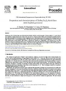
2.1 Separation of standard proteins Seven standard proteins (10 g/L, respectively), including cytochrome-c, myoglobin, RNase A, lysozyme, α-chymotrypsin, α-amylase and insulin, were completely separated by the HIC-type chromatographic cake (10 mm × 20 mm i.d.). The chromatogram is shown in Fig. 1. The good resolution indicates that the chromatographic cake can be
LI Ying et al. / Chinese Journal of Chromatography, 2008, 26(3): 331–334
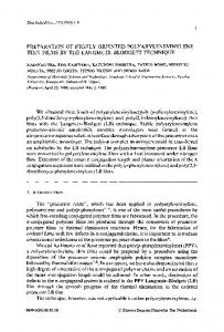
in their application. Thus, it is expected that the sample loading can be much more increased, and the fast separation and preparation of the active intact proteins from the low abundance ones in the human serum can be realized using HIC-type chromatographic cake. Fig. 2 shows the chromatograms of human serum separated by HIC-type chromatographic cake with different sample sizes. It is found that when the sample with volume more than 1.5 mL was loaded on the cake, the resolution was not satisfactory. Therefore, the largest sample loading of serum was 1.0 mL. Fig. 1
Chromatogram of seven standard proteins separated by HIC-type chromatographic cake Chromatographic cake: 10 mm × 20 mm i.d.; mobile phase: solution A. 3.0 mol/L (NH4)2SO4 + 0.050 mol/L KH2PO4 (pH 7.0), solution B. 0.050 mol/L KH2PO4 (pH 7.0); linear gradient: 0→30 min, 100% A→100% B; flow rate: 2.0 mL/min; detection wavelength: 280 nm. Peaks: 1. solvent; 2. Cyt-c, 10 μL; 3. Myo, 10 μL; 4. RNase A, 12 μL; 5. Lys, 8 μL; 6. α-Chy, 10 μL; 7. α-Amy, 10 μL; 8. Ins, 10 μL.
applied for the separation of the complex samples. 2.2 Separation and enrichment of four standard proteins from the diluted solution To test the enrichment of the low abundance proteins with the HIC-type chromatographic cake, 100 pmol–10 nmol of four standard proteins, including cytochrome-c, myoglobin, lysozyme and insulin, were separately added into 10 mL phosphate buffer solution. Then, the standard samples at low concentration were separated and enriched by the chromatographic cake with the progressive injection method, and then these HIC fractions were separated and desalted off-line with RPLC, and the lyophilized RPLC fractions were identified with MALDI-TOF MS. The results showed that these proteins at low concentration were enriched indeed, and their MS detection limits were 1 pmol/μL for cytochrome-c and myoglobin, and 0.1 pmol/μL for lysozyme and insulin. Therefore, it indicated that the chromatographic cake could really be used for the separation and enrichment of low abundance protein from 10 mL solution as the first dimensional liquid chromatography. 2.3 Separation and enrichment of human serum As mentioned above, the analytical scale HIC-type chromatographic cake has the same good resolution for the biopolymers as that of common chromatographic column. With the capabilities of low column pressure, large sample loading, and maintenance of the bioactivity of these biopolymers, it is especially available for the separation and preparation of complex biological samples with higher viscosity. In proteomics, 2-DE and MDLC are widely used for the separation of the complex samples, but the former needs gel digestion for identification and the latter is limited of the small sample size
Fig. 2 Chromatograms of human serum samples separated by HIC-type chromatographic cake with different sample size The chromatographic conditions are the same as in Fig. 1. Sample size: a. 0.1 mL; b. 0.3 mL; c. 0.5 mL; d. 0.7 mL; e. 1.0 mL; f. 1.5 mL; g. 2.0 mL. 1, 2, 3 and 4 denote the range of collected fractions.
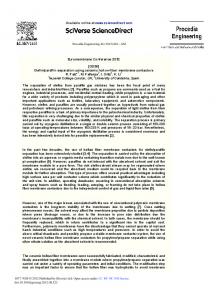
Four fractions denoted with numbers in Fig. 2 were collected after the HIC separation, and they were separated and desalted off-line with RPLC. The MALDI-TOF MS showed that the high abundance large molecular weight proteins, such as albumin, were present in Fraction 2, and the low abundance small species (Mr < 15000) were present in Fractions 3 and 4. The result indicated that not only the high abundance large molecular weight proteins were removed, but also numerous fractions can be detected with MALDI-TOF MS with the increase in loading sample volume (Table 1). Moreover, the MALDI-TOF MS intensity of the fraction peaks also increased sharply (shown in Fig. 3). From Table 1, it can be seen that a total of 285 fractions (Mr < 15000) were found with 1.0 mL sample size, whereas only 95 fractions were found with 50 μL sample size. To precisely and quantitatively evaluate the function of enrichment by the chromatographic cake, 1 μg cytochrome-c was added into 0.5 mL serum sample, and then it was separated and enriched with the novel method presented above. The MALDI-TOF MS spectrum is shown in Fig. 4.
LI Ying et al. / Chinese Journal of Chromatography, 2008, 26(3): 331–334
detected by MS. Therefore, the detection efficiency of the low abundance proteins/peptides in human serum with MALDI-TOF MS also significantly increased
3
Fig. 3
MALDI-TOF MS spectra of human serum separated by HIC-RPLC with different sample size Sample size: a. 500 μL; b. 50 μL.
Table 1 Number of proteins/polypeptides in the different sample sizes of healthy human serum separated with HIC-RPLC and detected by MALDI-TOF MS Injection volume/μL Number of proteins/polypeptides
50
100
300
500
700
1000
95
133
156
204
215
285
Conclusion
The chromatographic cake has the advantages of good resolution, low column pressure and large sample loading volume with high viscosity, and maintenance of the bioactivity of the biopolymers in the separation process. The active intact proteins in serum proteomics sample were isolated and enriched with HIC-type chromatographic cake, and then off-line separated by RPLC in 2D-LC. The MALDI-TOF MS indicated that the chromatographic cake could be applied not only for the fast separation and preparation of the complex samples in the removal of the high abundance proteins, but also for the isolation and enrichment of the low abundance proteins/peptides with large sample loading. In addition, the detection efficiency of the low abundance proteins/peptides in human serum with MALDI-TOF MS also significantly increased. A conclusion can be drawn that the novel approach of the multi-dimensional liquid chromatography based on the chromatographic cake coupled with MALDI-TOF MS will initiate an important access to the deep going proteomics research.
References [1] Scheepers P T J. Trends Anal Chem, 2006, 25(9): 841 [2] Merrell K, Southwick K, Graves S W, et al. J Biomol Tech, 2004, 15(4): 238 [3] Tanaka Y, Akiyama H, Kuroda T, et al. Proteomics, 2006, 6: 4845 [4] Li L, Ying W T, Yang H Y, et al. Chinese Journal of Chromatography, 2003, 21(1): 27 [5] Lilley K S, Razzaq A, Dupree P. Curr Opin Chem Biol, 2001, 6: 46 [6] Kashino Y, Harayama T, Pakrasi H B, et al. J Chromatogr B, 2007, 849: 282 [7] Zhang Y J, Cai Y, Wang J L, et al. Chinese Journal of Chromatogra, 2003, 21(1): 20 [8] Adkins J N, Varnum S M, Auberry K J, et al. Mol Cell Proteomics, 2002, 1: 947 [9] Sheng S, Chen D, van Eyk J E. Mol Cell Proteomics, 2006, 5: 26 [10] Gu X, Qu Q S, Yan C. Chinese Journal of Chromatography, 2007, 25(2): 157
Fig. 4 MALDI-TOF MS spectrum of 1 μg Cyt-c added into 0.5 mL original serum after separation with HIC-RPLC
The result showed that the low abundance cytochrome-c was separated and enriched from the complex serum sample and
[11] Dekker L J, Bosman J, Burgers P C, et al. J Chromatogr B, 2007, 847: 65 [12] Yao W B, Wu D, Geng X D. Chinese Journal of Chromatography, 2004, 22(2): 121 [13] Bradford M M. Anal Biochem, 1976, 72: 248