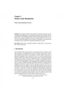
Journal of the Korean Physical Society, Vol. 66, No. 10, May 2015, pp. 1489∼1494
Feasibility of Using Geant4 Monte Carlo Simulation for IMRT Dose Calculations for the Novalis Tx with a HD-120 Multi-leaf Collimator Hyunuk Jung Department of Health Sciences and Technology, Sungkyunkwan University, Seoul 135-710, Korea
Jungsuk Shin, Kwangzoo Chung,∗ Youngyih Han,† Jinsung Kim and Doo Ho Choi Department of Radiation Oncology, Samsung Medical Center, Sungkyunkwan University School of Medicine, Seoul 135-710, Korea (Received 22 January 2015, in final form 25 March 2015) The aim of this study was to develop an independent dose verification system by using a Monte Carlo (MC) calculation method for intensity modulated radiation therapy (IMRT) conducted by using a Varian Novalis Tx (Varian Medical Systems, Palo Alto, CA, USA) equipped with a highdefinition multi-leaf collimator (HD-120 MLC). The Geant4 framework was used to implement a dose calculation system that accurately predicted the delivered dose. For this purpose, the Novalis Tx Linac head was modeled according to the specifications acquired from the manufacturer. Subsequently, MC simulations were performed by varying the mean energy, energy spread, and electron spot radius to determine optimum values of irradiation with 6-MV X-ray beams by using the Novalis Tx system. Computed percentage depth dose curves (PDDs) and lateral profiles were compared to the measurements obtained by using an ionization chamber (CC13). To validate the IMRT simulation by using the MC model we developed, we calculated a simple IMRT field and compared the result with the EBT3 film measurements in a water-equivalent solid phantom. Clinical cases, such as prostate cancer treatment plans, were then selected, and MC simulations were performed. The accuracy of the simulation was assessed against the EBT3 film measurements by using a gamma-index criterion. The optimal MC model parameters to specify the beam characteristics were a 6.8-MeV mean energy, a 0.5-MeV energy spread, and a 3-mm electron radius. The accuracy of these parameters was determined by comparison of MC simulations with measurements. The PDDs and the lateral profiles of the MC simulation deviated from the measurements by 1% and 2%, respectively, on average. The computed simple MLC fields agreed with the EBT3 measurements with a 95% passing rate with 3%/3-mm gamma-index criterion. Additionally, in applying our model to clinical IMRT plans, we found that the MC calculations and the EBT3 measurements agreed well with a passing rate of greater than 95% on average with a 3%/3-mm gamma-index criterion. In summary, the Novalis Tx Linac head equipped with a HD-120 MLC was successfully modeled by using a Geant4 platform, and the accuracy of the Geant4 platform was successfully validated by comparisons with measurements. The MC model we have developed can be a useful tool for pretreatment quality assurance of IMRT plans and for commissioning of radiotherapy treatment planning. PACS numbers: 42.40.Ht, 42.30.Kq Keywords: Monte Carlo simulation, Novalis Tx, HD-120 MLC, IMRT, Geant4, Radiotherapy DOI: 10.3938/jkps.66.1489
I. INTRODUCTION Radiation therapy is one of the major modalities used for cancer treatment. The most important advantage of radiation therapy is that it is non-invasive: thus its usage ∗ E-mail:
[email protected]; Tel: +82-2-3410-1353; Fax: +82-2-3410-2619 † E-mail:
[email protected]; Tel: +82-2-3410-2604; Fax: +82-23410-2619
is rising across the world [1]. The basic strategy of radiation therapy entails maximally applying radiation to a focal lesion while minimizing damage to the surrounding healthy tissues. Many radiation therapy techniques have been developed based on this principle, and among them, intensity modulated radiation therapy (IMRT) has been considered one of the most exciting developments since the introduction of computed tomography imaging into treatment planning [2–4]. Monte Carlo (MC) simulations are widely used in ra-
-1489-
-1490-
Journal of the Korean Physical Society, Vol. 66, No. 10, May 2015
diation therapy research, from micro dosimetry at the cell level to radiation shielding design [5,6]. Specifically, the MC algorithm is considered to be the most accurate way to calculate the dose in heterogeneous media, such as bone, soft tissue, muscle, and other tissues found in patients [7]. The Geometry And Tracking 4 (Geant4) code is an MC toolkit that has diverse research applications in medical physics. This toolkit offers physics models that can be applied over a wide energy range for different physics interactions, and it contains a variety of tools for modeling complex detector systems [8]. Therefore, applications of the Geant4 toolkit in radiation physics can cover different radiation sources (e.g., X-ray and charged particles) because of its superiority in simulating charged-particle interactions over a wide range of energies [8]. It can also be used as in various high-energy physics models for calculating hadronic interactions with great reliability and flexibility [9,10]. Considerable efforts have been made to improve our understanding of dosimetric properties, such as dose distributions in homogeneous/heterogeneous materials, dose distributions across build-up areas, and dose calculations with actual patient data by using the Geant4 toolkit [7,8,11–14]. Monte Carlo modeling of multi-leaf collimators (MLCs) has also been an important study because MLCs are critical components in the evaluation of the radiation dose when considering IMRT field with diverse and irregular shapes [15–19]. However, there has been no comprehensive report on the exact Geant4 modeling of the Novalis Tx system equipped with a highdefinition (HD)-120 MLC. The HD-120 MLC provides better dose conformities around treatment targets and spares critical organs better than any other MLC type because of its fine pitch. In this study, we have modeled the Novalis Tx linear accelerator head equipped with the HD-120 MLC by using the Geant4 code. We have also identified the tongue-and-groove effects of the MLC by comparing the calculated MLC fields with there measured using EBT3 films, thereby verifying the MLC’s leaf movement and dose conformity. Lastly, we have assessed the accuracy of the developed Geant4 models in clinical cases by comparing the Monte Carlo calculations with the film measurements.
II. MATERIALS AND METHODS 1. Geant4 Modeling - Novalis Tx with a HD-120 MLC
We modeled the gantry head of the Novalis Tx system equipped with a HD-120 MLC (Varian Medical Systems, Palo Alto, CA, USA) by using the Geant4 MC code (ver. 4.9.5.p02). The Novalis Tx head was modified based on the “Medical Linac Advanced Example” built-in Geant4
Fig. 1. (Color online) Schematic overview of the simulated Novalis Tx head, which included a target, primary collimator, flattening filter, ion chamber, mirror, X-Y jaws, and HD-120 MLC.
code and was improved using the data of the ‘Monte Carlo Data Package’ provided by Varian. Components implemented in the MC code included a target, primary collimator, flattening filter, ion chamber, mirror, X-Y jaws, and HD-120 MLC, as presented in Fig. 1. The constructed solid geometry (CSG) solids that are implemented options in the Geant4 toolkits were used for modeling all components except the HD-120 MLC, which has a complicated form. The CSG solids are basic figures predefined by Geant4. An appropriate CGS solid was selected for each head component, and the shape and the dimensions of the solid were modified according to the specifications provided by the Monte Carlo Data Package for implementation. The HD-120 MLC consists of two banks with 60 leaves in each bank. Each leaf is made of a tungsten alloy. The leaves are divided into two types, quarter leaves and half leaves. Thirty-two quarter leaves were positioned in the central part of the MLC and had widths of 2.5mm. Twenty-eight half leaves were positioned in the outer part of the MLC and had widths of 5.0-mm. Each leaf was spaced 0.0047 cm away from the adjacent leaf. The MLC was positioned 50.8-cm below the target. For dose calculations, we used a Livermore low-energy electromagnetic (EM) model. The range cut value, which is used to limit the occurrence of particles influencing scattering effects, was set at 0.1-cm. The MC dose cal-
Feasibility of Using Geant4 Monte Carlo Simulation · · · – Hyunuk Jung et al.
culation was divided into two phases, space calculation and dose calculation, to improve the calculation speed and to reduce calculation errors. The phase space had a size of 1.2 gigabytes and stored approximately 2 × 10⁷ photons and electrons in total. This phase space, which had dimensions of 30 × 30 cm2 , was above the y-jaw and 45 cm below the target. This was used to calculate the 10 × 10, 5 × 5, and 3 × 3 cm2 radiation fields shaped by the x-y jaws and the MLC. Dose calculations were performed with a voxel size of 1 × 1 × 1 mm3 in a 40 × 40 × 40 cm3 solid water phantom. Each calculation was performed by using parallel computations on two nodes with 40 core clusters (Intel Server Systems, Intel Corp., Santa Clara, CA, USA). Calculations of the phase space and the dose took approximately 12 hours and 4 hours, respectively.
2. Validation of the MC Model
Major beam modeling variables such as the mean energy, energy standard deviation, and radius of the incident electron beam were tuned by repeatedly calculating the percent depth dose (PDD) and the beam profiles of a 6-MV X-ray beam generated by the Novalis Tx accelerator head. Using the study by Grevillot et al. [20] as a reference, was optimized the variables as follows: The mean energy, which has the largest effect on the depth dose profile, was changed from 5.5-MeV to 7-MeV. The energy standard deviation was changed from 0.1-MeV to 0.5-MeV in 0.1-MeV steps. In addition, the radius of the electron beam was changed from 1-mm to 5-mm. For each set of tuning parameters, the PDD for a 10 × 10 cm2 radiation field defined by the jaws and the lateral profiles at 1.6-, 5-, 10-, and 20-cm depths of water were calculated. The resulting values were compared with test values, thereby optimizing the beam parameters. The depth dose and the lateral profile measurement were made by employing the CC13 ionization chambers and edge detectors within a Blue Phantom (IBA, Schwarzenbruck, Germany) with gantry of 0◦ at a 100-cm source-to-surface distance (SSD) by using the Novalis Tx 6-MV X-ray beam, which is the standard condition for an absolute output calibration. By matching the PDD curves in the calibration condition, we could interpret the output of the Monte Carlo simulation as an absolute output with a scale factor that depended on the number of simulation histories. In addition, the accuracy of the values calculated by using the MC code was assessed at fields of 3 × 3 and 5 × 5 cm2 in size by using the optimized beam parameters. The MC calculation was performed for a 10 × 10 cm2 field defined by the jaws with the even leaves of the MLCs closed to identify the tongue-and-groove effects of the HD-120 MLC. In addition, the two-dimensional dose distributions at a 10-cm depth on the water phantom was calculated for simple shapes of the MLC fields to
-1491-
identify the size and the dose conformity of the MLC fields. The computed results were compared with those of measurements using EBT3 films. The scanning of the irradiated films was performed using the DosimetryPRO Advantage Red system (VIDAR Systems Corp., USA), which meets all specific requirements for radiochromic film dosimetry.
3. EBT3 Film Measurement
Measurements with Gafchromic EBT3 film were performed by using 6-MV X-rays from a Novalis Tx linear accelerator at a SSD of 100-cm. The methods were for film measurements and analyses were published elsewhere [21]. A film calibration was conducted by creating a calibration curve to match the optical densities (ODs) of the film to the corresponding absorbed doses. For the collection of the calibration data, the film was positioned at a 5-cm depth in the water-equivalent solid phantom (Solid Water RMI 457, Gammex, Middleton WI, USA) perpendicular to the axis of the central beam. Each film was then irradiated in a 5 × 5 cm2 open field with dose levels of 20, 40, 60, 80, 120, 160, 200, 260, 320, 350, and 400 cGy. All exposed EBT3 films were scanned at a resolution of 178 dpi by using a DosimetryPRO Advantage Red system 20 hours after irradiation. Calibration data were imported into RIT113 (Radiological Imaging Technology, Inc., USA) version 6.1 analysis software and were used for gamma evaluations of the scanned images.
4. Clinical IMRT Cases
A dose assessment was conducted by using actual treatment cases for five patients who received radiation therapy for prostate cancer. Four to five static IMRT fields were examined at gantry 0◦ and a SSD of 100 cm by irradiating each static field to a single EBT3 film positioned at a 10-cm depth in a solid water phantom. For each static field, we assigned from 60 to 70 monitor units (MU) to deliver approximately 300 MU to the film in total because EBT film dosimetry has been calibrated very accurately for this dose range. In the MC simulation, each IMRT MLC field was virtually shaped by using the coded HD-120 MLC according to the treatment plan. The dose delivered by each MLC field was then calculated. The calculated results were then integrated using Matlab (MathWorks, Natick, MA, USA), and the integrated dose distribution was converted to a dose mapping form that could be imported into the RIT113 software. A gamma analysis using RIT113 software was used in the comparison of the MC and the EBT3 results. The comparison was made for over 10% of the maximum dose region due to the inherent inaccuracy of film dosimetry for low doses. The
-1492-
Journal of the Korean Physical Society, Vol. 66, No. 10, May 2015
Fig. 2. Comparison of the percent depth dose distribution between the results from MC simulations and from measurements with an ionization chamber in a water phantom for 3 × 3, 5 × 5, and 10 × 10 cm2 field sizes normalized at 60%, 80%, and 100% on the relative dose, respectively.
criteria of 3% of the local dose difference and a 3-mm distance to agreement (DTA) were used.
Fig. 3. Comparison of the lateral dose profiles at 1.6-, 5-, 10-, and 20-cm depths from MC simulations to there from measurements with an ionization chamber in a water phantom for 3 × 3, 5 × 5, and 10 × 10 cm2 field sizes normalized at 60%, 80%, and 100% on the relative dose, respectively.
III. RESULTS 1. Optimized Beam Parameters
Figure 2 presents the percent depth doses (PDDs) and the lateral profiles of the optimized Novalis Tx 6-MV X-rays. Through the process of beam optimization, we determined a mean energy of the incident electron beam (Emin) of 6.8-MeV, an energy standard deviation (Estd) of 0.5-MeV, and a spot size (re) of 3-mm. For a clearer presentation in Fig. 2, the maximum of the percent depth dose of the radiation fields for 3 × 3, 5 × 5, and 10 × 10 cm2 were normalized to 60%, 80%, and 100%, respectively. The MC calculation agreed with the measurements on average within a 1% difference and varied from 0.136% (3 × 3 cm2 ) to 0.371% (5 × 5 cm2 ). Comparisons of lateral profiles were made at 1.6-, 5-, 10-, and 20-cm depths in water, as shown in Fig. 3. The average difference was 2%, which varied from 0.791% (1.6-cm) to 1.183% (10-cm) for a 10 × 10 cm2 field size. For 3 × 3 and 5 × 5 cm2 field sizes, the MC calculations agreed with the measurements to within 2%. Based on these findings, we consider the Geant4 beam model for Novalis Tx linear accelerator 6-MV X-rays to have been properly optimized.
Fig. 4. Comparison of the offset axis profiles for static MLC fields. The shape of the MLC pattern was a repeated arrangement of single leaves opened and closed in alternation and bounded by the 10 × 10 cm2 jaw field and was used to identify the tongue-and-groove effects of the HD-120 MLC.
arrangement of single leaves opened and closed in alternation and bounded by the 10 × 10 cm2 jaw field. Lateral profiles were obtained along the line at 5 cm away from the center axis. Differences between MC calculations and film measurements were found to be below 3% on average, as shown in Fig. 4.
3. Gamma Analysis of Simple MLC Fields 2. Tongue-and-groove Effect
To evaluate the accuracy of the tongue-and-groove effect of the HD-120 MLC, we measured a static pattern of the MLC. The shape of the MLC pattern was a repeated
We compared simple MLC-shaped fields in order to assess the movement and the position of each MLC leaf had been defined accurately. Figure 5 shows our comparisons of the measured and the simulated results for two MLC fields. The pass rates for these cases using a 3%/3-mm
Feasibility of Using Geant4 Monte Carlo Simulation · · · – Hyunuk Jung et al.
-1493-
Table 1. Results of gamma evaluation with the 3%/3-mm criteria for comparisons between MC simulations and EBT3 measurements for IMRT patient plans. Passing Gamma Index Patient 1 95.28% Patient 2 95.46% Patient 3 95.71% Patient 4 95.53% Patient 5 93.73% Average 95.14%
Fig. 5. (Color online) Comparison of the simple MLCshaped fields from MC simulations and with there from EBT3 measurements. The boundary of the MC calculated field exhibits a blurred field boundary due to the calculation pixel size (1 × 1 mm2 ).
Fig. 6. (Color online) Comparison of IMRT patient plans obtained with MC simulations to there obtained from EBT3 measurement.
gamma-index criterion were 96.45% and 95.05%, respectively.
4. Validation with Clinical IMRT Plans
For clinical case validation, as shown in Fig. 6, we found strong agreement between the MC calculations and the EBT3 measurements. Pass rates were higher than 95%, on average, when using a 3%/3-mm gammaindex criterion. The details of our findings are summarized in Table 1.
IV. DISCUSSION In this study, we developed an MC model using Geant4 code for a Novalis Tx system equipped with a HD-120
MLC and applied this model to complicated IMRT clinical cases. Repeated calculations in our optimization process of the 10 × 10 cm2 field revealed beam parameters of a 6.8-MeV Emin, a 0.5-MeV Estd, and a 3-mm re. The average energy of the primary electron was approximately 1 MeV larger than that in a previous study by Grevillot et al. [20], which assessed the simulation of an Elekta Precise 6-MV Linac by using GATE v6.0 release. This difference can be attributed to the different target geometries of each vendor’s machine and to the different linac tunings used at the time of commissioning. Another factor that could account for this difference is the difference in physics models, low-energy or standard, used in the MC simulations. We found good agreement between our findings obtained by using MC calculations with an optimized 6-MV X-ray and the measurements obtained with an ionization chamber, less than 2% difference in PDD/Profile, and this was applied to 3 × 3 and 5 × 5 cm2 as well for comprehensive verification. Radiotherapy techniques such as IMRT or SRS require highly accurate dose conformity to make full use of their state-of-the-art advantages. For this reason, modeling the MLC with a MC simulation has been extensively studied over the past decade. Borges et al. [17] found differences in alternated leaf patterns and in the dynamic mode between film measurements and MC simulations; both differences were within 4%. Likewise, Okamoto et al. [19] demonstrated good agreement between the tongue-and-groove effects determined by using the MC calculations and the film measurements. For simple MLC fields and dynamic MLC-based fields, the pass rates were 98.5 and 97%, respectively, when using a gamma-index criterion (3%/3-mm) method of evaluation. Compared with previous studies, our MLC modeling showed comparable accuracy for simple MLC fields because the gamma pass rate was over 95%, suggesting correct modeling of the HD-120 MLC. Furthermore, the discrepancy for the tongue-and-groove effect was found to be below 3% for the quarter leaves, which required more accurate definition because of the high usage of the center leaves for clinical cases. We used prostate IMRT cases to validate our methods.
-1494-
Journal of the Korean Physical Society, Vol. 66, No. 10, May 2015
All tested cases exhibited a pass rate of over 95% with a 3%/3-mm gamma analysis. Although a volume comparative evaluation was not possible due the limitation of the film dosimeters, our two-dimensional comparison demonstrated the precision of our MC model and validated its usefulness as an IMRT verification tool. One drawback of this method for use in clinical applications could be the calculation times. Generating phase space files, for example, takes a relatively long time (12 hours), as do dose calculations in a homogeneous water phantom (4 hours). However, these calculation speeds can be improved with more powerful computing resources [22]. With current computing speeds and the validation of the code, this MC model could serve as an independent quality assurance tool for selected IMRT cases in comparison with two-dimensional dosimetry such as with a film or real-time two-dimensional arrays. In validating the dose calculations in a homogeneous water phantom, we have demonstrated the feasibility of using the Geant4 linear accelerator model for clinical IMRT cases. The developed MC model can be extended to making dose calculations with patient CT images. Thus, the MC model can function not only as a means for independent quality assurance but also as a validation of the dose calculation algorithm used in the treatment planning system, especially for heterogeneous media.
V. CONCLUSION In conclusion, the Novalis Tx accelerator head equipped with a HD-120 MLC has been successfully modeled by using the Geant4 toolkit. Implementation of the HD-120 MLC was verified, and the performance was tested through a simulation of the tongue-and-groove effect. The developed MC model will be useful as a pretreatment quality assurance tool for clinical IMRT cases.
ACKNOWLEDGMENTS This work was supported by a National Research Foundation of Korea (NRF) grant funded by the Korea government’s Ministry of Science, ICT and Future
Planning (MSIP, No.: 2013M2A2A7043507 and No.: 2012M3A9B6055201) and by a Samsung Medical Center grant (GFO1130081).
REFERENCES [1] G. Delaney, S. Jacob, C. Featherstone and M. Barton, Cancer 104, 1129 (2005). [2] Intensity Modulated Radiation Therapy Collaborative Working Group, Int. J Radiat. Oncol. Biol. Phys 51, 880 (2001). [3] L. E. Gaspar and M. Ding, Curr. Oncol. Rep. 10, 294 (2008). [4] V. T. DeVita, Jr. and S. A. Rosenberg, N. Engl. J. Med. 366, 2207 (2012). [5] D. W. Rogers, Phys. Med. Biol. 51, R287 (2006). [6] E. Spezi and G. Lewis, Radiat. Prot. Dosimetry 131, 123 (2008). [7] I. J. Chetty et al., Med. Phys. 34, 4818 (2007). [8] S. Agostinelli et al., Nucl. Instr. Meth. Phys. Res. A 506, 250 (2003). [9] C. Z. Jarlskog and H. Paganetti, IEEE 55, 1018 (2008). [10] J. Apostolakis et al., Radiat. Phys. Chem. 78, 859 (2009). [11] D. W. O. Rogers, Med. Phys. 22, 503 (1995). [12] E. Poon and F. Verhaegen, Med. Phys. 32, 1696 (2005). [13] W. Abdel-Rahman, J. P. Seuntjens, F. Verhaegen, F. O. Deblois and E. B. Podgorsak, Med. Phys. 32, 286 (2005). [14] C. M. Ma et al., Phys. Med. Biol. 45, 2483 (2000). [15] N. Tyagi, B. H. Curran, P. L. Roberson, J. M. Moran, E. Acosta and B. A. Fraass, J. Phys. Conf. Series 102, 012025 (2008). [16] M. K. Fix, W. Volken, D. Frei, D. Frauchiger, E. J. Born and P. Manser, Med. Phys. 38, 5311 (2011). [17] C. Borges, M. Zarza-Moreno, E. Heath, N. Teixeira and P. Vaz, Med. Phys. 39, 415 (2012). [18] L. A. Vazquez-Quino, B. Massingill, C. Shi, A. Gutierrez, C. Esquivel, T. Eng, N. Papanikolaou and S. Stathakis, J. Appl. Clin. Med. Phys. 13, 3960 (2012). [19] H. Okamoto, Y. Fujita, K. Sakama, H. Saitoh, T. Kanai, J. Itami and T. Kohno, Radiol. Phys. Technol. 7, 246 (2014). [20] L. Grevillot, T. Frisson, D. Maneval, N. Zahra, J. N. Badel and D. Sarrut, Phys. Med. Biol. 56, 903 (2011). [21] H. Jung, O. Kum, Y. Han, B. Park and K. H. Cheong, J. Korean Phys. Soc. 65, 1829 (2014). [22] K. Sutherland, S. Miyajima and H. Date, J. Phys.: Conf. Ser. 74, 021020 (2007).