Feedback in Hypothesis Testing: An ERP Study David Papo1 , Pierre-Marie Baudonnie`re2 , Laurent Hugueville 2 , and Jean-Paul Caverni 1,2
Abstract & We used event-related potentials (ERPs) to probe the effects of feedback in a hypothesis testing (HT) paradigm. Thirteen college students serially tested hypotheses concerning a hidden rule by judging its presence or absence in triplets of digits and revised them on the basis of an exogenous performance feedback. ERPs time-locked to performance feedback were then examined. The results showed differences between responses to positive and negative feedback at all cortical sites. Negative feedback, indicating incorrect performance, was associated to a negative deflection preceding a P300-like wave. Spatiotemporal principal component analysis
INTRODUCTION In real-world situations, people are often confronted with problems that they must solve relying only on incomplete information. To find a solution to these problems, hypotheses must be generated and modified in the light of their adequacy to either internally or externally generated feedback. Hypothesis testing (HT) can be thought of as a complex process consisting of inductive and deductive phases during which people generate and select new rules or apply previously tested ones, respectively. Feedback can be used to switch between these two phases in an adaptively advantageous manner. In this study, we provide a functional description of the role of feedback in HT at the cognitive level, sketch a computational model that we map onto a putative set of anatomical substrates, and then use the model to predict event-related potentials (ERPs) timelocked to feedback in a modified Wason’s (1960) 2-4-6 HT paradigm. Feedback and Its Consequences One way to study the modulation of HT by feedback is to think of feedback as a control parameter inducing jumps from one phase to another as values of feedback are varied. Within this framework, feedback itself is studied in an indirectly via its consequences on activity following its occurrence. In an early study, Elliott and
1
Universite´ de Provence, 2 CNRS
© 2003 Massachusetts Institute of Technology
(PCA) showed the interplay between early frontal components and later central and posterior ones. Lateralization of activity was selectively detectable at frontal sites, with a left frontal dominance for both positive and negative feedback. These results are discussed in terms of a proposed computational model of trial-to-trial feedback in HT in which the cognitive and emotive aspects of feedback are explicitly linked to putative mediating brain mechanisms. The properties of different feedback types and feedback-related deficits in depression are also discussed. &
colleagues used positron emission tomography to assess the impact of performance feedback on subsequent performance of cognitive tasks, including planning, guessing, and HT, in healthy subjects and patients with unipolar depression (Elliott, Frith, & Dolan, 1997; Elliott & Dolan, 1998; Elliott, Sahakian, Paykel, & Dolan, 1998). Neuropsychological results previously had shown greater detrimental impact of negative feedback on subsequent performance in depressed patients (Beats, Sahakian, & Levy, 1996). Neuroimaging data showed that response to both positive and negative feedback, indicating correct and incorrect performance, respectively, was associated in a task-dependent manner with a ventro-cortico-limbic network including the medial caudate nucleus and the orbitofrontal cortex and that activation of these structures was attenuated in depressed patients (Elliott et al., 1998). More recently, Monchi, Petrides, Petre, Worsley, and Dagher (2001) used event-related functional magnetic resonance to identify neural circuits mediating positive and negative feedback in the Wisconsin Card Sorting Test. Positive and negative feedback were both associated with dorsolateral prefrontal cortex increased activity with respect to a control feedback condition, coherently with a role of this area in monitoring new information in working memory. By contrast, a loop involving the midventrolateral prefrontal cortex, the caudate nucleus, and the mediodorsal thalamic nucleus increased activity specifically after reception of negative feedback, and was associated to the planning of a response to feedback. Bilateral increases in a rostral region of the anterior Journal of Cognitive Neuroscience 15:4, pp. 508– 522
cingulate area by comparison to the control feedback with negative feedback were interpreted as reflecting a conflict detection role of this area. The ventro-cortico-limbic circuit associated in both studies to feedback is implicated in reward-related mechanisms (Koepp et al., 1998; Thut et al., 1997) coherently with an emotive component of feedback and with a reward-related deficit in unipolar depression (Henriques & Davidson, 1997). Elliott, Friston, and Dolan (2000) further demonstrated dissociable neural responses to rewards and penalties. Activity in ascending dopaminergic systems projecting from midbrain to ventrostriatal areas reflected absolute, context-independent financial reward, whereas activity of projecting sites of this system, including the subgenual cingulate cortex, responded in a context-dependent manner to reward. By contrast, higher levels of financial losses were associated in a context-independent manner with bilateral hippocampal activation. The authors suggested a nonmnemonic hippocampal role in inhibiting the experience of reward, but an alternative explanation in terms of a behavioral inhibition system and a hippocampal role in mediating punishment in humans was also advanced (Gray, 1982). Positive and negative feedback should affect subsequent performance in a way that reflects this asymmetry in reward-related patterns of neural activity. Differences in regional cerebral blood flow associated with positive compared to negative feedback in control subjects were found in a corticobasal ganglia loop originating in the midventrolateral prefrontal cortex (Monchi et al., 2001) and in some parietal, temporal, and thalamic regions of the brain (Monchi et al., 2001; Elliott et al., 1997). Functional imaging studies have consistently demonstrated that posterior cortical areas are coactivated with the prefrontal cortex in cognitive tasks that involve active representation of information in working memory (Miller, 2000). Feedback should be able to modulate a top-down, reverberatory, fronto-parieto-temporal activity presumably involved in the generation – maintenance – modification of hypotheses in HT. Given neuroimaging techniques’ poor temporal resolution, these results cannot conclusively separate consequences of feedback on subsequent behavior from its most direct effects. Thus, normal and pathological action of positive and negative feedback could not be described in terms of dynamic patterns of brain activity, whereby different feedback values mediate subsequent performance in regions carrying out HT operations, through asymmetric, reward-related mechanisms. A different approach to feedback altogether, and therefore different analytical strategies and techniques, are needed to provide a mechanistic role of cognitive and emotive processes of feedback in HT. In a complementary theoretical framework, feedback is represented as a process in its own right that can be separated from its consequences and endowed with specific properties. Given the subsecond timescale of
this phenomenon, a good time resolution technique needs to be chosen. Miltner, Braun, and Coles (1997) used ERPs to assess feedback-related activity in a time production task in which subjects were given exogenous performance feedback by the experimenter. Miltner et al. observed a negative shift in the ERP, peaking between 230 and 330 msec, when subjects received feedback indicating incorrect performance, and compared it to a negative wave consistently found when subjects make errors in choice reaction time and go/nogo tasks (Dehaene, Posner, & Tucker, 1994; Gehring, Goss, Coles, Meyer, & Donchin, 1993; Falkenstein, Hohnsbein, Hoormann, & Blanke, 1991). This potential, termed error-related negativity (ERN), starts developing at around the time of the erroneous response and peaks about 100 msec later, while being unremarkable or less pronounced in correct response trials. Based on a putative common morphology of electrophysiological scalp activity and on dipole localization analysis, Miltner et al. concluded that ‘‘the same neural process involving the anterior cingulate cortex or the supplementary motor area is associated with incorrect response execution in a reaction time (RT) task and with error feedback in a time estimation task,’’ going on to suggest that ‘‘the ERN component may reflect the activity of a generic mechanism . . . specifically invoked to deal with errors of choice (as in choice RT tasks), errors of action (in Go – NoGo tasks), and time estimation errors’’ ( p. 795). However, exactly what computations this mechanism could carry out was left as an open question. Objectives The present experiment had two main goals. The first was to provide a representation of feedback in HT as a process separable from its consequences. We used a task similar to that used in existing neuroimaging experiments to take advantage of information on the relevant functional circuits deriving from these studies, while at the same time benefiting from ERPs’ optimal temporal resolution. We speculated that, due to their poor temporal resolution, neuroimaging techniques might not be suited to capture separable task-related activities and that ERPs would be able to deconvolute feedbackspecific properties in HT from their consequences. To use the evidence from neuroimaging studies to constrain our ERP results, we described the computational and implementation properties of the task in a comparable way on the anatomo-functional spaces defined by each technique. The second goal was to discuss the suggestion of a common mechanism for ‘‘trial-to-trial’’ and ‘‘in-flight’’ error-feedback activity (Miltner et al., 1997). These error-related mechanisms differ in intrinsic computational terms (Lawrence, 2000). In a general way, whereas trial-to-trial feedback resets an internal, taskrelated model, ERN-eliciting tasks have no workingmemory-resetting role and only involve some form of Papo et al.
509
evaluation of a previous response. Dissociations have been demonstrated in motor learning studies in patients with Huntington’s disease (Smith, Brandt, & Shadmehr, 2000; Gabrieli, Stebbins, Singh, Willingham, & Goetz, 1997). The dorso-striatal-cortical circuit commonly activated by error-feedback control in choice reaction time motor tasks known to elicit ERNs may be dissociable from the ventral striato-cortico-limbic circuit implicated by trial-to-trial feedback processing in a host of executive tasks (Monchi et al., 2001; Bush, Luu, & Posner, 2000; Elliott et al., 1997). Moreover, different brain circuits and therefore ERPs differing in both chronometric and topographic characteristics should be associated in a taskspecific manner to the consequences of the two types of feedback on further performance. We used a task very similar to Elliott et al.’s, which in turn is computationally equivalent to that by Miltner et al.; thus, both our feedback and that of Miltner et al. should engage brain circuits neurophysiologically dissociable from those eliciting ERNs. If Miltner et al.’s and our feedback elicit comparable electrophysiological responses, then the suggestion of a unique mechanism mediating different types of feedback should be reexamined. We describe the cognitive task that we employed and show how a computational account of this task can help interpret the electrophysiological response to feedback in terms of putative anatomo-functional correlates. Feedback in Hypothesis Testing We measured the electrophysiological response timelocked to exogenous performance feedback in a modified Wason’s (1960) 2-4-6 HT task. Subjects serially tested hypotheses concerning a hidden rule by judging its presence or absence in triplets of digits and revised
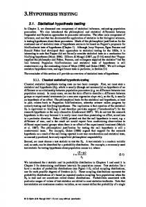
them on the basis of an exogenous performance feedback, following each answer that told them whether their answer was correct or not. Triplets were presented one at a time and blocks of 10 triplets corresponded to one single rule (cf. Figure 1). After each block, subjects were told that the target rule had been changed. Crucially, unbeknownst to the subjects there was no rule and feedback was preestablished; feedback frequency was manipulated so that an equal number of blocks had 8:2, 5:5, and 2:8 positive to negative feedback ratio, respectively. Contrary to the original Wason task, in the present context subjects could not propose triplets, but rather were confined to judging triplets proposed by the experimenter. This crucially constrained the range of possible testing strategies, and subjects, always in principle, responded according to what they thought was the correct answer. One viable way to carry out the task avoiding overloading working memory capacities was to test one hypothesis at a time, maintaining it until it received negative feedback while at the same time keeping track of all the hypotheses previously tested within the same block of trials. Thus, subjects were encouraged to act as minimal adaptive predictors and process only a relevant cue subset to generate a hypothesis while treating all other cues as irrelevant (Elliott & Dolan, 1998; Grossberg, 1984). Feedback in our HT paradigm was of the same trial-to-trial type as that in Miltner et al.’s. Moreover, in both cases feedback only informed subjects of the presence of a performance error but not of the nature of the error. Fractionating Feedback In our task, positive and negative feedback serve two different functions. Positive feedback should help in
Figure 1. Example of a trial with positive feedback.
510
Journal of Cognitive Neuroscience
Volume 15, Number 4
maintaining the current hypothesis active while keeping competing hypotheses in the foreground. Negative feedback should facilitate the subsequent shutting down of the discredited rule, inhibiting the past rule – feedback association, while promoting search for a new rule within the rule space. In both positive and negative feedback, a record of previously falsified rules must also be kept to avoid retesting. If feedback can be used to guide and monitor performance, it also has an emotional component due to the motivational consequences of success and failure (Elliott et al., 1997). A complete mechanistic representation of feedback should therefore include not only intrinsic HT computational aspects but also driving conditions. Accordingly, HT can be portrayed as an associative learning process wherein subjects acquire knowledge about a hidden rule through continuously updated response– reward associations. HT can be seen as instrumental in gaining positive rewarding feedback and minimizing stressful negative feedback. In this context, feedback is an abstract reinforcer the motivational effects of which trigger switches between sustained activity and behavioral inhibition followed by exploratory phases. Mapping Cognition into Anatomy Feedback is a dynamic gating mechanism regulating the maintenance or updating of rule-related representations. A complete description of feedback should include both this gating mechanism and its consequences. To understand how feedback can be used to guide behavior it is necessary to show how activity in brain regions mediating the performance of HT can be biased by feedback-related signals. The functional requirements of the HT process can be associated to identifiable anatomo-functional neural networks (Monchi, Taylor, & Dagher, 2000) and to formal properties of neurotransmitter systems (Grossberg, 1984). Prior to the reception of exogenous feedback, a tested rule is being kept in working memory via frontal biasing of posterior cortex (Cohen, Braver, & O’Reilly, 1998). Feedbackinduced refreshing of working memory ultimately entails modifying this pattern of fronto-posterior activity. Positive performance feedback has been shown to activate a limbico-cortico-striatal circuit originating in the medial prefrontal cortex (Monchi et al., 2001; Elliott et al., 1997), which has been directly implicated in the mediation of reward, via dopaminergic modulation (Koepp et al., 1998; Thut et al., 1997). Phasic dopaminergic signals show bistable activation– depression after reward or omitted reward stimuli (Schultz, 1998). Medial prefrontal excitatory input to the ventral striatum would be sustained/amplified by phasic ventral – tegmental (VTA) dopamine (DA) input, which would also compete with hippocampal stress-related signals (Behr, Glovell, Schmitz, & Heinemann, 2000; Gray, 1982). DA modu-
lation would ultimately reinforce thalamocortical reverberation implicated in working memory maintenance and sharpen the tuning of prefrontal pyramidal cells by increasing the excitability of GABA-ergic interneurons. Moreover, direct VTA projections to superficial layers in the prefrontal cortex can sustain long-range corticocortical fronto-posterior reverberatory activity as this is protected from competing rule-related activations (Durstewitz, Seamans, & Sejnowski, 2000; Miller, 2000). Negative feedback may shut down the global ongoing pattern of activation via stress-induced bursts of noradrenaline (NA) release in the hippocampus, which facilitate the consolidation of memory traces linked with negative feedback (Gray, 1982). Whereas the absence of reward would result in VTA DA phasic reward-related response inhibition (Schultz, 1998), stress-related release of hippocampal neurons would result in prefrontal selective inhibition of ventrostriatal DA release and incentive salience attribution to stimuli, thereby avoiding response perseveration ( Jackson & Moghaddam, 2001). The NA ascending system may promote subsequent exploratory behavior by disinhibiting basal forebrain cholinergic projections to both medial prefrontal and parietal cortices (Sarter & Bruno, 1999). At the cortical level, medial prefrontal stress-mediating activity (Drevets, 2000) would be coupled with increased activity in lateral orbitofrontal areas, as these areas are implicated in overriding behavioral choices based on previous reward values of stimuli (Monchi et al., 2001; Elliott, Dolan, & Frith, 2000). Stress may suppress neural activity in some cognitive processing areas during intense emotional states. During emotion-related tasks, blood flow increases in areas implicated in emotional processing, including the posteromedial orbital cortex and the ventral anterior cingulate cortex, but decreases during performance of attentionally demanding cognitive tasks. The opposite pattern occurs in dorsolateral prefrontal and dorsal anterior cingulate areas (Mayberg et al., 1999; Drevets & Raichle, 1998). As the switching of a flip-floplike right medial – left dorsolateral prefrontal stressrelated mechanism (Mayberg et al., 1999), resulting from hippocampal disinhibition, shuts down the previous activation pattern in the ventrostriatal loop, a similar mechanism in the orbitofrontal cortex would modulate the fronto-posterior rule-related pattern of activation, either directly via GABA-ergic gating (Northoff et al., 2002) or indirectly through modulation of basal forebrain activity and of a parallel but separable ventrostriatal circuit (O’Doherty, Kringelbach, Rolls, Hornak, & Andrews, 2001; Cavada, Compan ˜ y, Tejedor, Cruz-Rizzolo, ¨ ngu & Reinoso-Sua´rez, 2000; O ¨ r & Price, 2000). Thus, lateral – orbitofrontal – dorsolateral prefrontal cortical interactions would result in the inhibition of activity in populations responding to no-longer-rewarding hypotheses (Elliott, Dolan, et al., 2000), and would make new resources available for subsequent rule-searching activity in more posterior parieto-temporal areas (Frith, 2000). Papo et al.
511
A Computational Model The functional properties of the task used in the present study can be described in terms of Grossberg’s (1984) adaptive resonance theory (ART). The theory describes neural mechanisms capable of self-organizing and selfstabilizing learning in response to arbitrarily complex input environments via the interaction of an attentional subsystem, which learns precise representations and builds up top-down expectations, and an orienting subsystem overcoming rigidities when unfamiliar events occur. Familiar events activate a recognition code, which reads out a top-down template; this template is then matched against the input: A successful match amplifies the activity pattern initially activated by the input. Amplified short-term memory (STM) activity inhibits the orienting subsystem and ultimately leads to longterm memory (LTM) consolidation. A sufficiently large mismatch within the attentional subsystem activates the orienting subsystem, which resets representations within the attentional subsystem, inhibiting the most active representations while unblocking previously weakly activated representations. Performance feedback’s motivational valence is incorporated by adding a drive component to the model, so that conditioning of the feedback event to a feedback-related drive representation endows its sensory representation with conditioned reinforcer properties. The drive component inhibits output signals from the orienting component, preventing unwanted representation resetting and orienting responses, as a conditioned signal is being read-out. From Computation to Electrophysiology: Predictions Within ART theory, ERP component waves can be thought of as emerging from distributed STM/LTM patterns (Banquet & Grossberg, 1987; Grossberg, 1984). The anatomo-functional and electrophysiological levels can then be mapped onto each other via putative generators of the component ERP waves. Moreover, the computational requirements of the HT process are naturally fulfilled by formal properties of neurotransmitter systems (Grossberg, 1984). Thus, ERPs can describe feedback-related behavior in terms of interactions at the network level of ART’s gated dipoles’ opponent processes (Grossberg, 1984) reflecting flip-flop-like hippocampal (Gray, 1982) and cortical mechanisms (O’Doherty et al., 2001; Mayberg et al., 1999; Drevets & Raichle, 1998), mirrored by bistable activity devices at the cellular level (Durstewitz et al., 2000; Lewis & O’Donnell, 2000). Predictions concerning the electrophysiological response to feedback in our HT task follow naturally. The proposed anatomical model of feedback suggests that a hippocampal modulation of various corticobasal ganglia loops would gate the pattern of HT-related cortico-cortical fronto-posterior activation. The ART theory can readily account for the proposed hippocam512
Journal of Cognitive Neuroscience
pal– cortical interactions. The model predicts that in tasks requiring iterative hypothesis testing the search process should be reflected by a succession of N200 arising from early activation of the orienting system, merging with successive enduring reset-contingent inhibition (Banquet & Grossberg, 1987; Grossberg, 1984). P300 occurs whenever STM is reset by a main antagonistic rebound within catecholaminergic arousal systems. P300 may be generated when STM reset in frontal regions indirectly causes an STM reset at midbrain reinforcement circuits. Such a secondary reset discontinues the motivational bias that guided previous cognitive representations. This type of reset may involve circuits including the hippocampus and could be associated to the stressing effects of negative feedback (Elliott, Friston, et al., 2000). Rebound can be due either to the offset of a phasic cue or to the onset of an arousal burst. Whereas evaluation of both positive and negative feedback signals should equally engage the attentional subsystem, only negative feedback should give rise to an antagonistic rebound, due to the onset of an arousal burst. Therefore, positive and negative feedback should be associated to dissociable electrophysiological signals so that the profile associated to negative feedback would not merely be an amplitude or latency modulation of positive feedback’s one. Regardless of its value, feedback offset should be associated with a P300-like wave, and negative feedback would be associated with N200 – P300 complexes, preceded by an earlier positivity reflecting hippocampal mismatch. The model predicts at least two early sources of activity in frontal and fronto-central regions of the scalp, both promoted by an early hippocampal switch. A frontocentral ERN-like signal could serve an alerting function that mobilizes affective systems (Tucker, Hartry-Speiser, McDougal, Lure, & deGrandpre, 1999), and would rely on information necessary to distinguish positive from negative feedback. This should require coupling with a more anterior prefrontal component, in analogy with the finding of an ERN following both correct and incorrect performance in a letter-discrimination task in patients with lateral prefrontal damage (Gehring & Knight, 2000), and the coexistence of a fronto-polar and a fronto-central component in a speeded response task (Luu & Tucker, 2001). Finally, our reward-related account of feedback predicts ERPs morphologically similar to those elicited by monetary gains and losses (Gehring & Willoughby, 2002). Feedback modulation of subsequent HT activity predicts an anterior-to-posterior latency gradient as the STM reset wave elicited by the nonspecific arousal burst from an orienting generator is being distributed to further processing levels. We predicted an enduring early frontocentral complex in the 200 – 240 poststimulus time window, arising from hippocampal– cortical interactions, and a prominent posterior P300, reflecting long range Volume 15, Number 4
cortico-cortical modulation of activity leading to working memory resetting. An effective test of the model’s predictions with ERPs requires analyses to go beyond merely morphological aspects of the elicited component waves. To obtain detailed information about the ERPs’ time-varying topography and componential structure, we submitted our data to a spatiotemporal decomposition by principal component analysis (PCA) (Spencer, Dien, & Donchin, 2001). A ‘‘spatial’’ PCA using electrode sites as variables can identify spatial factors, that is, spatially highly correlated clusters in the data. The contribution of each spatial factor to each observation is then plotted as a time series for each experimental condition. The timeseries are then submitted to a ‘‘temporal’’ PCA to identify characteristic temporal patterns, the resulting scores of which are finally used to measure the activities of the ERP components of interest. In this vein, although predicting electrophysiological profiles morphologically similar to those found in Miltner et al. (1997) on the grounds of equally similar computational properties of the feedback used, we propose a qualitatively different interpretation of these results. We show how describing feedback in terms of a computational model and of spatiotemporal patterns of activity can help in incorporating information from different instrumental techniques and ultimately fully exploit ERPs’ temporal resolution. We discuss the suggestion of a functional equivalence between different types of error-related feedback along these lines.
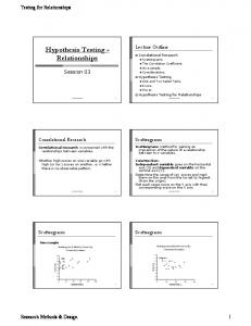
RESULTS Behavioral Performance Figure 2 shows mean response times for the first and last five trials in each block, for each block type. A feedback frequency (three levels: 8:2, 2:8, 5:5) by within block position (two levels: beginning, end) ANOVA revealed that response times increased as a function of the
Figure 2. Response times of the first and last five trials in each block of trials by block type.
frequency of negative feedback, F(2,18) = 4.93, p < .001, with a significant Feedback Frequency £ Block Position interaction, F(1,9) = 15.308, p < .001 between 8:2 and 2:8 feedback ratios. Recalling that feedback was preestablished and thus no correct answer existed, response times provided the only indirect behavioral criterion for correct performance. Within a single trial, subjects’ response precedes the experimenter’s feedback. Thus, in principle, response time is independent of feedback type. The fact that response times varied according to feedback frequency indicated the meaningfulness of this experimental manipulation. Moreover, as expected, response times relative to the first five trials in each block did not significantly differ across blocks with different positive-to-negative feedback ratios. ERP Analysis Mean amplitudes for all positive and negative feedback trials, irrespective of block type, were measured relative to a 200-msec prestimulus baseline at all electrodes. Figure 3 shows the grand averaged ERPs elicited by feedback stimuli at all electrodes. Separate waveforms are shown for feedback indicating correct and incorrect performance. Before collapsing all trials with the same feedback condition, we made sure that for each feedback condition the first five trials of each block did not significantly differ from the last five ones; that feedback-related activity for each feedback condition did not differ according to block type, that is, feedback type frequency; and that no significant differences could be found between the first and the last 15 blocks of trials, ruling out order effects or learning across blocks of trials. As in Miltner et al.’s (1997), early deflections elicited by the sensory properties of feedback were identical for positive and negative feedback. Positive feedback elicited a parieto-central P300, whereas negative feedback was characterized by P200 – N240 complexes with a frontal –central onset and lasting longer than in posterior regions (cf. Figure 4). In both feedback conditions, t tests revealed lateralization of activity selectively at frontal sites, with a left frontal dominance in the 210to 400-msec time window, Af7 vs. Af8: t(1,9) = 4.144, p < .01, for positive feedback, t(1,9) = 4.129, p < .01, for negative feedback; Af3 vs. Af4: t(1,9) = 3.63, p < .01 for positive feedback, t(1,9) = 5.97, p < .001, for negative feedback). To analyze the different ERP components as well as their spatial and temporal distribution, we performed a two-stage PCA (the method is described in detail in Spencer et al., 2001). In the first stage, PCA was applied to an association matrix representing the covariance between each pair of the 62 electrodes, computed over all subjects, experimental conditions (positive and negative feedback), and all time points in the 150- to 650-msec time window following feedback. Five spatial Papo et al.
513
Figure 3. Grand averaged ERPs (n = 10) for the positive and negative feedback at all 62 electrodes.
factors (accounting for ¹95% of the variance), each describing patterns of spatial variance in the data set, were extracted for rotation with the Varimax procedure. Topographic maps of the spatial factor loadings are presented in Figure 5. In Figure 6 we visualize the relationship between the activity represented by the spatial factors and the two feedback conditions by showing the spatial factor scores as time-series for each feedback condition, averaged across subjects. SF1 (posterior), SF2 (fronto-central), SF3 (left frontal), and SF4 (left fronto-temporal) all show differential responses to positive and negative feedback, albeit with different amplitudes and latencies. Each spatial pattern can represent several different patterns of brain activity during the epoch. To reduce dimensionality in the temporal domain, we submitted 514
Journal of Cognitive Neuroscience
the data to a temporal PCA, using the same procedure as in the previous phase. In this second step, the spatial factor scores associated with the time-points in the 150to 650-msec time window became the variables for the PCA, with spatial patterns, subjects, and feedback type as observations. Temporal PCA reduced the temporal dimensionality to five temporal factors, accounting for ¹93% of the variance. The temporal factor loadings and the percentage of variance accounted for by each factor after rotation are shown in Figure 7. The factor scores, representing the contribution of each temporal factor to the data, were the dependent variables for statistical analysis. We performed an ANOVA of the temporal factor scores associated with SF1 (posterior), SF2 (fronto-central), and SF3 (left frontal – polar) for TF1 (peak: 476 msec), TF2 (400 msec), TF3(620 msec), TF4 Volume 15, Number 4
Figure 4. Top: grand averaged ERPs (n = 10) for the positive and negative feedback at FCz (left) and POz (right). Bottom: grand averaged ERPs (n = 10) for positive (left) and negative feedback (right) at FCz and POz.
(312 msec), and TF5 (244 msec). Table 1 summarizes the results of these analyses. The results show a ubiquitous impact of feedback type in the 200 – 370 postfeedback
epoch, but a persistent effect in the 440 – 650 epoch only in SF1, a spatial pattern of activity heavily loading in posterior scalp areas.
Figure 5. Topographic maps of the factor loadings for the spatial factors. The percentage of variance accounted for by each factor after rotation is indicated.
Papo et al.
515
Figure 6. Grand average spatial factor scores for positive (F+) and negative (F¡) feedback. The value of the factor scores (y-axis) is a unitless dimension.
To further analyze our data in the temporal domain, we calculated for each subject and at each electrode the latency of the positive-to-negative uncoupling leading to the N240. The method consists of a standard linear interpolation technique and works as follows. For each 516
Journal of Cognitive Neuroscience
feedback condition (for each subject), identify at each electrode the peak of the negativity in the 100- to 140-msec range. Interpolate the descending ramus down to the 210- to 240-msec range (to minimize initial error, move a few points away from the negative peak). Volume 15, Number 4
central and late centroposterior feedback-related activity, with comparable morphological characteristics.
DISCUSSION
Figure 7. Temporal factor loadings. The percentage of variance accounted for by each factor after rotation is shown next to the factor number.
Take the first point 2 standard deviations away from the interpolating line as your reference point. The spatial PCA carried out on the resulting matrix using electrodes as variable yielded four components, accounting for ¹90% of the variance: SFT1 (frontal), SFT2 (posterior), SFT3 (central), and SFT4 (fronto-polar). We then used PCA factors to define four regions of interest (ROIs). ROIs were identified with electrodes associated in each spatial factor to scores ¶ .70. An ROI £ Hemisphere ANOVA revealed a main effect of regions, F(3,27) = 23.80, p < .0001, but no laterality effect. Follow-up tests revealed that while SFT1 and SFT4 did not differ between each other, there was an anteroposterior latency gradient, with SFT1 having significantly shorter latencies than SFT3, F(1,9) = 22.23, p < .0001, which in turn differed from SFT2, F(1,9) = 5.26, p < .05. Taken together with the results from PCA analyses, these results show early left frontal and midline fronto-
Table 1. Paired t Tests (9 df ) for the Correct and Incorrect Feedback Conditions for Each Combination of Temporal and Spatial Factor SF1
SF2
SF3
SF4
TF1
2.29*
ns
ns
ns
TF2
ns
ns
ns
ns
TF3
3.05*
ns
ns
2.67*
TF4
¡5.31***
¡5.16***
¡3.54**
¡4.89***
¡3.61**
¡3.65**
¡3.44**
¡380**
TF5 *p < .05.
**p < .01. ***p < .0001.
We used ERPs to study the electrophysiological response of subjects receiving exogenous feedback in an HT task of known computational aspects (Grossberg, 1984) and gross neurophysiology (Monchi et al., 2001; Elliott et al., 1997, 1998). Results showed that negative feedback was associated to an N240 delaying a P300-like wave. Spatiotemporal PCA analysis showed that the activity essentially resulted from the interplay of early-onset left frontal and midline fronto-central components and of two lagging central and posterior ones. Major learning effects could be discounted, so that results largely appear to reflect phasic reactions to the current feedback value, although our analyses cannot conclusively rule out within-block intertrial learning. We discuss morphological, spatiotemporal, and lateralization patterns and show their implications for the interpretation of previous studies of feedback. Morphology Positive and negative feedback were characterised by two waves of distinctive morphologies. A P300-like wave always accompanied feedback cue offset, regardless of the nature of feedback, whereas only negative feedback was accompanied by a negative N200-like deflection. ART theory predicts that negative feedback should induce early hippocampal mismatch, which in our data produced a P200. This would cause disinhibition of the orienting response resulting in wave complexes in the N200 – P300 range (Grossberg, 1984). The P300 buildup may reflect STM resetting of both the sensory and reinforcement values of feedback (Donchin & Isreal, 1980). The dimorphic patterns of electrophysiological activity elicited by positive and negative feedback can be represented in terms of specific catecholaminergic neuromodulation and mediating neural circuitry (Grossberg, 1984). The reinforcement effects of positive feedback could be mediated by midbrain DA neurons, which are involved in processing reward information and learning approach behavior (Schultz, 1998). Phasic depression of DA-related activity following negative feedback (Schultz, 1998) would shut down ongoing activity, as noradrenergic and cholinergic stress-related release promotes further search for a new rule via hippocampal resetting of reward-related ventral corticostriatal loops (Elliott, Friston, et al., 2000; Grossberg, 1984). Our reward-related account of feedback is corroborated by the similarity between the morphology of our feedback-related ERPs and those elicited by monetary gains and losses (Gehring & Willoughby, 2002). Our experiment extends their results to a larger class of Papo et al.
517
abstract rewards. The reward-related account of our type of feedback also dovetails with psychopharmacological studies showing dopaminergic P300 amplitude and latency modulation (Wang et al., 2000; Hansenne et al., 1995), with findings associating response to feedback to reward-related ventrostriatal-cortico-limbic neural circuits (Elliott et al., 1997) and with increased reward-related DA binding to ventral – striatal receptors in subjects playing a financially rewarded video game (Koepp et al., 1998; Thut et al., 1997). Our data are morphologically isomorphic to those found when subjects received feedback in a timeestimation task (Miltner et al., 1997) as well as to a negative frontal potential associated to inhibitory motor control in a stop-signal paradigm (Van Boxtel, van der Molen, Jennings, & Brunia, 2001). However, a complete description of feedback-related activity through ERPs should complement morphological characteristics with information concerning dynamic topographic patterns of activity (Spencer et al., 2001). For both studies, although frontal morphology and chronometry were comparable, scalp topography clearly was not. Spatiotemporal Patterns We discuss the patterns of task-related activity across the scalp, showing how these patterns can result from the activity of the two most prominent frontal components and their mutual relationship. Feedback can be thought of as a device switching on and off the cortico-cortical fronto-posterior workingmemory-related activity (Cohen et al., 1998). With positive feedback, the current pattern of activation should be kept active, sustained, and protected from interference. This is reflected by the fronto-posterior latency gradient of the P300. It is tempting to view the processes of selection and maintenance as arising from the cooperation of different dopaminergic reward signals for selectively reinforcing behavior: a phasic global reward DA signal, reinforced by cortico-striato-thalamic activity (Schultz, 1998), and a DA-mediated stabilization of neural representations in prefrontal cortex and the anteroposterior reverberatory activity (Durstewitz et al., 2000). Negative feedback should shut down neural activity associated to the invalidated rule while releasing new resources for subsequent rule searching, thus explaining the lagged posterior onset and resolution. The earlyonset, long-lasting frontal P200 –N240 complex together with the general pattern of anteroposterior activation can be interpreted in terms of a frontal role in making resources available for further posterior rule-related processing once a hypothesis is invalidated by negative feedback (Frith, 2000). Accordingly, differences between positive and negative feedback in parieto-temporal regions found in neuroimaging studies (Monchi et al., 2001; Elliott et al., 1997) can be understood as resulting from more anterior activity. 518
Journal of Cognitive Neuroscience
Our model predicted that a hippocampal feedbackinduced switch should promote activity in two parallel but separable networks: a ventrostriatal – orbitofrontal network directly involved in the mediation of the rewarding or stressing value of feedback, and a midline medial– prefrontal more connected to the modulation of fronto-posterior patterns of rule-related activity (Monchi et al., 2001; Elliott et al., 1997). Our results closely resemble the coexisting responselocked fronto-polar and midline prefrontal components arising from anterior cingulate and orbital frontal sources, respectively, found in a speeded response task (Luu & Tucker, 2001). Our model suggests a complex relationship between these two components. The fronto-central component may depend on the fronto-polar component for information necessary to interpret the current value of feedback, as it serves an alerting function mobilizing the affective systems; the midline fronto-central component may either directly or indirectly feed the frontopolar component with the information necessary to promote appropriate compensatory behavior (Gehring & Knight, 2000). Deficits in the brain circuits from which the two frontal components arise should have differential impacts on the overall pattern of activity across the scalp. Decreased orbitofrontal GABA-mediated gating should result in a general decreased ability to use feedback to reorganize the pattern of HT-related posterior activity (Northoff et al., 2002), whereas deficits in the medial prefrontal cortico-striato-thalamic circuit should be associated with erroneous evaluation of feedback value (Elliott et al., 1997). Frontal Activity, Lateralization, and Depression: Some Implications The fronto-polar component revealed by spatial PCA was the only component showing marked hemispheric lateralization. Left frontal lateralization of response to feedback fits nicely with a reward-related account of feedback in the light of reported left frontal and striatal mediation of approach behavior and reward (Miller & Tomarken, 2001; Delgado, Nystrom, Fissell, Noll, & Fiez, 2000; Koepp et al., 1998). Left lateralization may also reflect the activity of a hippocampal– dorsolateral prefrontal circuit mediating encoding of new information (Fletcher, Shallice, & Dolan, 1998). Taken together these interpretations may suggest a direct role of feedback rewarding properties in promoting new memory encoding. Unipolar depression is associated with behavioral oversensitivity to negative feedback (Beats et al., 1996), right prefrontal hyperactivation (Henriques & Davidson, 1997), left prefrontal task-related hypoactivation (Bench, Frackowiak, & Dolan, 1995), and left subgenual prefrontal cortex abnormalities (Drevets, 2000). Moreover, high frequency excitatory and low frequency Volume 15, Number 4
inhibitory repetitive transcranial magnetic stimulation respectively of the left and right prefrontal cortex were shown to induce antidepressant effects (Klein et al., 1999; Pascual-Leone, Pino, Pallordo, & Catala, 1996). Stress following incorrect performance feedback should shut down ongoing cognitive activity while promoting subsequent search and selection of new rules. However, to prevent enduring inhibition of new search for hypotheses following negative feedback, stress needs to be regulated and coping strategies elaborated. Insofar as pathological, feedback-related behavior in depression arises from inappropriate activation of mechanisms that are adaptive in normal circumstances (Elliott et al., 1998), the etiology of abnormal lateralization in depression can help in interpreting both the behavioral oversensitivity to negative feedback in depression and the pattern of lateralization in the present study. Hypersensitivity to negative feedback in depression may be linked to an increased propensity of the hippocampus to shut down ongoing activity in response to stress-induced NA bursts. The observed left – right frontal imbalance may reflect increased sympathetic arousal and corticosterone responses to restraint stress, resulting from a dysfunction of the left medial prefrontal cortex (Drevets, 2000). This would result in a tendency to overrespond to the emotional component of aversive stimuli, as defective hippocampal serotonergic release would impede coping toward aversion, failing to decrease the consequences of negative feedback’s aversive effects (Mongeau, Blier, & de Montigny, 1997; Gray, 1982). Our model predicts that NA-driven hippocampalmediated disinhibition of the orienting response and serotonergic control of prefrontal stress-related glutamatergic reward-suppressing response may be control parameters determining the morphological and chronometric characteristics of the P200 and N240. Trial-to-Trial Feedback versus Online Error Correction Another important goal of this study was to explain Miltner et al.’s (1997) conclusions on the equivalence between on-line error-correction mechanisms and trialto-trial feedback. Miltner et al. used ERPs to study the effects in a time estimation task of a trial-to-trial type of feedback computationally equivalent to that in the present study. The authors considered that their results concerning negative feedback could be explained by the interplay of a positive P300 and an N200-like component. The negative component associated to incorrect performance feedback was assimilated to the ERN associated to error detection in choice reaction time tasks on the grounds of dipole analysis indicating a common medial frontal localization. However, given its poor spatial resolution, dipole analysis cannot conclusively exclude that the ERN following an error and the negativity following
feedback arise from two separate subdivisions of the anterior cingulate cortex (Bush et al., 2000). The present study, Miltner et al.’s (1997), and Elliott et al.’s (1997, 1998) all used the same trial-to-trial feedback. Our results closely resembled Miltner et al.’s in ERPs’ morphology and chronometry at prefronto-central scalp sites. Our results, and therefore Miltner et al.’s, could be coherent with an N200 interpretation in terms of a ventrostriatal-cingulate start and stop system, as opposed to a premotor-dorsal anterior cingulate – lateral prefrontal circuit likely tapped into by ERN-eliciting tasks. Miltner et al. also acknowledged that the computational properties of their task were underspecified. The lack of theoretical embedding of feedback rendered the authors unable to interpret their data in any psychologically precise way. Mapping of the cognitive into the anatomo-functional space via some neuroimaging technique and the extent to which evidence from studies using different techniques can be used as a constraint are severely limited by underspecification of these two spaces. Miltner et al.’s description in terms of segregated brain activity focuses on the poor spatial resolution of ERPs, portraying feedback as a purely pointwise process, failing to use the excellent temporal resolution to account for the mechanisms through which feedback affects further cognitive processing. We showed that ERPs can be used to fractionate feedback into psychologically meaningful phases and to interpret results from different techniques when theory-driven hypotheses are interpreted in terms of integrated brain activity, via analyses of spatiotemporal patterns of activity across the scalp. We propose that computationally different types of error-related signals serve the same self-monitoring function, evaluating the motivational significance of events including errors and conflict (Bush et al., 2000), making resources available for further processing (Frith, 2000), albeit through separable neural circuits dedicated to the control of action or to the reward-driven selection of new responses (Elliott, Dolan, et al., 2000). A common basis for the different types of feedback may be that of a catecholaminergic context detection system, the properties of which would reflect those of the networks called upon in the fulfillment of different tasks, each resulting in a specific spatiotemporal pattern of activity. Our negativity could also be part of the frontal midline theta rhythm as was shown for the ERN (Luu & Tucker, 2001). Both signals could reflect a mechanism for action regulation in which hippocampal– medial prefrontal interactions modulate the outflow to the relevant corticostriatal control circuits, ensuring consistency of parallel computations in distributed networks. Caveats and Conclusion From the present experiment alone it is not possible to conclusively clarify the relation between the effects of Papo et al.
519
feedback as a warning signal from preparatory and more lasting effects on subsequent performance. If feedbackrelated activity is a mere reward context evaluation process, positive feedback’s profiles in our design should be equivalent to negative feedback’s in a context in which subjects can propose triplets of numbers and resort to a wide range of testing strategies, for example, falsification strategies; in such a context, negative feedback would allow subjects to falsify a given hypothesis and would therefore be more informative than a positive feedback. If, however, feedback-related activity also incorporates preparation for subsequent activity, this equivalence should not hold. In conclusion, the present study described the response to feedback and its consequences in an HT task, complementing existing results from different techniques. A reward-related cortico-striatal circuit and its stress-driven resetting by medial temporal brain structures were proposed to fulfill the regulatory properties of feedback in HT by modulating cortico-cortical fronto-parieto-temporal interactions. The specific neurophysiological brain structures that control the stress response are central to the capacity to adaptively recombine information from different sources and to generate novel hypotheses, arguably the hallmark of creative behavior. Analyses in the frequency domain can potentially unify findings concerning the cognitive processes underlying prefronto-hippocampal generation of the theta rhythm and the pattern of lateralization and of behavioral feedback-related deficits in depression. Our model should also be tested in pathologies including obsessive– compulsive disorder, anxiety, and psychopathy.
METHODS Subjects Thirteen right-handed graduate and undergraduate students volunteered in the experiment. Subjects had normal or corrected-to-normal vision and no history of neurological or psychiatric disease. Two of the subjects could not be kept for further analyses due to excessive rates of recording artefacts while one subject was excluded because she was not naõ¨ve as to the task’s manipulations. The 10 remaining subjects (five women and five men; mean age = 25.2, range 21 – 30 years) were all blind as to the experiment’s objectives. Stimuli and Procedure Subjects sat in front of a computer screen and judged, using a double-button press device, whether triplets of numbers were instances of a hidden rule chosen by the experimenter. At the beginning of each trial one triplet was presented. Subjects were asked to respond only when confident about their response. Subjects’ responses appeared on the screen underneath the 520
Journal of Cognitive Neuroscience
triplet as an O (‘‘Yes, the proposed triplet is an instance of the hidden rule’’) or an N (‘‘No, the proposed triplet is not an instance of the hidden rule’’). Following a time interval varying between 800 and 1200 msec, subjects received the experimenter’s feedback on the computer screen. A green square superimposed on the subjects’ O/N response signaled a correct response while a red one signaled a wrong response. Triplet, subjects’ responses, and feedback stayed on the screen till ±1 sec after feedback onset. The screen was then offset before the following trial could start. There was a 3- to 5-sec intertrial interval. There were 30 blocks of 10 trials each, corresponding to 30 different hidden rules. Subjects were informed that the rule was changed at the end of each block of trials. Successive blocks were separated by time intervals of the order of 30 sec/1 min. Unbeknownst to the subjects, the feedback was controlled by the experimenter and not by subjects’ performance, as there were no rules behind the triplets. Feedback frequency was manipulated, so that an equal number of blocks had 8:2, 5:5, and 2:8 positive-to-negative feedback ratios, respectively. In the (8:2) and (2:8) blocks the last five trials were paired to five consecutive positive and negative feedbacks, respectively, while in all blocks the first five trials comprised either two or three positive/negative feedback responses. The order of presentation of blocks was quasi-randomized across subjects. ERP Recording System Brain electrical activity was recorded from 62 electrodes positioned according to the extended 10-20 System location, with a nasion reference. The electro-oculogram (EOG) was also recorded for blink, vertical, and horizontal eye movement correction (Gratton, Coles, & Donchin, 1983). The EEG was amplified (0.05 – 100 Hz) and low-pass filtered with a 25- to 30-Hz transition band (12 dB/octave roll-off). The signals were digitized at 500 Hz over a 900-msec epoch, including a 200-msec prestimulus baseline. Acknowledgments We thank Florence Bouchet and Jean-Paul Bourzeix for technical support; Laurent Pezard, Clay Holroyd, and an anonymous referee for invaluable contributions in improving the manuscript; Manuela Piazza for very useful discussions and comments; and Diego Gil and Federico Bonaglia for statistical support. Reprint requests should be sent to David Papo, LPC Universite´ de Provence—Case 66, 3, place Victor Hugo; 13331 Marseille Cedex 3, France, or via e-mail:
[email protected].
REFERENCES Banquet, J.-P., & Grossberg, S. (1987). Probing cognitive processes through the structure of event-related potentials Volume 15, Number 4
during learning: An experimental and theoretical analysis. Applied Optics, 26, 4931 – 4946. Beats, B. C., Sahakian, B. J., & Levy, R. (1996). Cognitive performance in tests sensitive to frontal lobe dysfunction in elderly depressed. Psychological Medicine, 26, 591 –603. Behr, J., Glovell, T., Schmitz, D., & Heinemann, U. (2000). Dopamine depresses excitatory synaptic transmission onto rat subicular neurons via presynaptic D1 -like dopamine receptors. Journal of Neurophysiology, 84, 112 –119. Bench, C. J., Frackowiak, R. S. J., & Dolan, R. J. (1995). Changes in regional cerebral blood flow on recovery from depression. Psychological Medicine, 25, 247 –251. Bush, G., Luu, P., & Posner, M. I. (2000). Cognitive and emotional influences in anterior cingulate cortex. Trends in Cognitive Sciences, 4, 215 – 222. ˜ y, T., Tejedor, J., Cruz-Rizzolo, R. J., & Cavada, C., Compan Reinoso-Sua´rez, F. (2000). The anatomical connections of the macaque monkey orbitofrontal cortex. A review. Cerebral Cortex, 10, 220 – 242. Cohen, J. D., Braver, T. S., & O’Reilly, R. C. (1998). A computational approach to prefrontal cortex, cognitive control, and schizophrenia: Recent developments and current challenges. In A. C. Roberts, T. W. Robbins, & L. Weiskrantz (Eds.), The prefrontal cortex. Oxford: Oxford University Press. Dehaene, S., Posner, M. I., & Tucker, M. D. (1994). Localization of a neural system for error detection and compensation. Psychological Science, 5, 303 – 305. Delgado, M. R., Nystrom, L. E., Fissell, C., Noll, D. C., & Fiez, J. A. (2000). Tracking the hemodynamic responses to reward and punishment in the striatum. Journal of Neurophysiology, 84, 3072 – 3077. Donchin, E., & Isreal, J. B. (1980). Event related brain potentials and psychological theory. In H. H. Kornhuber & L. Deecke (Eds.), Motivation, motor, and sensory processes of the brain: Progress in brain research (pp. 697 – 715). Amsterdam: Elsevier. Drevets, W. C. (2000). Neuroimaging studies of mood disorders. Biological Psychiatry, 48, 813 – 829. Drevets, W. C., & Raichle, M. E. (1998). Reciprocal suppression of regional cerebral blood flow during emotional versus higher cognitive processes: Implications for interactions between emotion and cognition. Cognition and Emotion, 12, 353 –385. Durstewitz, D., Seamans, J. K., & Sejnowski, T. J. (2000). Neurocomputational models of working memory. Nature Neuroscience, 3 (Suppl.), 1184 – 1191. Elliott, R., & Dolan, R. J. (1998). Activation of different anterior cingulate foci in association with hypothesis testing and response selection. Neuroimage, 8, 17 – 29. Elliott, R., Dolan, R. J., & Frith, C. D. (2000). Dissociable functions in the medial and lateral orbitofrontal cortex: Evidence from human neuroimaging studies. Cerebral Cortex, 10, 308 – 317. Elliott, R., Friston, K., & Dolan, R. J. (2000). Dissociable neural responses in human reward systems. Journal of Neuroscience, 20, 6159 – 6165. Elliott, R., Frith, C. D., & Dolan, R. J. (1997). Differential neural response to positive and negative feedback in planning and guessing tasks. Neuropsychologia, 35, 1395 – 1404. Elliott, R., Sahakian, B. J., Paykel, E. S., & Dolan, R. J. (1998). Abnormal neural response to feedback on planning and guessing tasks in patients with unipolar depression. Psychological Medicine, 28, 559 –571. Falkenstein, M., Hohnsbein, J., Hoormann, J., & Blanke, L. (1991). Effects of cross-modal divided attention on late ERP
components. II. Error processing in choice reaction tasks. Electroencephalography and Clinical Neurophysiology, 78, 447 –455. Fletcher, P. C., Shallice, T., & Dolan, R. J. (1998). The functional roles of the prefrontal cortex in episodic memory. I. Encoding. Brain, 121, 1239 – 1248. Frith, C. D. (2000). The role of dorsolateral prefrontal cortex in the selection of action, as revealed by functional imaging. In S. Monsell & J. Driver (Eds.), Control of cognitive processes: Attention and performance XVIII. Cambridge: MIT Press. Gabrieli, J. D., Stebbins, G. T., Singh, J., Willingham, D. B., & Goetz, C. G. (1997). Intact mirror-tracing and impaired rotary pursuit skill learning in patients with Huntington’s disease: Evidence for dissociable memory systems in skill learning. Neuropsychology, 11, 272 – 281. Gehring, W. J., Goss, B., Coles, M. G. H., Meyer, D. E., & Donchin, E. (1993). A neural system for error detection and compensation. Psychological Science, 4, 385 – 390. Gehring, W. J., & Knight, R. T. (2000). Prefrontal –cingulate interactions in action monitoring. Nature Neuroscience, 3, 516 –520. Gehring, W. J., & Willoughby, A. R. (2002). The medial frontal cortex and the rapid processing of monetary gains and losses. Science, 295, 2279 – 2282. Gratton, G., Coles, M. G., & Donchin, E. (1983). A new method for off-line removal of ocular artifact. Electroencephalography and Clinical Neurophysiology, 55, 468 – 484. Gray, J. A. (1982). The neuropsychology of anxiety: An enquiry into the functions of the septo-hippocampal system. Oxford: Oxford University Press. Grossberg, S. (1984). Some psychophysiological and pharmacological correlates of a developmental, cognitive, and motivational theory. Brain and Information: Event Related Potentials, 425, 58 – 151. Hansenne, M., Pitchot, W., Gonzalez Moreno, A., Papart, P., Timsit-Berthier, M., & Ansseau, M. (1995). Catecholaminergic function and P300 amplitude in major depressive disorder (P300 and catecholamines). Electroencephalography and Clinical Neurophysiology, 96, 194 – 196. Henriques, J. B., & Davidson, R. J. (1997). Brain electrical asymmetries during cognitive task performance in depressed and nondepressed subjects. Biological Psychiatry, 42, 1039 – 1050. Jackson, M. E., & Moghaddam, B. (2001). Amygdala regulation of nucleus accumbens dopamine output is governed by prefrontal cortex. Journal of Neuroscience, 21, 676 – 681. Klein, E., Kreinin, I., Chistyakov, A., Koren, D., Mecz, L., Marmur, S., Ben-Shachar, D., & Feinsod, M. (1999). Therapeutic efficacy of right prefrontal slow repetitive transcranial magnetic stimulation in major depression. Archives of General Psychiatry, 56, 313 – 320. Koepp, M. J., Gunn, R. N., Lawrence, A. D., Cuningham, V. J., Dagher, A., Jones, T., Brooks, D. J., Bench, C. J., & Grasby, P. M. (1998). Evidence for striatal dopamine release during a video game. Nature, 393, 266 – 268. Lawrence, A. D. (2000). Error correction and the basal ganglia: Similar computations for action, cognition and emotion? Trends in Cognitive Sciences, 4, 365 – 367. Lewis, B. L., & O’Donnell, P. (2000). Ventral tegmental area afferents to the prefrontal cortex maintain membrane potential ‘‘up’’ states in pyramidal neurons via D1 dopamine receptors. Cerebral Cortex, 10, 1168 – 1175. Luu, P., & Tucker, D. M. (2001). Regulating action: Alternating activation of midline frontal and motor cortical networks. Clinical Neurophysiology, 112, 1295 – 1306. Mayberg, H. S., Liotti, M., Brannan, S. K., McGinnis, S., Mahurin, R. K., Jerabek, P. A., Silva, J. A., Tekell, J. L., Martin, Papo et al.
521
C. C., Lancaster, J. L., & Fox, P. T. (1999). Reciprocal limbic-cortical function and negative mood: Converging PET findings in depression and normal sadness. American Journal of Psychiatry, 156, 675 – 682. Miller, A., & Tomarken, A. J. (2001). Task-dependent changes in frontal brain asymmetry: Effects of incentive cues, outcome expectancies, and motor responses. Psychophysiology, 38, 500 –511. Miller, E. K. (2000). The prefrontal cortex and cognitive control. Nature Reviews, 1, 59 – 65. Miltner, W. H. R., Braun, C. H., & Coles, M. G. H. (1997). Event-related brain potentials following incorrect feedback in a time estimation task: Evidence for a ‘‘generic’’ neural system for error correction. Journal of Cognitive Neuroscience, 9, 788 – 798. Monchi, O., Petrides, M., Petre, V., Worsley, K., & Dagher, A. (2001). Wisconsin Card Sorting revisited: Distinct neural circuits participating in different stages of the task identified by event-related functional magnetic resonance imaging. Journal of Neuroscience, 21, 7733 – 7741. Monchi, O., Taylor, J. G., & Dagher, A. (2000). A neural model of working memory processes in normal subjects, Parkinson’s disease and schizophrenia for fMRI design and predictions. Neural Networks, 13, 953 – 973. Mongeau, R., Blier, P., & de Montigny, C. (1997). The serotonergic and noradrenergic systems of the hippocampus: Their interactions and the effects of antidepressant treatments. Brain Research Reviews, 23, 145 – 195. Northoff, G., Witzel, T., Richter, A., Gessner, M., Schlagenhauf, F., Fell, J., Baumgart, M., Kaulisch, T., Tempelmann, C., Heinzel, A., Ko ¨ tter, R., Hagner, T., Bargel, B., Hinrichs, H., Bogerts, B., Scheich, H., & Heinze, H.-J. (2002). GABA-ergic modulation of prefrontal spatio-temporal activation pattern during emotional processing: A combined fMRI/MEG study with placebo and Lorazepam. Journal of Cognitive Neuroscience, 14, 348 – 370. O’Doherty, J., Kringelbach, M. L., Rolls, E. T., Hornak, J., & Andrews, C. (2001). Abstract reward and punishment representations in the human orbitofrontal cortex. Nature Neuroscience, 4, 95 – 102.
522
Journal of Cognitive Neuroscience
¨ ngu O ¨ r, D., & Price, J. L. (2000). The organization of networks within the orbital and medial prefrontal cortex of rats, monkeys and humans. Cerebral Cortex, 10, 206 –219. Pascual-Leone, A., Rubio, B., Pallordo, F., & Catala, M. D. (1996). Beneficial effect of rapid-rate transcranial magnetic stimulation of the left dorsolateral prefrontal cortex in drug resistant depression. Lancet, 348, 233 –237. Sarter, M., & Bruno, J. P. (1999). Cortical cholinergic inputs mediating arousal, attentional processing and dreaming: Differential afferent regulation of the basal forebrain by telencephalic and brainstem afferents. Neuroscience, 95, 933 – 952. Schultz, W. (1998). Predictive reward signal of dopamine neurons. Journal of Neurophysiology, 80, 1 – 27. Smith, M. A., Brandt, J., & Shadmehr, R. (2000). Motor disorder in Huntington’s disease begins as a dysfunction in error feedback control. Nature, 403, 544 –549. Spencer, K. M., Dien, J., & Donchin, E. (2001). Spatiotemporal analysis of the late ERP responses to deviant stimuli. Psychophysiology, 38, 343 –358. Thut, G., Schultz, W., Roelcke, U., Nienhusmeier, M., Missimer, J., Maguire, R. P., & Leenders, K. L. (1997). Activation of the human brain by monetary reward. NeuroReport, 8, 1225 – 1228. Tucker, D. M., Hartry-Speiser, A., McDougal, L., Lure, P., & deGrandpre, D. (1999). Mood and spatial memory: Emotion and the right hemisphere contribution to spatial cognition. Biological Psychiatry, 50, 103 – 125. Van Boxtel, G. J. M., van der Molen, M. W., Jennings, J. R., & Brunia, C. H. M. (2001). A psychophysiological analysis of inhibitory motor control in the stop-signal paradigm. Biological Psychology, 58, 229 – 262. Wang, L., Kuroiwa, Y., Li, M., Kamitani, T., Wang, J., Takahashi, T., Suzuki, Y., Ikegami, T., & Matsubara, S. (2000). The correlation between P300 alterations and regional cerebral blood flow in non-demented Parkinson’s disease. Neuroscience Letters, 282, 133 – 136. Wason, P. C. (1960). On the failure to eliminate hypotheses in a conceptual task. Quarterly Journal of Experimental Psychology, 12, 129 – 140.
Volume 15, Number 4