Neuroscience Letters 254 (1998) 65–68
Feeding-induced changes in temporal patterning of muscle activity in the lobster stomatogastric system Stefan Clemens1, Pierre Meyrand, John Simmers* Laboratoire de Neurobiologie des Re´seaux, Universite´ Bordeaux I and CNRS, UMR 5816, Place du Dr. Peyneau, F-33120 Arcachon, France Received 17 March 1998; received in revised form 22 June 1998; accepted 22 June 1998
Abstract In the lobster Homarus gammarus, rhythmic masticatory movements of the foregut gastric mill are generated by a small neural network in the stomatogastric ganglion. We have used EMG recordings from intact animals to analyse gastric network output in relation to cycle period before and after feeding. In pre-prandial conditions, muscles controlling lateral teeth closure and medial tooth protraction (driven by MG and GM motor neurons, respectively) express relatively constant, return stroke-like burst durations, but change to a variable-duration power stroke-like phenotype after feeding. In contrast, the LPG neuron-innervated lateral teeth opener muscle switches from power stroke to return stroke-like behavior. Thus alternate phases within a single motor program may invert their temporal properties according to the behavioral situation. 1998 Elsevier Science Ireland Ltd. All rights reserved
Keywords: Stomatogastric nervous system; Neural network; Rhythmicity; Modulation; Feeding behavior; Power stroke; Return stroke; Crustacea
Rhythmic behavior is an important feature of all animals, requiring the generation of stereotyped motor patterns that can be adapted to environmental stimuli and changing behavioral requirements [2]. Underlying most rhythmic behaviors are networks of neurons that drive functionally antagonistic muscles, and which therefore are often mutually inhibitory with opposing phase relations [6]. Within such motor programs, one can often distinguish a main load bearing or power stroke phase that varies with cycle period, alternating with a relatively constant-duration return stroke phase (e.g. [3,7,8,14]). Can this asymmetrical organization of motor output, like other features of central network activity, also vary with changing behavioral demands? One of the best known models of motor rhythm generation is the gastric network in the stomatogastric nervous * Corresponding author. Tel.: +33 5 56223920; fax: +33 5 56830350; e-mail:
[email protected] 1 Present address: Georgia State University, Department of Biology, Atlanta, GA 30302-4010, USA.
system (STNS) of decapod Crustacea [9,16]. A major advantage of studying this network, which drives rhythmic masticatory movements of the two lateral and one medial teeth of the foregut gastric mill, is that it is comprised almost entirely of a few identified motor neurons (Fig. 1A). Thus any change in motor output to gastric muscles directly reflects changes within the central network itself. Although distinct power stroke and return stroke phases in the gastric cycle have been recognized from previous studies on spiny lobster, the identity of these phases remains unclear. For example, an earlier in vitro study reported that the power stroke phase corresponded to lateral teeth closure and medial tooth protraction [15], while endoscopic observations in vivo suggested that teeth opening and retraction corresponded more to the power stroke [10]. To shed light on this discrepancy, we have compared gastric network activity in vitro with network operation in intact unrestrained H. gammarus in two different behavioral situations, before and after feeding. For in vitro experiments, the STNS was dissected from the stomach and pinned out on a Sylgard-lined Petri dish as
0304-3940/98/$19.00 1998 Elsevier Science Ireland Ltd. All rights reserved PII S0304- 3940(98) 00511- 4
66
S. Clemens et al. / Neuroscience Letters 254 (1998) 65–68
described previously [16]. Intrasomatic and extracellular axonal recording and stimulation techniques were also conventional. For in vivo experiments, silver wire electrodes were implanted into three of the four main gastric muscles of animals which were left for 2 days to recover [4]. EMG recordings were then made before and after feeding using standard techniques for amplification, display and data analysis. The gastric network (Fig. 1A) consists of two neuronal subgroups, one controlling lateral teeth opening/closing, and the other, controlling medial tooth protraction/retraction. When the STNS is isolated in vitro, the lobster gastric network can generate a motor pattern normally responsible for these movements, as illustrated in Fig. 1B. Here, GM and LG neurons which control protractor and closer muscles of the medial and lateral teeth, respectively, fire in a relatively small fraction of each cycle, while LPG neurons, that normally drive lateral teeth retraction, express long intense bursts. In the same spontaneously-active preparations, altogether different temporal patterns of network output can be expressed in response to central modulatory influences [9,11–13]. As shown in Fig. 1C, following a brief electrical stimulation of the single input nerve to the STG, GM and LG neurons now fire in long bursts during a given gastric cycle, while LPG neurons are strongly active for a much smaller fraction of the cycle. Such alterations in the temporal structure of gastric patterning in vitro can be correlated with different behavioral states in vivo. Before feeding (Fig. 2A), the lobster gastric network is already active with a mean cycle period (±SE, as for subsequent data) of 30 ± 6 s [4]. The MG neuron-innervated muscle responsible for closing the lateral teeth, and the GM neuron-innervated muscle that protracts the medial tooth, are active for mean durations of 12.5 ± 0.3 and 13 ± 0.3 s per cycle, respectively. In contrast, burst durations of the lateral teeth opener muscle, innervated by the two LPG neurons, are 25 ± 2 s. After feeding however (Fig. 2B), the network accelerates to mean periods of 18 ± 1 s and the duty cycles of MG and GM neurons increase (cf. MG bars in Fig. 2A,B), while those of the LPG neurons decrease. Importantly, despite this variability in gastric network expression, which is similar to that observed in vitro (cf. Fig. 1B,C), the sequence of neuronal bursting within the motor pattern remains unchanged. In the next step we examined the phase-relationships of gastric muscle activity before and after feeding (n = 7), then assessed the temporal relationship with ongoing cycle period (Fig. 3). Before feeding (white bars in Fig. 3A), MG and GM neuron bursts occupy 45 ± 2 and 58 ± 1%, respectively, of the gastric cycle period. Moreover, the phase lag between MG and GM neuron bursting is 6 ± 0.5% cycle length. The mean duty cycle of the LPG neurons occupies about 68 ± 1% of the gastric cycle and expresses a considerable overlap (ca. 18% cycle length) with its functional antagonist, the MG neuron. After feeding (black bars in Fig. 3A), several changes in gastric network performance
occurs: firstly, MG and GM neurons enhance (although not significantly) their relative burst durations to 60 ± 1 and 65 ± 1% cycle length, respectively. In contrast, LPG neuron duty cycle is significantly decreased to 35 ± 1% cycle length. Secondly, whereas the phase lag between MG and GM neurons does not change significantly, the overlap between MG and LPG neuron bursts becomes significantly decreased to 8 ± 1% cycle length. Under pre-prandial conditions, no significant correlation
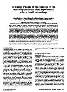
Fig. 1. The lobster gastric mill network and its activity patterns in vitro. (A) Synaptic wiring diagram of the 16 neuron network in the stomatogastric ganglion (STG). Boxes enclose motor neuron subgroups responsible for opening (LPG neurons) and closing (MG/LG neurons) the lateral teeth, and protracting (GM) and retracting (DG) the medial tooth. MG and LG neurons are electrically coupled and operate as a unit. Numbers of each cell type are indicated. Stick and ball connections denote inhibitory synapses, resistor symbols represent electrical connections, diode indicates rectifying electrical coupling. (B) Intra- and extracellular recordings of spontaneous activity in vitro. In this typical pattern, bursting of LPG neurons occupies the major fraction of each cycle while LG neuron expresses relatively short bursts. Note that the medial tooth protractor motor neurons (GM) fire in time with LG. (C) Gastric activity from the same preparation after a 1 s electrical stimulation of the STG input nerve. In this second pattern, LG and GM neuron bursts now predominate in the gastric cycle and LPG fires relatively shorter bursts. Int 1, Interneuron 1; LPG, lateral posterior gastric neuron; MG, medial gastric neuron; LG, lateral gastric neuron; GM, gastric mill neuron; DG, dorsal gastric neuron.
S. Clemens et al. / Neuroscience Letters 254 (1998) 65–68
67
LPG-innervated lateral teeth opener muscle corresponds to classical power stroke behavior, such as the stance phase of terrestrial locomotion [3,7,14], while the closer and protractor muscles innervated by MG and GM, respectively, operate as fixed-duration return stroke. After feeding however, the relationship of bursting in all three gastric neuron types with period length becomes inverted (Fig. 3B2); MG and GM neuron activity durations now increase linearly with cycle period (MG: r = 0.60, r2 = 0.85; GM: r = 0.61, r2 = 0.95) while LPG neuron bursts remain relatively constant (r = 0.27, r2 = 0.70). Therefore,
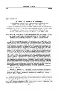
Fig. 2. Influence of feeding on gastric network output in vivo. (A) Simultaneous EMG recordings of three gastric muscles before feeding. Gastric cycle period is ca. 30 s. Note the relatively long-lasting bursts of the LPG neuron-innervated muscle. In the top trace, the faster rhythm is that of the pyloric network recorded from a neighboring lateral pyloric (LP) muscle. (B) Same animal 15 min after feeding: the gastric network expresses significantly shorter cycles (ca. 20 s) and MG and GM neuron duty cycles are now longer than the LPG neurons.
is apparent between the burst durations of MG (gray circles, dashed line) and GM neurons (open squares, dotted line) and ongoing cycle period (MG: r = 0.15, r2 = 0.38; GM: r = 0.27, r2 = 0.29; Fig. 3B1). In contrast, a clear correlation exists between the duration of LPG neuron bursts (black triangles, solid line) and cycle period (r = 0.74, r2 = 0.95). Functionally therefore, this variable duration activity of the
Fig. 3. Phase-relationships and burst durations versus gastric cycle period before and after feeding. (A) Before feeding (white boxes), LPG neuron bursts occupy a major fraction of the cycle, with a considerable overlap with the end of each MG neuron burst. After feeding (black boxes), the duty cycles of MG and GM neurons have slightly increased, whereas the relative duration of LPG neuron bursts has significantly decreased. The beginning and end of each box represent the mean (±SE) onset and offset times of bursts in the indicated neuron-innervated muscle. *Significant difference between pre- and post-prandial values (P , 0.01, Mann–Whitney). Note that onset of LPG bursts varied over a similar phase range in pre- and post-prandial conditions. (B) Before feeding (B1), MG and GM neuron burst durations are 5–15 s and are weakly correlated (r = 0.15 and 0.27, respectively) with cycle period. LPG neuron bursts, however, vary from 10 to >60 s and are closely correlated (r = 0.74) to cycle period. After feeding (B2), MG and GM bursts have lengthened to 5–30 s, and now express a strong relationship (r = 0.60 and 0.61, respectively) with cycle period. Although LPG bursts have also shortened, they are no longer closely correlated (r = 0.27) with cycle period. Pooled data in (B) from same seven experiments as in (A).
68
S. Clemens et al. / Neuroscience Letters 254 (1998) 65–68
lateral teeth closer (MG) and medial tooth protractor (GM) muscles now correspond to a variable-duration power stroke phase of gastric cycling, whereas the opener muscle, innervated by LPG neurons, now acts more like the constantduration return stroke. Thus, alternate phases of the gastric motor program may switch their temporal relationships with cycle period depending on the expression of feeding behavior. It is interesting that this switch in intact Homarus corresponds to two different gastric patterns reported separately from in vitro [15] and in vivo [10] studies on spiny lobster. That we can observe both patterns under standardized in vivo conditions suggests that these different phenotypes of gastric motor output are more likely to be related to behavioral changes than to different experimental approaches or differences between species. What is the functional significance of this power stroke/ return stroke switch in network function? It could be that the gastric teeth are called upon to chew food after feeding (and so teeth closure and protraction act as the load-bearing power stroke phase) whereas in unfed conditions, rhythmic teeth opening and retraction movements may serve as the power stroke phase in a different task such as mixing and pumping [5]. A similar bifunctional capability has been observed in lobster walking [3] where certain leg muscles can switch between power stroke- and return stroke-like behavior, depending on forward or backward stepping. In this case however, the switch is restricted to a muscle subset only [1] rather than involving the entire muscle ensemble as in the gastric system. The feeding-related switch in cycle period-dependence of gastric muscle activity necessarily reflects fundamental changes in gastric network properties, and involves all network elements since in both motor patterns, their overall firing sequence is maintained. A likely possibility is that the switch derives from changes in intrinsic excitability of gastric neurons [15] due to extrinsic modulatory instruction [11]. For example, induction of an oscillatory capability could allow individual neurons to produce regenerative constant duration, return stroke-like bursts, while at other times they are less inherently rhythmic and exhibit proportional power stroke-like discharge according to general levels of synaptic excitation. While this idea awaits further direct testing, it is interesting that such variability in neuronal behavior has already been found to dramatically alter gastric network performance in vitro [13]. Whether other motor systems similarly achieve power/return stroke changes by altering the balance between extrinsic synaptic drive and the expression of intrinsic membrane properties also remains to be seen.
Much of the impetus for the initial work came from the late Professor M. Moulins. S.C. was a Marie-Curie Research Fellow in the 3rd and 4th Framework program of the European Union. This work also benefited from an HFSP grant. [1] Ayers, J.L. Jr. and Davis, W.J., Neuronal control of locomotion in the lobster Homarus americanus. I. Motor programs for forward and backward walking, J. Comp. Physiol. A, 115 (1977) 1–27. [2] Chiel, H.J. and Beer, R.D., The brain has a body: adaptive behavior emerges from interaction of nervous system, body and environment, Trends Neurosci., 20 (1997) 553–557. [3] Clarac, F. and Chasserat, C., Quantitative analysis of walking in a decapod crustacean, the rock lobster Jasus lalandii. I. Comparative study of free and driven walking, J. Exp. Biol., 107 (1983) 189–201. [4] Clemens, S., Combes, D., Meyrand, P. and Simmers, J., Long term expression of two interacting motor pattern generating networks in the stomatogastric system of freely behaving lobster, J. Neurophysiol., 79 (1998) 1396–1408. [5] Clemens, S., Massabuau, J.-C., Legeay, A., Meyrand, P. and Simmers, J., In vivo modulation of interacting central pattern generators in lobster stomatogastric ganglion: influence of feeding and partial pressure of oxygen, J. Neurosci., 18 (1998) 2788–2799. [6] Getting, P.A., Emerging principles governing the operation of neuronal networks, Annu. Rev. Neurosci., 12 (1989) 185–204. [7] Grillner, S., Control of locomotion in bipeds, tetrapods, and fish. In J.M. Brookhart (Ed.), Handbook of Physiology: The Nervous System II, American Physiological Society, Mountcastle, VB, Bethesda, MD, 1981, pp. 1179–1236. [8] Grillner, S. and Dubuc, R., Control of locomotion in vertebrates: spinal and supraspinal mechanisms. In S.G. Waxman (Ed.), Advances in Neurology: Functional Reviews in Neurological Disease, Vol. 47, Raven Press, New York, 1988, pp. 425–453. [9] Harris-Warrick, R.M., Marder, E., Selverston, A.I. and Moulins, M., Dynamic Biological Networks, MIT Press, Cambridge, MA, 1992, 328 pp. [10] Heinzel, H.G., Gastric mill activity in the lobster. I. Spontaneous modes of chewing, J. Neurophysiol., 59 (1988) 528–550. [11] Heinzel, H.G., Gastric mill activity in the lobster. III. Effects of proctolin on the isolated central pattern generator, J. Neurophysiol., 59 (1988) 566–585. [12] Meyrand, P., Simmers, J. and Moulins, M., Dynamic construction of a neural network from multiple pattern generators in the lobster stomatogastric nervous system, J. Neurosci., 14 (1994) 630–644. [13] Nagy, F., Dickinson, P.S. and Moulins, M., Control by an identified modulatory neuron of the sequential expression of plateau properties of, and synaptic inputs to, a neuron in a central pattern generator, J. Neurosci., 8 (1988) 2875–2886. [14] Pearson, K.G., The control of walking, Sci. Am., 235 (1976) 72–86. [15] Russell, D.F., Pattern and reset analysis of the gastric mill rhythm in a spiny lobster, Panulirus interruptus, J. Exp. Biol., 114 (1985) 71–98. [16] Selverston, A.I. and Moulins, M., The crustacean stomatogastric system, Springer, Berlin, 1987, 338 pp.