Zeremski et al. BMC Gastroenterology 2014, 14:118 http://www.biomedcentral.com/1471-230X/14/118
RESEARCH ARTICLE
Open Access
FibroSURE as a noninvasive marker of liver fibrosis and inflammation in chronic hepatitis B Marija Zeremski1*, Rositsa B Dimova2, Samantha Benjamin1, Jessy Makeyeva1, Rhonda K Yantiss3, Maya Gambarin-Gelwan1,4 and Andrew H Talal1,5
Abstract Background: Noninvasive markers of liver fibrosis have not been extensively studied in patients with chronic hepatitis B virus (HBV) infection. Our aim was to evaluate the capacity of FibroSURE, one of the two noninvasive fibrosis indices commercially available in the United States, to identify HBV infected patients with moderate to severe fibrosis. Methods: Forty-five patients who underwent liver biopsy at a single tertiary care center were prospectively enrolled and had FibroSURE performed within an average interval of 11 days of the biopsy. Results: Of the 45 patients, 40% were Asian, 40% were African American, and 13% were Caucasian; 27% were coinfected with HIV and 67% had no or mild fibrosis. We found FibroSURE to have moderate capacity to discriminate between patients with moderate to high fibrosis and those with no to mild fibrosis (area under receiver operating characteristic [AUROC] curve = 0.77; 95% confidence interval [CI] [0.61, 0.92]). When we combined the fibrosis score determined by FibroSURE with aspartate aminotransferase (AST) measurements and HIV co-infection status, the discriminatory ability significantly improved reaching an AUROC of 0.90 (95% CI [0.80, 1.00]). FibroSURE also had a good ability to differentiate patients with no or mild from those with moderate to high inflammation (AUROC = 0.83; 95% CI [0.71, 0.95]). Conclusions: FibroSURE in combination with AST levels has an excellent capacity to identify moderate to high fibrosis stages in chronic HBV-infected patients. These data suggest that FibroSURE may be a useful substitute for liver biopsy in chronic HBV infection. Keywords: Hepatitis B virus, Liver biopsy replacement, Liver fibrosis assessment, Liver histology
Background Chronic hepatitis B virus (HBV) infection affects 350 to 400 million people worldwide [1,2]. The disease can progress to cirrhosis and hepatocellular carcinoma in approximately 15% to 40% of patients. The long-term complications are more frequent in patients with active viral replication as well as in those with significant intrahepatic inflammation and fibrosis [3]. Therefore, assessment of liver pathology is very important clinically as it identifies patients most likely to benefit from antiviral treatment.
* Correspondence:
[email protected] 1 Division of Gastroenterology and Hepatology, Department of Medicine, Weill Cornell Medical College, New York, NY, USA Full list of author information is available at the end of the article
Currently, the “gold standard” for the assessment of hepatic fibrosis is liver biopsy. Liver biopsy is recommended by the most recent AASLD guidelines on HBV in order to determine the need for antiviral therapy for those with normal liver enzymes who meet treatment criteria based on the viral load [4,5]. However, the procedure has several drawbacks including morbidity, mortality, significant sampling error, and high cost. It is associated with marked inter- and intra- observer/pathologist variability, and the use of different scoring systems to assess the degree of intrahepatic inflammation and fibrosis further complicates comparisons between pathologists. These limitations are important issues, particularly when patients seek a second opinion [6]. Finally, the invasive nature of the biopsy makes it unsuitable for serial hepatic sampling. In an attempt to overcome these limitations, several
© 2014 Zeremski et al.; licensee BioMed Central Ltd. This is an Open Access article distributed under the terms of the Creative Commons Attribution License (http://creativecommons.org/licenses/by/4.0), which permits unrestricted use, distribution, and reproduction in any medium, provided the original work is properly credited. The Creative Commons Public Domain Dedication waiver (http://creativecommons.org/publicdomain/zero/1.0/) applies to the data made available in this article, unless otherwise stated.
Zeremski et al. BMC Gastroenterology 2014, 14:118 http://www.biomedcentral.com/1471-230X/14/118
indices that incorporate parameters associated with inflammation or fibrogenesis, such as FibroTest/FibroSURE [7,8], Forns’ index [9], or the aspartate aminotransferase (AST)/platelet ratio index [10], have been proposed as noninvasive alternatives. FibroSURE (Laboratory Corporation of America (LabCorp), Burlington, NC) and FIBROSpect II (Prometheus Laboratories, San Diego, CA) are the only two commercially available noninvasive indices used in the United States. Both of these tests have been developed and extensively validated in patients with hepatitis C [11,12], but their utility in hepatitis B has not been adequately evaluated. In this study, we prospectively assessed the performance characteristics of FibroSURE as a noninvasive test for measuring liver inflammation and fibrosis in patients with chronic hepatitis B infection.
Methods Patients
All HBV-infected patients who underwent a liver biopsy at our institution between 2005 and 2012 were prospectively approached and offered participation in this study. We enrolled 45 HBV-infected patients, 12 (26.7%) of whom were co-infected with HIV. Patients with HCV co-infection and other co-existing liver diseases were excluded. The study was approved by the Weill Cornell Medical College institutional review board and was performed in accordance with the Declaration of Helsinki. After signing an informed consent, each patient’s blood was drawn and sent to LabCorp for performance of the FibroSURE. Alanine aminotransferase (ALT) and AST levels, as well as HBV DNA measurements, were extracted from the electronic medical record and were performed, on average, within 1 month (ALT and AST), or within 2 months (HBV DNA), of the liver biopsy or FibroSURE. All liver biopsies were evaluated according to the Scheuer classification [13] by the same hepatopathologist (RKY).
Page 2 of 8
to estimate the linear combination of the available variables, which detects fibrosis stage and inflammation grade, we used logistic regression. Comparison of the ROC curves was based on the methods described by DeLong et al. [14]. The significance level in all tests (two-sided) was set to 0.05.
Results Patients’ characteristics
A total of 45 patients with chronic HBV infection, 12 of whom were HIV/HBV co-infected, were included in this study. Patients’ mean age was 42.4 years, 58% were male, 40% were Asian, 40% were African American, and 13% were Caucasian (Table 1). Among the twelve HIV/HBV co-infected patients, 83.3% were African American and 16.7% were Caucasian. Ten patients, nine of whom were HIV co-infected, were on treatment for chronic HBV infection during the time of the liver biopsy/FibroSURE. One patient was taking Lamivudine, two were on Tenofovir, four patients were on Truvada, and three patients, one of whom was HBV mono-infected, were on Entecavir. Most of Table 1 Patients characteristics Parameter Gender Ethnicity
Grade
Statistical analysis
Statistical analysis was performed using SAS (SAS Institute Inc., Cary, NC, USA) and R (http://www.r-project.org/). Aminotransferase levels were logarithmically transformed for the analysis. Associations between categorical variables were assessed through Fisher’s exact test and logistic regression. For continuous variables, comparisons between groups were performed using Wilcoxon rank-sum or Kruskal-Wallis tests. Diagnostic abilities of FibroSURE were expressed through the estimated area under the receiver operating characteristic (AUROC) curve and by calculation of the respective sensitivity, specificity, positive and negative predictive values estimated for appropriate cut-off values. The AUROC was estimated by the trapezoidal rule and represents the probability that a randomly selected pair of subjects will be classified correctly. In order
Patients (N = 45)
Mean (SD) age, years
Stage
26 (58%)
Female
19 (42%)
Caucasian
6 (13%)
African American
18 (40%)
Asian
18 (40%)
Hispanic
1 (2%)
Indian
2 (4%)
0
4 (9%)
1
20 (44%)
2
14 (31%)
3
6 (13%)
4
1 (2%)
0
18 (40%)
1
12 (27%)
2
8 (18%)
3
4 (9%)
4 HIV co-infected Mean ALT (U/L) (range) Mean AST (U/L) (range) Mean HBV DNA (IU/ml) (range) Positive HBeAg
42.4 (12.15)
Male
3 (7%) 12 (27%) 53 (10–240) 46 (13–302) 33,476,858 ( =2 (n = 15) n (%) or Mean (SD)
Stage < 2 (n = 30) n (%) or Mean (SD)
46.87 (13.93)
40.17 (10.72)
1.05
0.993; 1.110
0.089
11 (42.31)
15 (57.69)
2.75
0.713; 10.605
0.1418
OR
95% CI
Univariable logistic regression
Multivariable logistic regression***
p-value
p-value
Gender, N (%)
Male Female*
4 (21.05)
15 (78.95)
Race, N (%)
Caucasian*
1 (16.67)
5 (83.33)
AA
9 (50.00)
9(50.00)
5
0.483; 51.769
Asian
4 (22.22)
14 (77.78)
1.429
0.127; 16.025
Other
1 (33.33)
2 (66.67)
2.5
0.1; 62.602
0.135
0.031; 0.581
0.0072**
0.0156**
9.333
2.110; 41.278
0.0032**
0.0066**
3.015§
1.426; 6.374
0.0039**
0.0158**
§
Diagnosis Inflammation
HBV
7 (21.21)
26 (78.79)
HIV/HBV*
8 (66.67)
4 (33.33)
Grade > 1
12 (57.14)
9 (42.86)
Grade < =1* FibroSURE
3 (12.50)
21 (87.50)
0.462 (0.297)
0.212 (0.167)
0.2845
log10 ALT (U/L)
1.81 (0.25)
1.52 (0.28)
3.314
1.393; 7.883
0.0067**
log10 AST (U/L)
1.70 (0.21)
1.51 (0.25)
2.455§
1.065; 5.658
0.0350**
4.60 (2.39)
§
0.674; 2.520
0.4303
log10 HBV DNA (IU/ml)
5.24 (2.98)
0.0641
Zeremski et al. BMC Gastroenterology 2014, 14:118 http://www.biomedcentral.com/1471-230X/14/118
Table 2 Logistic regression analysis on moderate to severe stage of fibrosis
1.304
*Reference group for the reported odds ratios. **p-value < 0.05. ***Based on the best selected model. § OR of 1 SD increase. Abbreviations: ALT alanine aminotransferase, AST aspartate aminotransferase, HBV hepatitis B virus, SD standard deviation, OR odds ratio, CI confidence interval, AA African-American, HIV human immunodefiency virus.
Page 5 of 8
Zeremski et al. BMC Gastroenterology 2014, 14:118 http://www.biomedcentral.com/1471-230X/14/118
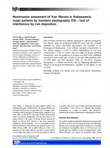
Figure 2 Receiver operating characteristic (ROC) curves of the combined model (as described in Results), FibroSURE and log10 (AST) for the detection of moderate to severe fibrosis (stage ≥ 2). Values of the area under ROC curves for the individual variables are indicated in parenthesis at the bottom of the image (A). Sensitivity and specificity of the model score for a range of cut-off values (B).
In the United States, FibroSURE is one of the two commercially available noninvasive indices of hepatic fibrosis and inflammation. FibroSURE is a multicomponent index comprised of six serum markers including α2-macroglobulin, haptoglobin, apolipoprotein A-1, total bilirubin, γ-glutamyltransferase, and ALT that are combined with patients’ age and gender to calculate separate scores for fibrosis and necroinflammatory activity. It is
Page 6 of 8
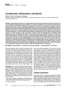
Figure 3 Receiver operating characteristic (ROC) curves of the FibroSURE inflammation score, log10(AST) and log10(ALT) for the detection of moderate to severe inflammation (grade ≥ 2) (A). Sensitivity and specificity of the FibroSURE inflammation score for a range of cut-off values (B).
currently marketed for liver histology assessment in chronic HCV-infected patients, and only a few studies evaluated the utility of this test in chronic HBV infection [15-20]. However, in all of these studies, as opposed to performing FibroSURE in a commercial laboratory where the reproducibility of the test is closely monitored and subject to performance measures, serum marker measurements in the prior studies were performed in local laboratories and calculation of the fibrosis score was performed
Zeremski et al. BMC Gastroenterology 2014, 14:118 http://www.biomedcentral.com/1471-230X/14/118
individually by study authors. To our knowledge, the commercially available FibroSURE test has not been evaluated previously in chronic HBV-infected patients from the United States. Our data showed that FibroSURE had reasonable ability to discriminate between HBV-infected patients with moderate to high fibrosis and those with mild or no fibrosis. Moreover, the performance of FibroSURE test in our HBV-infected patients was very similar to results previously published for patients infected with HCV [11]. The addition of AST and HIV co-infection status to the FibroSURE score significantly improved its discriminatory capacity, with an AUROC of 0.90, making the combination of these three parameters an excellent index for identification of HBV-infected patients with advanced fibrosis. As a marker of intrahepatic inflammation, FibroSURE showed good ability to discriminate between patients with moderate to high inflammation and those with mild or no inflammation. However, its discriminatory ability did not differ significantly from either ALT or AST individually. Excellent performance characteristics are only the initial attribute required for the adaptation in the clinic of any diagnostic test. In addition, tests must be conveniently obtainable and readily interpretable in the clinical setting with a high degree of reproducibility as demonstrated through laboratory performance characteristics. Reproducibility is particularly important for serial application of a test, such as might be required of FibroSURE if it were to be used as a benchmark for determining treatment outcome or if it were to be performed repeatedly to assess progression of inflammation or fibrosis. We [21] and others [22] have previously shown that the commercially available FibroSURE has the utility to identify fibrosis progression in patients with HCV. Whether FibroSURE has similar prognostic significance in HBV infection remains to be determined. A limitation of this study is the small sample size. Although all HBV-infected patients who underwent a liver biopsy at our institution between 2005 and 2012 were consecutively approached, their number was small and not all who were approached agreed to study participation. However, the racial diversity of our subjects is representative of the U.S. population of HBV-infected individuals. In contradistinction to other countries, HBV infection prevalence in the United States is estimated to be ~2% among Asians and 0.73% among non-Hispanic Blacks, the ethnicities with the highest HBV prevalence in the U.S. [23]. These two groups account for 39% and 31% of individuals, respectively, in the U.S. with chronic HBV infection. The ethnicity of patients in this study (40% Asian, 40% Black) is reflective of that found in the general U.S. population and can be considered a strength of our study population. An additional limitation of the study is the heterogeneous patient population
Page 7 of 8
consisting of unequal numbers of HBV mono-infected and HIV/HBV co-infected patients.
Conclusions In chronic HBV infection, liver biopsy is usually performed in patients with active disease in order to identify those who might be expected to benefit the most from treatment. However, many patients are reluctant to undergo the procedure due to its invasiveness and high costs. Our study shows that performance of FibroSURE in combination with AST levels can be a good substitute for liver biopsy for estimation of hepatic fibrosis in HBVinfected patients. The test can be performed on one tube of blood easily obtained at the point of care. Based upon these results, we believe that the FibroSURE can be used to identify patients in whom HBV treatment should be considered. Competing interests This study was funded in part through the Fellows Research Program sponsored by Bristol-Myers Squibb and Laboratory Corporation of America for performance of the Fibrosure test. Authors’ contributions MZ and AHT designed the study. MZ, SB, JM, and MG-G acquired the data. MZ, RBD, RKY, and AHT analyzed and interpreted the data. RBD performed statistical analysis of the data. MZ and AHT wrote the manuscript. All authors read and approved the final version of the manuscript. Acknowledgments We acknowledge Laboratory Corporation of America for defraying the costs of FibroSURE. We also acknowledge the patients for their participation in the study. Author details 1 Division of Gastroenterology and Hepatology, Department of Medicine, Weill Cornell Medical College, New York, NY, USA. 2Department of Biostatistics, State University of New York at Buffalo, Buffalo, NY, USA. 3 Department of Pathology and Laboratory Medicine, Weill Cornell Medical College, New York, NY, USA. 4Gastroenterology and Nutrition Service, Memorial Sloan-Kettering Cancer Center, New York, NY, USA. 5Division of Gastroenterology, Hepatology and Nutrition, Department of Medicine, State University of New York at Buffalo, Buffalo, NY, USA. Received: 19 March 2014 Accepted: 19 June 2014 Published: 3 July 2014 References 1. Lai CL, Ratziu V, Yuen MF, Poynard T: Viral hepatitis B. Lancet 2003, 362(9401):2089–2094. 2. Kuo A, Gish R: Chronic hepatitis B infection. Clin Liver Dis 2012, 16(2):347–369. 3. Jury E: EASL International Consensus Conference on Hepatitis B. 13–14 September, 2002: Geneva, Switzerland. Consensus statement (short version). J Hepatol 2003, 38(4):533–540. 4. Rockey DC, Caldwell SH, Goodman ZD, Nelson RC, Smith AD: Liver biopsy. Hepatology 2009, 49(3):1017–1044. 5. Lok AS, McMahon BJ: Chronic hepatitis B: update 2009. Hepatology 2009, 50(3):661–662. 6. Zeremski M, Talal AH: Noninvasive markers of hepatic fibrosis: Are they ready for prime time in the management of HIV/HCV co-infected patients? J Hepatol 2005, 43(1):2–5. 7. Rossi E, Adams L, Prins A, Bulsara M, De Boer B, Garas G, MacQuillan G, Speers D, Jeffrey G: Validation of the FibroTest biochemical markers score in assessing liver fibrosis in hepatitis C patients. Clin Chem 2003, 49(3):450–454.
Zeremski et al. BMC Gastroenterology 2014, 14:118 http://www.biomedcentral.com/1471-230X/14/118
8.
9.
10.
11.
12.
13. 14.
15.
16.
17.
18.
19.
20.
21.
22.
23.
Imbert-Bismut F, Ratziu V, Pieroni L, Charlotte F, Benhamou Y, Poynard T: Biochemical markers of liver fibrosis in patients with hepatitis C virus infection: a prospective study. Lancet 2001, 357(9262):1069–1075. Forns X, Ampurdanes S, Llovet JM, Aponte J, Quinto L, Martinez-Bauer E, Bruguera M, Sanchez-Tapias JM, Rodes J: Identification of chronic hepatitis C patients without hepatic fibrosis by a simple predictive model. Hepatology 2002, 36(4 Pt 1):986–992. Wai CT, Greenson JK, Fontana RJ, Kalbfleisch JD, Marrero JA, Conjeevaram HS, Lok AS: A simple noninvasive index can predict both significant fibrosis and cirrhosis in patients with chronic hepatitis C. Hepatology 2003, 38(2):518–526. Poynard T, Imbert-Bismut F, Munteanu M, Messous D, Myers RP, Thabut D, Ratziu V, Mercadier A, Benhamou Y, Hainque B: Overview of the diagnostic value of biochemical markers of liver fibrosis (FibroTest, HCV FibroSure) and necrosis (ActiTest) in patients with chronic hepatitis C. Comp Hepatol 2004, 3(1):8. Patel K, Gordon SC, Jacobson I, Hezode C, Oh E, Smith KM, Pawlotsky JM, McHutchison JG: Evaluation of a panel of non-invasive serum markers to differentiate mild from moderate-to-advanced liver fibrosis in chronic hepatitis C patients. J Hepatol 2004, 41(6):935–942. Scheuer PJ: Classification of chronic viral hepatitis: a need for reassessment. J Hepatol 1991, 13(3):372–374. DeLong ER, DeLong DM, Clarke-Pearson DL: Comparing the areas under two or more correlated receiver operating characteristic curves: a nonparametric approach. Biometrics 1988, 44(3):837–845. Poynard T, Ngo Y, Munteanu M, Thabut D, Ratziu V: Noninvasive Markers of Hepatic Fibrosis in Chronic Hepatitis B. Curr Hepat Rep 2011, 10(2):87–97. Mbaye PS, Sarr A, Sire JM, Evra ML, Ba A, Daveiga J, Diallo A, Fall F, Chartier L, Simon F, Vray M: Liver stiffness measurement and biochemical markers in Senegalese chronic hepatitis B patients with normal ALT and high viral load. PLoS ONE 2011, 6(7):e22291. Bonnard P, Sombie R, Lescure FX, Bougouma A, Guiard-Schmid JB, Poynard T, Cales P, Housset C, Callard P, Le Pendeven C, Drabo J, Carrat F, Pialoux G: Comparison of elastography, serum marker scores, and histology for the assessment of liver fibrosis in hepatitis B virus (HBV)-infected patients in Burkina Faso. Am J Trop Med Hyg 2010, 82(3):454–458. Bottero J, Lacombe K, Guechot J, Serfaty L, Miailhes P, Bonnard P, Wendum D, Molina JM, Lascoux-Combe C, Girard PM: Performance of 11 biomarkers for liver fibrosis assessment in HIV/HBV co-infected patients. J Hepatol 2009, 50(6):1074–1083. Sebastiani G, Vario A, Guido M, Alberti A: Sequential algorithms combining non-invasive markers and biopsy for the assessment of liver fibrosis in chronic hepatitis B. World J Gastroenterol 2007, 13(4):525–531. Myers RP, Benhamou Y, Imbert-Bismut F, Thibault V, Bochet M, Charlotte F, Ratziu V, Bricaire F, Katlama C, Poynard T: Serum biochemical markers accurately predict liver fibrosis in HIV and hepatitis C virus co-infected patients. Aids 2003, 17(5):721–725. Zeremski M, Dimova R, Astemborski J, Thomas DL, Talal AH: CXCL9 and CXCL10 chemokines as predictors of liver fibrosis in a cohort of primarily African-American injection drug users with chronic hepatitis C. J Infect Dis 2011, 204(6):832–836. Wilson LE, Torbenson M, Astemborski J, Faruki H, Spoler C, Rai R, Mehta S, Kirk GD, Nelson K, Afdhal N, Thomas DL: Progression of liver fibrosis among injection drug users with chronic hepatitis C. Hepatology 2006, 43(4):788–795. Ioannou GN: Hepatitis B virus in the United States: infection, exposure, and immunity rates in a nationally representative survey. Ann Intern Med 2011, 154(5):319–328.
doi:10.1186/1471-230X-14-118 Cite this article as: Zeremski et al.: FibroSURE as a noninvasive marker of liver fibrosis and inflammation in chronic hepatitis B. BMC Gastroenterology 2014 14:118.
Page 8 of 8
Submit your next manuscript to BioMed Central and take full advantage of: • Convenient online submission • Thorough peer review • No space constraints or color figure charges • Immediate publication on acceptance • Inclusion in PubMed, CAS, Scopus and Google Scholar • Research which is freely available for redistribution Submit your manuscript at www.biomedcentral.com/submit