Figure S1. Multiple sequence alignment of membrane-bound carbonic ...
Recommend Documents
Figure S1: Multiple sequence alignment of the 3' half of snR80. Conservation of snR80 across diverse yeast, showing conserved sequence and structural ...
The alignment consists of transferrin sequences from organisms of various taxa. The disulphide bridges (Cys11-Cys49, Cys21-Cys45, Cys271-. Cys370 ...
Figure S1. Sequence alignment of a hemoglobin alpha subunit. Tyrosine C7 (Tyr 42, human) is highly conserved. P69905.
Figure S1 Sequence alignment of TAG from different organisms (S. aureus MSSA476; S. aureus MRSA252; S. typhi; E. coli; gi|152977981, Actinobacillus ...
IL HS FATHL LENGV DIRA I OQLLG SNL ST TO I. IL HS Y ATHL F E O G V N I K I I 99 L LG SNL STT I ... Ape-A L P PDSRI I E. AV Y KRL KSL A KRAGL. Tho-A I ...
Sequence alignment between the four PIIIA binding domains of the varied NaV subtypes. Loops with low sequence identify as well as the voltage sensing ...
Supplementary Figure 1. Multiple Sequence Alignment of HU from different species of Mycobacteria and actinobacteria. ... M. gilvum; 10. M. avium subsp. 11.
H Z ITT TIL TILL ATT TILLITI TILTI. C I I II || K || 1 || 1 | | | | | | LA ALIA A. LA O 9 b A A A A LA I. H I L M N M M. å³ AID1. Z LI. I I O. TI. |. II I I ALI I I II I M N M L H | I |.
XIII I II I II I III TILL LITT TIL A LI TIITLITI TIL LITT TIL TILT I. O Z ITT TILL LITT TIL TIL TIL TIL TITTI. > ALLT TIL TIL TILL MITT TIL TILL TILL. A AI I I I II I TIL I I I I II II.
server (at www. expasy.ch/tools/blast/) to gather and align themâ. Claverie J, Notredame C (2007). Bioinformatics for Dummies (2nd Edn). Wiley publishing, Inc.
Table 2. The robustness of noncontextual tables. The range, median, and standard deviation for the number of examples drawn on per substitution score. Table.
ten across the page and the other down the left-hand side. Whenever .... of Medicine, http://dot.imgen.bcm.tmc.edu:9331/multi-align/multi-align.html). Once the.
... EEY DSSYD- I DVD ELV I SMRTDL NRFMSQAEDR EKGNRCCLIC KIVHR I ... IVKIT YQFFA HD - AETILLL TKN ISGFMEG TIYHFPKV IN EANRPEDEYH ...
May 12, 2016 - sequence), identical (in a rigid local structural align or participate a ... orm.html. ALTAVIST www.bibiserv.techfak.uni bielefeld.de/altavist/. II. METHODS AND .... A horizontal cladogram, with the root to the left m Greek clados ...
(http://www.jalview.org/jvmmemoryparams.html) . Introduction to ... The File â Output To Textbox menu option allows the alignment to be copied and pasted into ...
May 5, 2006 - Multiple sequence alignment. Robert C Edgar. 1 and Serafim Batzoglou. 2. Multiple sequence alignments are an essential tool for.
The yellow color highlighted the amino acid sequences ... the conversed amino acid of MTHFR among monocot and dicot species. Amino acid ..... Pipecolic Acid.
Page 1. 23. 26. 54. 62 gp177(F-CphI)[Synechococcus_phage_S-PM2]. H. R. Q. C C . D DH TG. Q LC CN G. YL. EE .........NGV AI.. KGEGD.
W. Chen, . Edvardsen, F. Campagne, and G. Vriend. Gpcrdb: an information system for g protein-coupled receptors. Nu- cleic Acids Res., 26(1):277â291, 1998.
'twilight zone' of RNA sequence alignmentâthe homology range below which sequence alignment alone is unlikely to produce reliable results and researchers ...
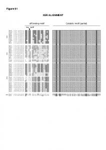
Figure S1. Multiple sequence alignment of membrane-bound carbonic ...
Figure S1. Multiple sequence alignment of membrane-bound carbonic anhydrase proteins: pufferfish CA. XII, human CA XII, human CA VI, and human CA IV.
Figure S1. Multiple sequence alignment of membrane-bound carbonic anhydrase proteins: pufferfish CA XII, human CA XII, human CA VI, and human CA IV. The red colored hollow boxes show the conserved cysteine residues that likely form a disulfide bond in the CA proteins.
Figure S2. mRNA expression of carbonic anhydrase CA XII in different tissues (brain, gill, heart, liver, intestine, kidney and blood) of pufferfish (Takifugu rubripes) quantified by reverse transcription polymerase chain reaction (RT-PCR). β-actin was used as internal control.