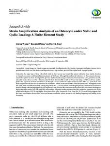
radius (µm) time = 100 msec theta in radian. Caâconcentration (. µM). (b). Figure 2. Radial and angular calcium distribution in a neuron cell for case I (a) t=10 ms ...
J. Appl. Math. & Informatics Vol. 31(2013), No. 5 - 6, pp. 695 - 709
http://dx.doi.org/10.14317/jami.2013.695
FINITE ELEMENT MODEL TO STUDY CALCIUM DIFFUSION IN A NEURON CELL INVOLVING JRYR, JSERCA AND JLEAK AMRITA TRIPATHI∗ AND NEERU ADLAKHA
Abstract. Calcium is well known role for signal transduction in a neuron cell. Various processes and parameters modulate the intracellular calcium signaling process. A number of experimental and theoretical attempts are reported in the literature for study of calcium signaling in neuron cells. But still the role of various processes, components and parameters involved in calcium signaling is still not well understood. In this paper an attempt has been made to develop two dimensional finite element model to study calcium diffusion in neuron cells. The JRyR, JSERCA and JLeak, the exogenous buffers like EGTA and BAPTA, and diffusion coefficients have been incorporated in the model. Appropriate boundary conditions have been framed. Triangular ring elements have been employed to discretized the region. The effect of these parameters on calcium diffusion has been studied with the help of numerical results. AMS Mathematics Subject Classification : 65H05, 65F10. Key words and phrases : Triangular ring elements, Receptors, Endoplasmic Reticulum, Pump, Leak, Variational form.
1. Introduction Intracellular calcium is an important second messenger in living cells. Ca2+ regulates a variety of intracellular processes, but its efficiency as a signaling agent depends on the degree of appropriation by cytoplasmic calcium buffers and influx and out flux of calcium due to receptors, leaks and pumps in neuron cells [6, 12]. The calcium is essential for almost every process in human organs like heartbeat, muscles contractions, bones activity and brain functionality etc. Ca2+ dynamics is the switch over of Ca2+ ions between intracellular Ca2+ stores and the cytosol, entering and leaving ions between the cells and binding activity of calcium and calcium binding proteins [18]. The most important calcium binding ∗
Received November 7, 2012. Revised January 7, 2013. Corresponding author. c 2013 Korean SIGCAM and KSCAM. ⃝ 695
Accepted February 4, 2013.
696
A. Tripathi and N. Adlakha
proteins are itself buffers that are located in Ca2+ stores. The binding of calcium molecules with buffer depends on calcium concentration in the cell [10, 21]. The significance and role of Ca2+ influx, although widely acknowledged have been a subject of controversial interpretations. On one hand, there is disagreement about the role of Ca2+ influx and on the other hand about how exactly Ca2+ influx is modulated during Ca2+ oscillations. It maintains that depletion of the ER Ca2+ causes increased entry of Ca2+ across the plasma membrane [7]. The concentration dependent binding of Ca2+ to buffers serves as an indicator of free calcium concentration in intracellular measurements. The active elements of the exchange processes are channels, receptors, serca and leaks in the membranes. They bound the intracellular Ca2+ that is stored in the endoplasmic reticulum and other storage devices. Serca Pumps transport the Ca2+ against its electro chemical gradient. Leak receives the Ca2+ that comes from pump and is stored in the endoplasmic reticulum (ER). Within the ER Ca2+ maintains the high capacity and low efficiency of Ca2+ binding proteins. They maintain the balances of Ca2+ ions through active (in) and passive (out) process [6]. The characteristics of single RyR channels are fundamental to understand the origin of many intracellular Ca2+ signaling phenomena. The amplitude of single RyR [23] channel current reveals the number of Ca2+ passing through the channel per second. The open probability (Po) of single RyR channels allows us to predict how certain agonists and antagonists regulate its activity. The Ca2+ -induced Ca2+ release process governs the activity of the other types of RyR channel. The distinguishing functional attributes of each RyR or IP3R channels likely underlie the spatiotemporal complexity of intracellular Ca2+ signaling in cells [15, 19]. Initial studies were the experimental investigations made with fruitful alternatives by many research workers. They obtained the results using the molecular modelling for receptor of Calcium profile and analysis on the linearized buffered Ca2+ diffusion [17]; Also attempts have been made by [9, 10] to find solutions to steady state problems of calcium diffusion in single neuronal cell. [11] modelled the above-mentioned phenomenon and solved steady state case of this equation for a spherically symmetric region to estimate rapid buffering approximation near an open Ca2+ channel in a neuron cell. Some theoretical investigations have also been carried out during the last few decades. [21, 22] developed the finite element model to study the cytosolic Ca2+ concentration in one and two dimensions rapid buffering approximation. The calcium distribution in neuron cell using finite element method and finite volume method for one and two dimensions in polar coordinates [1-3]. Jha et. al. developed one dimensional steady state finite element and finite volume models to study calcium distribution in astrocytes [4, 5]. Kotwani et. al. developed one dimensional unsteady state finite difference model for calcium diffusion in fibroblast [16]. In the present study an attempt has been made to model the calcium diffusion in neuron cell involving , and . The model has been developed for two dimensional unsteady state. Triangular ring based finite element method has been employed to obtain the solution. A computer program has been developed in MATLAB
Finite element model to study Calcium Diffusion
697
7.11 for the problem and simulated on Core i3 processor with 2.13 GHz processing speed, 64-bit machine with 320 GB memory. Numerical results have been used to study the relationships among various parameters. 2. Mathematical Formulation Calcium kinetics in neurons is governed by a set of reaction-diffusion equations which can be framed assuming the following bimolecular reaction between Ca2+ and buffer species [8,17]: [ 2+ ] k+ Ca + [Bj ] ↔ [CaBj ] (1) − k
where [Bj ] and [CaBj ] are free and bound buffer respectively , and j is an index over buffer species. The resulting partial differential equations in two dimensions for equation (1) using Fickian diffusion can be stated as [9,21]. [ ] [ ] ∂ Ca2+ = DCa ▽2 Ca2+ + Σj Rj (2) ∂t ∂ [Bj ] = DBj ▽2 [Bj ] + Σj Rj (3) ∂t ∂ [CaBj ] = DCaBj ▽2 [CaBj ] − Σj Rj (4) ∂t where Rj = −kj+ [Bj ][Ca2+ ] + kj− [CaBj ] (5) DCa ,DBj ,DCaBj are diffusion coefficients of free calcium, free buffer and Ca2+ bound buffer respectively. and are association and dissociation rate constants for buffer j respectively. Given equation (1-5) can be further simplified and expressed in polar cylindrical coordinates for a two dimensional unsteady state case as given below [19]: [ ] ∂ Ca2+ ∂t
(
( [ ( ]) [ ] )) ∂ Ca2+ 1 ∂ 1 ∂ 1 ∂ Ca2+ = DCa r + r ∂r ∂r r ∂θ r ∂θ ([ 2+ ] [ 2+ ] ) + − k [B]∞ Ca − Ca ∞
(6)
Introducing JRY R , JLEAK and JSERCA in equation (6) for unsteady state case we get: (
1 ∂ r ∂r
(
[ ]) ∂ Ca2+ rDCa ∂r
[ ] )) [ ] ∂ Ca2+ DCa ∂ Ca2+ + − r ∂θ ∂t ([ 2+ ] [ 2+ ] ) + − k [B]∞ Ca − Ca ∞ + J (7) 1 ∂ r ∂θ
(
where J = JRY R + JLEAK − JSERCA [ ]) ([ ] JRY R = vRY R P0 Ca2+ ER − Ca2+
698
A. Tripathi and N. Adlakha
JLEAK = vLEAK
([
Ca2+
and JSERCA = vSERCA
] ER
[ ]) − Ca2+
[ 2+ ]2 Ca 2
2 kSERCA + [Ca2+ ] where JSERCA is serca pump, JRY R is flux due to ryanodine receptor and JLEAK is the flux due to sink. KSERCA is dissociation rate of serca pump, uER is calcium concentration due to endoplasmic reticulum. vSERCA is pump rate, vLEAK is the leak rate and vRY R is receptor rate.Ca2+ is calcium concentration Po is the probability for channel to be open and close. Thus equation (7) can be put in the form:
(
− −
1 ∂ r ∂r
(
( ]) ] )) ] [ [ [ ∂ Ca2+ 1 ∂ 1 ∂ Ca2+ 1 ∂ Ca2+ r + − ∂r r ∂θ r ∂θ DCa ∂t
([ ] [ ]) k + [B]∞ ([ 2+ ] [ 2+ ] ) 1 Ca − Ca ∞ + vRY R P0 Ca2+ ER − Ca2+ DCa DCa [ 2+ ]2 ([ 2+ ] [ 2+ ]) Ca 1 1 vLEAK Ca ER − Ca − vSERCA 2 2 DCa DCa kSERCA + [Ca2+ ]
= 0
(8)
It is assumed that at time t=0 the cell maintains the background calcium concentration of 0.1 µM . Thus the initial condition along the time is taken as [8, 9]: [ 2+ ] Ca t=0 = 0.1µM (9) The point source of calcium concentration is assumed at r → 0, θ → π.The boundary condition is taken as [9, 10]:: [ ] ∂ Ca2+ lim (−2πrDCa ) = σCa (10) r→5,θ→π ∂n where n is normal to the surface. At the another boundary far from the source it is assumed to remain at background concentration of Ca2+ ∞ is 0.1 µ M. Thus we have: [ 2+ ] lim Ca = 0.1µM (11) r→5,θ→0
Ca2+ ∞
Here, is a background Ca2+ concentration, B∞ is the total buffer concentration, represents the flux due to Ca2+ [6]. The problem is solved for two cases [ ] (i)kSERCA >> Ca2+ [ ] (ii)kSERCA > Ca2+
then we have [ 2+ ]2 Ca 2 + [Ca2+ ] kSERCA
]2 [ 2+ ] Ca2+ Ca