VECTOR/PATHOGEN/HOST INTERACTION, TRANSMISSION
First Detection of Leishmania infantum in Phlebotomus (Larroussius) major (Diptera: Psychodidae) from Iran K. AZIZI,1 Y. RASSI,2 E. JAVADIAN,2 M. H. MOTAZEDIAN,3 Q. ASGARI,3 M. R. YAGHOOBI-ERSHADI2
AND
J. Med. Entomol. 45(4): 726Ð731 (2008)
ABSTRACT Ghir-Karzin district is one of the most important endemic foci of visceral leishmaniaisis (VL) in Fars province of southern Iran. To identify the vector(s) of Leishmania in this focus, a total of 2,539 sand ßies were collected during June to September 2005. Eleven species (six Phlebotomus and Þve Sergentomyia) were identiÞed, of which Phlebotomus papatasi Scopoli and Phlebotomus major Annandale were the Þrst (37.4%) and third (11.2%) most common species, respectively. Natural leptomonad infections were observed in two P. papatasi (4.25%) and three P. major (6.65%) specimens on dissection and microscopic examination. Using a Leishmania genusÐspeciÞc standard polymerase chain reaction (PCR; primers RV1-RV2) and a species-speciÞc nested-PCR (primers LINR4, LIN17, and LIN 19), Leishmania infantum and Leishmania major kinetoplast minicircle DNA was detected in 6 of 72 P. major (8.3%) and 4 of 65 P. papatasi (6.1%), respectively. This is the Þrst detection of L. infantum in P. major, implicating this sand ßy as a probable vector of VL in Iran. KEY WORDS visceral leishmaniasis, Leishmania infantum, Phlebotomus major, nested polymerase chain reaction, Iran
The Leishmaniases are a group of diseases caused by infection with protozoan parasites of the genus Leishmania (Kinetoplastida: Trypanosomatidae). They are distributed worldwide and affect at least 12 million people annually. More than 20 species of Leishmania have been described as causative agents of human leishmaniasis, and speciÞc clinical features are largely associated with each Leishmania species (Kato et al. 2005). Visceral leishmaniasis (VL), which is commonly caused by Leishmania infantum in the Mediterranean basin, the Middle East, and Latin America, affects approximately half a million new patients each year (Lachaud et al. 2002). Because of systemic parasite dissemination, VL is the most severe form of leishmaniasis, which is nearly always fatal if left untreated (Shyam and Rai 2002). Phlebotomine sand ßies (Diptera: Psychodidae) are the sole vectors of Leishmania, and species of the genus Phlebotomus are the only known vectors in the Old World (Alexander and Maroli 2003). Although ⬎700 sand ßy species have been described, only a few (⬇50) have been shown to be able to support the development of Leishmania species and thus are vectors of disease (Alexander 2000).
1 Corresponding author: Tropical and Infectious Diseases Research Center, Hormozgan University of Medical Sciences, PO Box 79199 Ð 16753, Bandar Abbas, Iran (e-mail:
[email protected]). 2 Department of Medical Entomology, School of Public Health, Tehran University of Medical Sciences, Tehran, Iran. 3 Department of Parasitology and Mycology, Faculty of Medicine, Shiraz University of Medical Sciences, Shiraz, Iran.
Although VL is seen sporadically throughout Iran, there are three important endemic foci: Ardebil and East-Azerbaijan in the northwest and Fars province in the south. Although wild and domestic carnivores are commonly considered the main reservoirs, rodents have also been reported as reservoirs in the Meshkinshahr district in northwestern Iran (Edrissian et al. 1999, Mohebali et al. 2005). Two sand ßy species, Phlebotomus (Paraphlebotomus) alexandri Sinton and Phlebotomus (Larroussius) kandelakii Shschurenkova, have been reported as the proven or probable vectors of L. infantum in Iran (Azizi et al. 2006, Rassi et al. 2005). Three other species, Phlebotomus (Larroussius) keshishiani Shschurenkova, Phlebotomus (Larroussius) perfiliewi Parrot, and Phlebotomus (Larroussius) major Annandale, have been found naturally infected with promastigotes and are suspected vectors of VL in the country (Sahabi et al. 1992, Seyyedi Rashti et al. 1995, Rassi 1997). Ghir-Karzin district is one of the most important endemic foci of VL in Fars province, with ⬎10 cases of disease (all aged ⬍10 yr) annually. Previously in this district, two sand ßy species (P. keshishiani and P. major) were reported to be naturally infected with promastigotes on dissection and microscopic examination. The aim of this study was to identify the vector(s) of VL in this focus using dissection and microscopic examination, as well as molecular techniques such as nested and standard polymerase chain reaction (PCR).
0022-2585/08/0726Ð0731$04.00/0 䉷 2008 Entomological Society of America
July 2008
AZIZI ET AL.: PCR-BASED DETECTION OF L. infantum IN P. major Materials and Methods
Study Area. The study was carried out in 2005 in Ghir-Karzin district, Fars province, in southern Iran. This district is situated in a hilly area, south of the Zagross mountain range (53⬚10⬘ E, 28⬚25⬘ N) at an altitude of 780 m. Ghir is a small town in this district with ⬎25,000 inhabitants. The average maximum and minimum temperatures in the summer are 42 and 19⬚C and in winter are 21 and 3⬚C, respectively. The relative humidity ranges from 27 to 85%, and the annual precipitation is ⬇204 mm. Sand Fly Collection. Sand ßies were caught at three stations, Shahrak-e-emam, Sekkeh-Ravan, and Ghir suburbs, where cases of VL had been reported, using CDC miniature light traps, sticky traps, and mouth aspirators. Collection of sand ßies was performed monthly for 3Ð5 consecutive d from June to September 2005. Sixty sticky traps and three light traps were used at each station per night. The male ßies were stored in 70% ethanol for subsequent mounting and species identiÞcation. The females were selected for dissection and DNA extraction. Dissection and Identification of Sand Flies. Sand ßies were washed in 1% detergent solution for 2 min and dissected in a drop of normal saline (pH 7.2) and examined for promastigotes. The head and last abdominal segments were mounted on a microscope slide in a drop of Puri medium (Smart 1965), so that each ßy could be identiÞed to species, according to the keys provided by Lewis (1982). The remaining portion of each unfed, parous female of the more common Phlebotomus species (P. major, Phlebotomus papatasi Scopoli, P. alexandri, and Phlebotomus sergenti Parrot) was used for DNA extraction and PCR. Parity was determined based on the presence or absence of yellowish pigments in the accessory glands. DNA Extraction. Total DNA was extracted from each sand ßy body as described elsewhere (Azizi et al. 2006). Brießy, each body was homogenized with 200 l lysis buffer (50 mM Tris-HCl [pH 7.6], 1 mM EDTA, and 1% Tween 20) and 12 l of a proteinase K solution (containing 19 l of the enzyme/ml) in a 1.5-ml microcentrifuge tube. The process continued by adding 300 l of a phenol:chloroform:isoamyl alcohol mixture (25:24:1 by volume). The mixtures were centrifuged, and the DNA was precipitated with 400 l cold, pure, ethanol, resuspended in 50 l double-distilled water (DDW), and stored at ⫺20⬚C until DNA was ampliÞed by PCR. For promastigote DNA extraction from specimens found positive by dissection and microscopic examination, the microscopic slides were washed three times with lysis buffer, and the lysates were transferred to microtubes and processed as described above. All experiments were performed in the Medical Parasitology Laboratory, Faculty of Medicine, Shiraz University of Medical Sciences. Leishmania Reference Strains. Reference strains of L. infantum (MCAN/IR/96/Lon 46), L. major (MHOM/IR/54/LV 39), and L. tropica (MHOM/IR/ 89/ARD 2) were used as standards. All were obtained
727
from the Medical Parasitology Laboratory, the School of Public Health and Institute of Health Research, Tehran University of Medical Sciences. Amplification of Kinetoplast Minicircle DNA from Sand Flies. An assay based on the seminested PCR and a slight modiÞcation of the protocol described by Aransay was used to amplify the variable area of the minicircle kDNA of any Leishmania present in the sand ßy bodies (Aransay et al. 2000). This method was changed to a two-step nested PCR, each step of which was carried out in a separate tube, with the product of the Þrst step being diluted with DDW (4:1) and used as a template for the second step (nested). The forward primer LIN R4 (5⬘-GGGGTTGGTGTAAAATAGGG-3⬘) was used for both steps, and primers LIN 17 (5⬘-TTTGAACGGGATTTCTG-3⬘) and LIN 19 (5⬘-CAGAACGCCCCTACCCG-3⬘) were used as reverse primers in the Þrst and second steps, respectively. These primers were designed within the conserved area of the minicircle and contained the conserved sequence blocks 3, 2, and 1, respectively (Aransay et al. 2000). The Þrst step (ampliÞcation reaction) was carried out in a total of 25 l containing 250 M of each dNTPs, 1.5 mM MgCl2, 1U Taq polymerase (Cinagene, Tehran, Iran), 1 M primer LIN R4, 1 M primer LIN 17, and 5 l of DNA extract in 1⫻ PCR buffer (Boehringer Mannheim, Mannheim, Germany). The mixture was incubated in a CG1Ð96 thermocycler (Corbett Research, Sydney, Australia) set to give 5 min at 94⬚C, followed by 30 cycles, each cycle consisting of 30 s at 94⬚C, 30 s at 52⬚C, and 1 min at 72⬚C, and a Þnal extension at 72⬚C for 10 min and held at 4⬚C. The second step (nested) was carried out in a separate tube containing a 20-l reaction mixture of 1⫻ Taq polymerase buffer, MgCl2, dNTPs, and Taq polymerase as described for the Þrst step, plus 1 M primer LIN R4, 1 M primer LIN 19, and 2 l of the ampliÞcation reaction mixture (diluted 4:1 with DDW). The PCR proÞle was the same as in the Þrst step except it was run for 33 cycles. The second method was a standard PCR based on the protocol described by Le Fichoux et al. (1999) (with some modiÞcation) using RV1 (forward) (5⬘CTTTTCTGGTCCCGCGGGTAGG-3⬘) and RV2 (reverse) (5⬘-CCACCTGGCCTATTTTACACCA-3⬘) primers to amplify a 145-bp sequence of LT1 in the conserved region of kinetoplast DNA minicircles (Le Fichoux et al. 1999). PCR was carried out using 5 l of DNA solution in a Þnal volume of a 25-l reaction mixture containing 0.2 mM each of dNTP, 1.5 mM MgCl2, 0.1 U of TaqDNA polymerase (Cinagene), 1 M of each primer, and 5 l of DNA extract in 1⫻ PCR buffer (Boehringer Mannheim). Each reaction mixture was overlaid with mineral oil before being transferred to a CG1Ð96 thermocycler (Corbett Research). After an initial denaturation (4 min at 94⬚C), 40 cycles (denaturation, 30 s at 94⬚C; annealing, 30 s at 59⬚C; polymerization, 30 s at 72⬚C) were carried out, and PCR was terminated by a Þnal extension at 72⬚C for 10 min and held at 4⬚C.
728
JOURNAL OF MEDICAL ENTOMOLOGY
Vol. 45, no. 4
Table 1. Species diversity and relative abundance of sand flies collected in Ghir-Karzin district in 2005 and their Leishmania infection rates by microscopic examination and PCR No. and % parous females examined for Leishmania DNA (PCR)
No. collected Species
P.(Phlebotomus) papatasi P.(Larroussius) major P.(Paraphlebotomus) alexandri P.(Paraphlebotomus) sergenti P.(Phlebotomus) bergeroti P.(Phlebotomus) salehi S.(Sergentomyia) dentata S.(Sergentomyia) sintoni S.(Sintonius) clydei S. (Parrotomyia) baghdadis S.(Sergentomyia) theodori Total
No. and % females dissected and examined for promastigotes
Males
Females
Total
% total
Number examined
Number infected
Number examined
Number infected
563 105 75 90 45 15 210 31 19 17 17 1,187
387 179 93 53 39 19 320 92 87 51 32 1,352
950 284 168 143 84 34 530 123 106 68 49 2,539
37.4 11.2 6.6 5.6 3.3 1.3 20.9 4.8 4.2 2.7 1.9 100
65 72 32 12 Ñ Ñ Ñ Ñ Ñ Ñ Ñ 181
4 (6.15) 6 (8.33) 0 (0) 0 (0) Ñ Ñ Ñ Ñ Ñ Ñ Ñ 10 (5.52)
47 45 39 10 Ñ Ñ Ñ Ñ Ñ Ñ Ñ 141
2 (4.26) 3 (6.65) 0 (0) 0 (0) Ñ Ñ Ñ Ñ Ñ Ñ Ñ 5 (3.54)
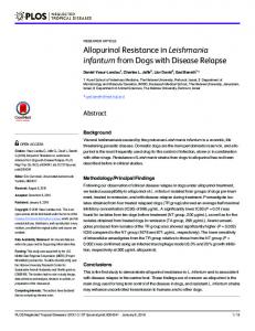
Negative controls (DNA extracted from a male sand ßy and an aliquot of distilled water) were included in each PCR run to detect contamination that could lead to false-positive results; all were found to be negative. A 5-l sample of each PCR product was subjected to electrophoresis in a 1.5% agarose gel. The bands were stained with ethidium bromide and visualized under UV trans-illumination. Parasites were identiÞed by comparing the size of the band produced from a test sample with those produced from the reference strains of L. infantum, L. major, and L. tropica. For example a band of 720 bp (in the nested PCR method) indicated that the parasite was L. infantum. Results A total of 2,539 sand ßies (1,187 males and 1,352 females) were collected from three different locations, among which 11 Phlebotomine species were identiÞed, including six species of Phlebotomus and Þve species of Sergentomyia (Table 1). P. (Phlebotomus) papatasi Scopoli and P. (Larroussius) major Annandale were the Þrst and third most prevalent species in the collection, representing 37.4 and 11.2% of the total sand ßies caught, respectively. Altogether, 47 P. papatasi, 45 P. major, 39 Phlebotomus alexandri Sinton, and 10 Phlebotomus sergenti Parrot were dissected and examined microscopically for promastigotes. The results of the dissection are presented in Table 1. Promastigotes were observed only in two specimens of P. papatasi and three specimens of P. major (Fig. 1). The promastigote-positive P. papatasi specimens were collected outdoors from rodent, burrows on sticky traps, but all of the infected specimens of P. major were caught in and around the VL patients, dwellings using sticky and CDC light traps (Þve and one specimens, respectively). Altogether, 65 P. papatasi, 72 P. major, 32 P. alexandri, and 12 P. sergenti (including slides of dissected
specimens) were checked for Leishmania DNA using a genus-speciÞc PCR (RV1-RV2 primers) and a species-speciÞc nested PCR (primers LIN R4, LIN 17, and LIN 19). Four specimens of P. papatasi and six of P. major were found to be positive for Leishmania DNA using standard PCR. All of them had a 145-bp band indicative of the LT1 fragment in the conserved region of Leishmania kinetoplast DNA (kDNA) minicircles (Fig. 2). The nested PCR method could identify these parasites as Leishmania major and L. infantum with primers LIN R4, LIN 17, and LIN 19. The size of all ampliÞed products from P. papatasi specimens was ⬇560 bp, which was equal to the band size of the L. major standard strain. The visible bands obtained from P. major–infected specimens coincided with those of the L. infantum standard strain, equal to 720 bp. The obtained bands of standard strains of L. major, L. infantum, and L. tropica were 560, 720, and 760 bp, respectively (Fig. 3).
Fig. 1. Geimsa-stained promastigotes of L. infantum from a naturally infected, wild-caught, P. major female sand ßy collected in Ghir-Karzin district, Fars province, 2005.
July 2008
AZIZI ET AL.: PCR-BASED DETECTION OF L. infantum IN P. major
729
Fig. 2. The results of the standard PCR-based ampliÞcation of kinetoplast DNA. The bands correspond to molecular weight marker (lane 1), reference strains of L. infantum and L. major (lanes 2 and 15), samples of P. papatasi (lanes 3Ð7), samples of P. major (lanes 10Ð14), a male sand ßy as control (lane 8), and blank (lane 9).
Discussion Diagnosis of vector species and proper knowledge of their biology are absolutely essential for designing effective control programs for vector-borne diseases. Finding naturally infected, wild-caught specimens is essential evidence in incrimination of a sand ßy species as a vector. However, only parasite isolation and/or identiÞcation can conÞrm the vector. There are two classical procedures used for detection of Leishmania in suspected vectors: microscopic dissection, which is not deÞnitive because most ßagellate
protozoa are morphologically indistinguishable, and culture of isolated parasites, which often does not give results because of failure to grow or contamination (Aransay et al. 2000). Dissection of sand ßies to examine them microscopically for promastigote infections is time-consuming and requires highly skilled microscopists. Molecular methods using different PCR techniques are powerful and efÞcient tools for detection and identiÞcation of Leishmania DNA within sand ßies. The PCR technique is a highly sensitive and powerful
Fig. 3. The results of the nested PCR-based ampliÞcation of kinetoplast DNA. The bands correspond to molecular weight markers (lanes 1 and 8), reference strains of L. tropica (lane 2), L. major (lane 3), L. infantum (lane 4), two of the infected P. major specimens (lanes 6 and 7), and a male sand ßy as control (lane 5). The bands are shown on a 1.5% agarose gel stained with ethidium bromide.
730
JOURNAL OF MEDICAL ENTOMOLOGY
tool, which has been used by others for detecting Leishmania DNA in naturally infected sand ßies (De Bruijn and Barker 1992, Mukherjee et al. 1997, Rodriguez et al. 1999). One of the most suitable targets for PCR is minicircle kinetoplast DNA because of the high number of copies per parasite cell (10,000), and a well-known sequence of the variable region, which has high diversity in different species, making them easily distinguishable (Aransay et al. 2000). In this study, infection of P. major by L. infantum was conÞrmed using both microscopic and molecular methods. P. major has a wide distribution from Morocco to southeast China. It is a main vector of VL in Greece and is also considered to be a vector of VL in other countries in the Mediterranean basin (Hoogstral and Heyneman 1969, Leger et al. 1979). This species has been incriminated as a possible vector of L. infantum in the western Black Sea region of Turkey (Daldal et al. 1998). In Iran, P. major was Þrst reported from northern parts and later from other parts of the country, in all areas where human cases of VL have been reported. Several workers have suspected P. major as the main vector of VL in Iran, on the basis of epidemiological evidence (Javadian and Nadim 1975, Nadim et al. 1978). Sahabi et al. (1992) found 5 of 150 dissected P. major (3.3%) naturally infected with promastigotes in GhirKarzin district (our study area), further implicating this species as a probable vector of VL in Iran (Sahabi et al. 1992). The standard PCR-based assay used in this study was successfully used by Le Fichoux et al. (1999) to amplify Leishmania-kinetoplast minicircle DNA from blood samples of asymptomatic blood donors who lived in an endemic area of VL in southern France. This method ampliÞed the LT1 (145 bp) sequence in the conserved region of Leishmania-kDNA minicircles. Aransay et al. (2000) used the primers LINR4, LIN 17, and LIN 19 in a seminested PCR for ampliÞcation of the variable region of Leishmania-kDNA minicircles in wild populations of P. alexandri, P. tobbi, P. neglectus, P. simici, and P. papatasi in Greece. Their assay was carried out in a single tube, whereas we used two separate tubes for a truly nested PCR. The standard PCR assay was Leishmania genus speciÞc and ampliÞed a 145-bp band in all of our Leishmania reference strains (Fig. 2), whereas the nested PCR ampliÞed the different sized bands for these species and could clearly differentiate among them (The resulting bands were 560, 720, and 760 bp for L. major, L. infantum, and L. tropica, respectively; see Fig. 3). The two PCR methods used in this study have the same sensitivity and efÞciency for detecting Leishmania infections in naturally infected sand ßies and both Leishmania-kDNA in the same infected sand ßies (four specimens of P. papatasi and six specimens of P. major).
Vol. 45, no. 4
All P. major and P. papatasi specimens found infected in this study were parous, indicating longevity sufÞcient to complete the parasite cycle in their bodies. Furthermore, the infected ßies showed no sign of a blood meal, and therefore, when caught, probably harbored promastigotes that had developed from amastigotes ingested, within a blood meal, at least several days earlier (Cihakova and Volf 1997). Based on (1) observations of promastigotes in naturally infected, wild caught, specimens of P. (Laroussius) maor (which were collected from VL patients, homes); (2) identiÞcation of the parasites as L. infantum by species-speciÞc nested PCR; (3) high abundance of this man-biting sand ßy species in this endemic focus (11.2%); (4) absence the other suspected vectors of Leishmania; and (5) failure to Þnd other Phlebotomus species naturally infected with Leishmania parasites, we conclude that P. major should be considered as the primary vector of VL in this endemic focus in Fars province, southern Iran.
Acknowledgments We thank M. Jalali for assistance in sampling, M. Kalantari and M. Karamian for helping with the molecular assays, Shiraz University of Medical Sciences for providing the molecular biology facilities, and M. A. Oshaghi and E. Fazel for revising the article.
References Cited Alexander, B. 2000. sampling methods for Phlebotomine sandßies. Med. Vet. Entomol. 14: 109 Ð122. Alexander, B., and M. Maroli. 2003. Control of Phlebotomine sandßies. Med. Vet. Entomol. 17: 1Ð18. Aransay, A. M., E. Scoulica, and Y. Tselentis. 2000. Detection and identiÞcation of Leishmania DNA within naturally infected sand ßies by seminested PCR on minicircle kinetoplastic DNA. Appl. Environ. Microbiol. 66: 1933Ð 1938. Azizi, K., Y. Rassi, E. Javadian, M. H. Motazedian, S. Rafizadeh, M. R. Yaghoobi Ershadi, and M. Mohebali. 2006. Phlebotomus (Paraphlebotomus) alexandri: a probable vector of Leishmania infantum in Iran. Ann. Trop. Med. Parasitol. 100: 63Ð 68. Cihakova, J., and P. Volf. 1997. Development of different Leishmania major strains in the vector sandßies Phlebotomus papatasi and P. duboscqi. Ann Trop. Med. Parasitol. 91: 267Ð279. Daldal, N., Y. Ozbel, A. Babaoglu, N. Turgay, M. Z. Alkan, N. Babalioglu, and M. A. Ozcel. 1998. Phlebotomus major syriacus: a possible vector of visceral leishmaniasis in western black sea region of Turkey. J. Egypt. Soc. Parasitol. 28: 271Ð275. De Bruijn, M. H., and D. C. Barker. 1992. Diagnosis of new world leishmaniasis: speciÞc detection of species of the Leishmania braziliensis complex by ampliÞcation of kinetoplast DNA. Acta Trop. 52: 45Ð58. Edrissian, G. H., A. Nadim, A. V. Alborzi, and S. Ardehali. 1999. Visceral leishmaniasis: the Iranian experiences. Arch. Iran. Med. 1: 22Ð26. Hoogstral, H., and D. Heyneman. 1969. Leishmaniasis in the Sudan Republic, Þnal epidemiological report. Am Trop. J. Med. Hyg. 18: 1091Ð1210.
July 2008
AZIZI ET AL.: PCR-BASED DETECTION OF L. infantum IN P. major
Javadian, E., and A. Nadim. 1975. Studies on cutaneous leishmaniasis in Khuzestan, Iran. Part II: status of sandßies. Bull. Soc. Path. Ex. 68: 467Ð 471. Kato, H., H. Uezato, K. Katakura, M. Calvopina, J. D. Marco, P. Barroso, E. Gomez, T. Mimori, M. Korenaga, H. Iwata, S. Nonaka, and Y. Hashiguchi. 2005. Detection and identiÞcation of leishmania species within naturally infected sandßies in the Andean areas of Ecuador by a polymerase chain reaction. Am. J. Trop. Med. Hyg. 72: 87Ð93. Lachaud, L., S. Marchergui-Hammami, E. Chabbert, J. Dedet, and P. Bastien. 2002. Comparison of six PCR methods using peripheral blood for detection of canine visceral leishmaniasis. J. Clin. Microbiol. 40: 210 Ð215. Le Fichoux, Y., J. F. Quaranta, J. P. Aufeuvre, A. Lelievre, P. Marty, I. Suffia, D. Rousseau, and J. Kubar. 1999. Occurrence of Leishmania infantum parasitemia in asymptomatic blood donors living in an area of endemicity in southern France. J. Clin. Microbiol. 37: 1953Ð1957. Leger, M., A. Saratsiotis, B. Pesson, and P. Leger. 1979. La leishmaniose en Grece. Resultants dune enquet entomologic effectuee en Juine 1977. Ann. Parasit. Hum. Comp. 54: 11Ð29. Lewis, D. J. 1982. A taxonomic review of the genus Phlebotomus (Diptera: Psychodidae). Bull. Br. Mus. Nat. Hist. Entomol. 45: 121Ð209. Mohebali, M., H. Hajjaran, Y. Hamzavi, I. Mobedi, S. Arshi, Z. Zarei, B. Akhundi, K. Manouchehri, R. Avizeh, and M. Fakhar. 2005. Epidemiological aspects of canine visceral leishmaniasis in the Islamic Republic of Iran. Vet. Parasitol. 129: 243Ð251. Mukherjee, S., M. Q. Hassan, A. Ghosh, A. Bhattacharya, and S. Adhya. 1997. Leishmania DNA in Phlebotomus and
731
Sergentomyia species during a kala-azar epidemic. Am. J. Trop. Med. Hyg. 57: 423Ð 425. Nadim, A., A. Navid-Hamidi, E. Javadian, G. TahvildariBidruni, and H. Amini. 1978. Present status of kala-azar in Iran. Am. J. Trop. Med. Hyg. 27: 25Ð28. Rassi, Y. 1997. Natural promastigote infection of sand ßies and its Þrst occurrence in Sergentomyia dentata in Ardabil province, north west of Iran. Iran. J. Publ. Health. 26: 7Ð12. Rassi, Y., E. Javadian, A. Nadim, A. Zahraii, H. Vatandoost, M. H. Motazedian, K. Azizi and M. Mohebali. 2005. Phlebotomus (Larroussius) kandelakii the principle and proven vector of visceral leishmaniasis in north west of Iran. Pak. J. Biol. Sci. 8: 1802Ð1806. Rodriguez, N., C. M. Aguilar, M. A. Barrios, and D. C. Barker. 1999. Detection of Leishmania braziliensis in naturally infected individual sandßies by the polymerase chain reaction. Trans. Roy. Soc. Trop. Med. Hyg. 93: 47Ð 49. Sahabi, Z., M. A. Seyyedi-Rashti, A. Nadim, E. Javadian, M. Kazemeini, and M. R. Abai. 1992. A preliminary report on the natural leptomonad infection of Phlebotomus major in an endemic focus of V.L. in Fars province, south of Iran. Iran. J. Publ. Health. 21: 87Ð93. Seyyedi-Rashti, M. A., Z. Sahabi and A. Kanani-Notash. 1995. Phlebotomus (Larroussius) keshishiani; Shchurenkova 1936, another vector of visceral leishmaniasis in Iran. Iran. J. Publ. Health. 24: 23Ð30. Shyam, S., and M. Rai. 2002. Laboratory diagnosis of visceral leishmaniasis. Clin. Diag. Lab. Immunol. 9: 951Ð958. Smart, J. 1965. A handbook for the identiÞcation of insects of medical importance, 4th ed. British Museum (Natural History), London, United Kingdom. Received 27 May 2007; accepted 17 March 2008.