Journal of Electron Microscopy Advance Access published December 14, 2012 Journal of Electron Microscopy 0(0): 1–12 (2012) doi: 10.1093/jmicro/dfs083 ........................................................................................................................................................................................................................................................
Review
Fluorescence imaging of synapse formation and remodeling Shigeo Okabe* Department of Cellular Neurobiology, Graduate School of Medicine, The University of Tokyo, 7-3-1 Hongo Bunkyo-ku, Tokyo 113-0033, Japan *To whom correspondence should be addressed. E-mail:
[email protected] ..............................................................................................................................................................................................
Abstract
..............................................................................................................................................................................................
Keywords
live-cell imaging, fluorescent proteins, synapse, postsynaptic density, glutamate receptors, two-photon microscopy, super-resolution microscopy
..............................................................................................................................................................................................
Received
26 September 2012, accepted 9 November 2012
..............................................................................................................................................................................................
Introduction In the central nervous system (CNS) of vertebrates, neuronal precursors are differentiated from the neuroepithelial cells, migrate within the nervous tissue and settle within specific cortical layers or nuclei. After migration, neurons extend long axonal processes and start to connect with their target neurons. The contact sites between presynaptic axons and postsynaptic dendrites start to differentiate into synapses, which are the structures important in information processing and memory storage.
Electrophysiological and structural properties of mature synapses were studied extensively in the late 20th century. However, appropriate techniques for the reliable detection and measurement of nascent synapses during development had not been available until recently. Therefore, researchers initially focused on several types of model synapses in the peripheral nervous system, such as the neuromuscular junctions (NMJs), which were larger than synapses in the CNS and easier to manipulate experimentally [1]. Nascent NMJs could be identified
........................................................................................................................................................................................................................................................
© The Author 2012. Published by Oxford University Press [on behalf of Japanese Society of Microscopy]. All rights reserved. For permissions, please e-mail:
[email protected]
Downloaded from http://jmicro.oxfordjournals.org/ at University of Tokyo Library on December 16, 2012
Brain function is based on proper connectivity between neuronal cells. In the developing brain, neurons extend axons and form synaptic connections with appropriate postsynaptic neurons. Molecular mechanisms underlying establishment of proper synaptic connections are one of the most important topics in the field of developmental neurobiology. Dynamics of synaptic structure and local recruitment of synaptic molecules can be studied by live-cell imaging of neurons expressing fluorescent probes of synaptic molecules. In this review, examples of live-cell fluorescence imaging are presented and their contributions to our understanding about the molecular mechanisms of synapse formation and remodeling are discussed. Imaging of synaptic proteins in living neurons revealed rapid formation of individual synapses within hours and extensive remodeling of synaptic connections. Different types of neurons express unique protrusions from dendrites and axons, which play important roles in synapse formation and maturation. Rapid formation of synaptic structure is associated with continual assembly and disassembly of synaptic scaffolding proteins, which are essential building blocks of the presynaptic active zone and the postsynaptic density (PSD). Quantitative analyses of PSD scaffolding proteins further confirmed their essential roles in maintenance of the synaptic structure. These examples clearly indicate that fluorescence-based live-cell imaging is an indispensable technique in the research on synapse development and its impact will further increase in combination with development of new light microscopic techniques in the future.
2
J O U R N A L O F E L E C T R O N M I C R O S C O P Y , 2012
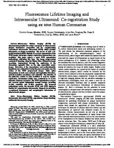
Structural components of synapses and molecules involved in the assembly of presynaptic and postsynaptic specializations Detailed morphological characteristics of synapses have been studied by using transmission electron microscopy of chemically fixed or quickly frozen samples of the nervous tissue (Fig. 1). Synapses are the sites of membrane adhesion between presynaptic axons and postsynaptic dendrites or cell bodies.
Axonal boutons are the cytoplasmic swellings formed at the contact sites between the axonal plasma membrane and the target neurons along the course of axonal trajectories. Axonal boutons contain synaptic vesicles, which are the storage sites of neurotransmitters, such as glutamate and γ-Aminobutyric acid (GABA). Arrival of action potentials to the presynaptic terminal activates voltage-gated calcium channels and triggers exocytosis of synaptic vesicles. Neurotransmitters released from the presynaptic membrane diffuse across the synaptic cleft and activate neurotransmitter receptors on the postsynaptic membrane. Most excitatory synapses in the forebrain pyramidal neurons are formed onto dendritic spines, small protrusions containing densely packed actin filaments [5]. Another morphological feature of excitatory synapses is the presence of the postsynaptic density (PSD), located at the plasma membrane of the dendritic spine and apposed to the presynaptic active zone [6]. The PSD is composed of a variety of proteins, including membrane proteins, such as glutamate receptors and cell adhesion molecules, together with PSD scaffolding proteins and signaling molecules. In addition to the PSD, the postsynaptic cytoplasm contains several other microstructures, such as spine apparatus, endosomal membranes and mitochondria. The sizes of both presynaptic axonal boutons and dendritic spines are in the order of several micrometers. The PSDs are disk-like structures with diameters of 200–500 nm and thicknesses of 30–60 nm [7]. Synapse morphology and distribution of synaptic proteins are difficult to resolve without the aid of fluorescence microscopy. Under optimized imaging conditions, structural distinction between different types of spines, such as thin, stubby and mushroom spines, can be reliably achieved and could be confirmed by retrospective electron microscopy [8,9]. In order to identify nascent synaptic structures, it is essential to develop techniques that enable expression of fluorescent protein (FP)tagged synaptic molecules and subsequent detection of fluorescence signals in living neurons. A number of FP-based probes for presynaptic and postsynaptic components have been developed and utilized for live-cell imaging. Formation and remodeling of presynaptic structures can be monitored
Downloaded from http://jmicro.oxfordjournals.org/ at University of Tokyo Library on December 16, 2012
using specific fluorescent probes, and the mechanisms of synaptic competition between multiple presynaptic axons targeted to the same postsynaptic muscle could be analyzed in detail. In the CNS, however, several factors prevented direct detection and analyses of developing synapses. First, synaptic structures in the CNS are much smaller than NMJs and the resolution of light microscopy is not sufficient for the detection of their detailed morphology. Second, the mammalian brain is covered by the cranium and direct access to the brain parenchyma requires invasive surgery. Because the brain tissue is fragile and easy to develop edema, highly sophisticated surgical procedures should be invented and applied to small animals. To circumvent this difficulty, isolated preparations of the nervous system, such as dissociated neuronal cultures and slice cultures, have been developed and widely used for the analyses of synapse development within the CNS [2,3]. Imaging experiments in these reduced preparations in combination with the development of green fluorescent protein (GFP)-based fluorescent probes provided essential information about the behavior of nascent synapses and the time course of their differentiation. Another possible approach toward the understanding of synapse development in the CNS is the histological analyses of brain sections taken from different stages of brain development. Analyses of synapse development in vivo by electron microscopy revealed structural details of nascent synapses quantitatively [4]. Fluorescence imaging of living neurons and ultrastructural studies of the fixed brain tissue provide us with complementary information and their integration has facilitated our understanding about CNS synapse development.
S. Okabe Fluorescence imaging of synapse formation
3
using GFP-tagged synaptophysin or synaptobrevin2/ VAMP2 [10,11]. Both of these probes show selective accumulation in axonal boutons. Synaptophysin is one of the most abundant proteins of synaptic vesicles. Synaptophysin contains four transmembrane domains with cytoplasmic N- and C-termini. Synaptophysin tagged with GFP at its C-terminus can be properly recruited to the synaptic vesicles and is utilized as a reliable marker of local synaptic vesicle accumulation [11]. Synaptobrevin2/VAMP2 is also an integral synaptic vesicle protein with a single transmembrane domain and a cytoplasmic N-terminal domain. In the process of exocytosis, the assembly of a SNARE (soluble N-ethylmaleimidesensitive factor attachment protein [SNAP] receptor) complex from SNAP-25, syntaxin and synaptobrevin2/VAMP2 is proposed to drive the formation of fusion pore between vesicles and the plasma membrane. Synaptobrevin2/VAMP2 tagged with GFP at its N-terminus is functional and does not perturb synaptic vesicle exocytosis. GFP-tagged synaptobrevin2/VAMP2 is localized at the sites of synaptic vesicle accumulation and can be utilized as a reliable presynaptic marker [10]. Detection of the postsynaptic specialization can be achieved using FP-tagged PSD molecules. 2-amino-3-(3-hydroxy-5-methyl-isoxazol-4-yl)propanoic
acid (AMPA)-type and N-methyl-D-aspartic acid (NMDA)-type glutamate receptors are enriched in the biochemically purified PSD fraction and their postsynaptic localization was confirmed by immunoelectron microscopy. AMPA receptors and NMDA receptors are essential components of glutamate-mediated synaptic transmission and their presence at the postsynaptic sites can be detected by expression of FP-tagged receptors. In the case of AMPA receptors, their distribution on dendritic membrane is diffuse and local density of AMPA receptors on the postsynaptic membrane is not extremely high compared with extrasynaptic receptors [12]. Furthermore, it is proposed that nascent synapses may lack AMPA receptors (silent synapses) and local activation of NMDA receptors triggers recruitment of AMPA receptors to silent synapses [13]. From these considerations, FP-tagged AMPA receptor subunits, such as GFP-GluA1 and GluA2, have not been utilized frequently to detect nascent synapses. However, these GFP-tagged AMPA receptors are useful in the detection of activity-dependent modifications of postsynaptic functions [14]. Recruitment of NMDA receptors to the postsynaptic sites takes place in the early stage of synaptogenesis. NMDA receptors are tetramers composed of two GluN1 subunits and two GluN2
Downloaded from http://jmicro.oxfordjournals.org/ at University of Tokyo Library on December 16, 2012
Fig. 1. Ultrastructure of the excitatory synapse. (a) Transmission electron micrograph of an excitatory synapse in the mouse hippocampus. Scale bar, 200 nm. (b) Major cytoplasmic components of the presynaptic and postsynaptic cytoplasm of the excitatory synapses.
4
J O U R N A L O F E L E C T R O N M I C R O S C O P Y , 2012
subunits. NMDA receptors tagged with GFP at their extracellular N-termini have been utilized for the detection of postsynaptic sites in cultured cortical neurons [15]. PSD scaffolding molecules are highly concentrated in the postsynaptic sites and are good candidates for postsynaptic markers. PSD-95 is one of the most abundant scaffolding molecules in the PSD [16]. PSD-95 can interact directly with NMDA receptors and also indirectly with AMPA receptors through its binding to transmembrane AMPA receptor regulatory proteins. PSD-95 interaction partners are not restricted to glutamate receptors, but include cell adhesion molecules, such as neuroligins and synCAMs, scaffolding molecules, such as guanylate kinase-associated protein (GKAP), and signaling molecules, such as synaptic GTPase-activating protein for Rac (SynGAP), neuronal nitric oxide synthase (nNOS) and spine-associated Rap-Gap (SPAR) [17]. Multiple binding partners of PSD-95 indicate its essential role in PSD organization. PSD-95 with its C-terminal tagged with GFP shows postsynaptic
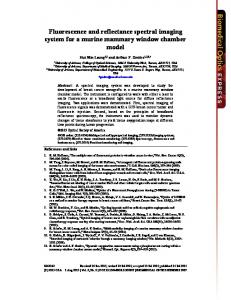
distribution indistinguishable from that of endogenous PSD-95 and has been utilized as a reliable fluorescent marker of the postsynaptic specialization (Fig. 2a) [18,19]. In addition to PSD-95, other GFP-tagged PSD scaffolding proteins, such as GFP-Homer and GFP-Shank, have also been utilized as probes for the PSD detection (Fig. 2b) [20,21]. The basic properties of these scaffolding proteins are similar, with quantitative difference in their local turnover rate and response to neuronal activity. Electron microscopic studies reported ultrastructural difference between asymmetrical (Gray type 1; correspond to excitatory glutamatergic synapses) and symmetrical (Gray type 2; correspond to inhibitory GABAergic synapses) synapses [22]. These structural differences are derived from the difference in thickness of the postsynaptic membrane specialization. Postsynaptic membranes of excitatory synapses develop thicker meshwork of protein assembly and are detected as prominent electrondense structures [6]. Typical PSD proteins, such as PSD-95, Homer and Shank, are specifically
Downloaded from http://jmicro.oxfordjournals.org/ at University of Tokyo Library on December 16, 2012
Fig. 2. Clustering of PSD scaffolding proteins tagged with GFP at the postsynaptic sites and their turnover in living hippocampal neurons in culture. (a) A cultured hippocampal neuron expressing PSD-95-GFP (green) stained with a lipophilic dye DiI (red). Arrows indicate colocalization of PSD-95-GFP puncta (green) and dendritic spines (red). Arrowheads indicate a single straight axon devoid of PSD-95 puncta. Scale bar, 10 μm for the main panel and 4 μm for the lower small images. (b) Formation and elimination of the PSD detected by expression of GFP-tagged Homer1c in cultured hippocampal neurons. Imaging of the same dendritic segments with an interval of 24 h for 6 days revealed continual remodeling of the PSDs, with gradual increase in the total number of GFP-Homer1c clusters. Scale bar, 4 μm. Reprinted from Okabe et al. [19].
S. Okabe Fluorescence imaging of synapse formation
preparations and could quantitatively explain the overall rate of synapse density (Fig. 2b) [18,19]. It is possible to determine the time course of molecular assembly at presynaptic and postsynaptic sites simultaneously by using multicolor imaging of FP-tagged synaptic proteins. We expressed PSD-95 tagged with yellow fluorescent protein (YFP) together with synaptophysin tagged with cyan fluorescent protein (CFP) to determine whether presynaptic and postsynaptic molecules appear at synaptic sites with different time courses [11]. We found a strong temporal correlation between clustering of PSD-95-YFP and that of synaptophysin-CFP at the synaptic contact sites (Fig. 3d and e).
Live-cell fluorescence imaging of synaptic molecules and structure Before GFP technology enabled us to visualize dynamics of synaptic molecules in living neurons, discussions on the time-course of synapse development had been based on the comparison of immunocytochemical and electron microscopic data of fixed preparations. Because the density of synapses and dendritic spines increases gradually in both culture preparations and in vivo, it was widely accepted that the differentiation of individual synapses is also a slow process which may take a few weeks. However, time-lapse imaging of PSD-95 tagged with GFP in hippocampal pyramidal neurons in culture revealed rapid establishment of PSD-95-GFP fluorescent clusters on time scales of several hours [19]. These PSD-95-GFP clusters were apposed to the presynaptic boutons and were selectively localized within dendritic spines, suggesting their identity as synaptic PSD structures [11]. A similar time course of postsynaptic molecular assembly was confirmed by several independent groups and has been taken as convincing evidence for rapid establishment of synaptic specialization [28,29]. If synapse assembly is a rapid process, how is relatively slow increase of overall synaptic density achieved? A possible explanation is that synapse addition and elimination take place simultaneously and the rate of addition is maintained to be moderately higher than the rate of elimination. Indeed, this difference in addition and elimination of PSD-95-GFP clusters existed in culture
Fig. 3. Simultaneous detection of multiple synaptic components by dual-color time-lapse imaging. (a–c) Imaging of the PSDs by expression of PSD-95-yellow fluorescent protein (YFP) (b) together with detection of spine structure by cyan fluorescent protein (CFP) as a volume marker. (a) Overlay of PSD-95-YFP (red) and CFP (green) was also presented. (c) Formation of a PSD-95-YFP cluster (arrows) is coordinated with the enlargement of the spine head. Scale bar, 3 μm. (d–e) Simultaneous imaging of synaptophysin-CFP (d) and PSD-95-YFP. (e) Accumulation of synaptic vesicles detected by synaptophysin-CFP is synchronized with clustering of PSD-95-YFP (arrows). Scale bar, 3 μm. Reprinted from Okabe et al. [11,21].
Downloaded from http://jmicro.oxfordjournals.org/ at University of Tokyo Library on December 16, 2012
accumulated at excitatory asymmetrical synapses and their GFP-tagged probes can be utilized as specific markers of this type of synapses. In turn, GFP-tagged scaffolding proteins showing specific localization at inhibitory postsynaptic sites, such as gephyrin-GFP, can be utilized for the detection of symmetrical synapses in living neurons [23,24]. By using GFP-tagged presynaptic and postsynaptic molecules, time-lapse imaging of living neurons revealed the process of synapse formation and timing of recruitment of synaptic molecules. These analyses revealed basic principles of synapse formation and mechanisms of molecular assembly at the synaptic junctions [11,25–27].
5
6
J O U R N A L O F E L E C T R O N M I C R O S C O P Y , 2012
Roles of dendritic and axonal protrusions in synaptogenesis Neuronal networks in the mammalian cortex are composed of two types of neurons, excitatory pyramidal-shaped neurons and inhibitory neurons. Although glutamatergic excitatory synapses are formed on dendrites of both pyramidal neurons and interneurons, their postsynaptic morphology and molecular composition are distinct. The most obvious difference is the absence of dendritic spines in excitatory synapses on interneuron dendrites [30,31]. Dendritic filopodia from pyramidal neurons are thought to be the precursors of spines and important in searching nearby axons. It has not yet been clarified whether interneuron dendrites
have any searching systems to contact nearby axons. Without such a mechanism to enhance the chance of contacting nearby axons, the ability of interneurons to increase synaptic density should be quite limited. However, previous electron microscopic studies reported that dendritic shafts of mature interneurons are densely covered with glutamatergic synapses, suggesting the presence of interneuron-specific strategy to increase synaptic contacts [32]. To solve this problem, time-lapse imaging of synapse formation on interneuron dendrites was performed and two important observations were made [33]. First, dendritic protrusive activity of interneurons was developmentally regulated. Although dendrites of mature interneurons had few protrusions, immature interneurons expressed numerous dendritic protrusions, which were longer than typical filopodia of pyramidal neurons. Second, PSD-95 clusters were frequently observed on these dendritic protrusions and showed slow translocation toward the base of protrusions (Fig. 4). These observations indicated that dendritic protrusions of interneurons serve as conduits for retrograde translocation of synaptic structure to the parental dendrites. This translocation was dependent on microtubules present within dendritic protrusions and was driven by dynein motor system. These experimental data indicate that the behavior of synaptic structure may differ between different types of synapses even within the same brain area. In the case of pyramidal neurons, the positions of individual synapses along dendritic shafts are precisely determined by the position of initial protrusive activity of dendritic filopodia. On the other hand, the positions of excitatory synapses along interneuron dendrites may be more flexible and the synaptic connectivity can be increased effectively by using long protrusions as synaptic conduits. Assembly of synaptic structure is a stochastic process and the behavior of a large number of synapses with different stages of maturation is difficult to be classified and characterized. If synapse maturation can be synchronized, the time course of synapse development may be determined more precisely and molecular markers and structural features for each developmental stage may be characterized more easily. In the cerebellum,
Downloaded from http://jmicro.oxfordjournals.org/ at University of Tokyo Library on December 16, 2012
Interestingly, appearance of synaptophysin-CFP clusters tends to precede clustering of postsynaptic PSD-95-YFP with time intervals of