Force Fields Including Charge Transfer and Local Polarization Effects: Application to Proteins Containing Multi/Heavy Metal Ions DMITRI V. SAKHAROV,1 CARMAY LIM1,2 1
Institute of Biomedical Sciences, Academia Sinica, Taipei 115, Taiwan 2 Department of Chemistry, National Tsing Hua University, Hsinchu 300, Taiwan Received 2 October 2007; Revised 3 April 2008; Accepted 4 May 2008 DOI 10.1002/jcc.21048 Published online 19 June 2008 in Wiley InterScience (www.interscience.wiley.com).
Abstract: The question whether molecular dynamics (MD) simulations can yield reliable structural and dynamical properties of metalloproteins depend on the accuracy of the force field, i.e., the potential energy function (PEF) and associated parameters modeling the interactions of the metal ion of interest with water and protein ligands. Previously, we had developed a CTPOL PEF for protein simulations of Zn21 bound to Cys2 and/or His0 that includes charge transfer and local polarization effects as well as metal van der Waals parameters that reproduce the structural and thermodynamical properties of 22 dications. Here, we evaluate if the CTPOL PEF and the new metal parameters (referred to as the CTPOLa force field) can be applied to proteins containing polynuclear metal-binding sites and heavy toxic metal ions, using the CdZn2-Cys9 b-domain of rat liver metallothionein-2 and the Hg21-bound 18-residue peptide from MerP as test systems. Using the CTPOLa force field, simulations of the b-domain of rat liver metallothionein-2 totaling 19 ns could preserve the experimentally observed CdZn2–Cys9 complex geometry and overall protein structure, whereas simulations neglecting charge transfer and local polarization effects could not. However, the CTPOLa force field cannot reproduce the experimentally observed linear bicoordination of Hg21 in the MerP peptide without adding an angular restraint to the CTPOL PEF to correct the angle distribution about Hg21. Thus, the force fields presented herein for the group IIB metal ions can be applied to simulation studies of proteins containing polynuclear metal-binding sites and heavy metal ions in aqueous solution. PEF neglecting charge transfer and local polarization effects in conjunction with vdW parameters adjusted to reproduce the structural and thermodynamical properties of only the metal ion in question could not yield an accurate representation of the metal-binding site and overall protein structure. q 2008 Wiley Periodicals, Inc.
J Comput Chem 30: 191–202, 2009
Key words: Zn21; Cd21; Hg21; metalloproteins; metallothionein-2; merP
Introduction The group IIB metal ions are of great interest because Zn21, one of the most abundant divalent metal ions in living organisms,1 is an essential cofactor in many metabolic enzymes and transcription factors.2–8 On the other hand, Cd21 and Hg21, which are in the same periodic group as Zn21, are known to be toxic to living organisms.9,10 One likely cause of cadmium and mercury’s toxicity stems from the high affinity of the ‘‘soft’’ (large, polarizable) Cd21 and Hg21 for the ‘‘soft’’ donor sulfur atoms of cysteines, enabling them to displace the ‘‘borderline’’5 native Zn21 cofactor from Cys-rich binding sites such as Cysrich Zn-finger cores.11,12 In replacing the native Zn21 cofactor, the heavier Cd21 and/or Hg21 may not maintain the active protein conformation (by destroying the native tetrahedral binding-
site geometry, for example), thus disrupting the critical function of essential Zn proteins.7,8,13–15 Notably, a theoretical study has provided a physicochemical basis for why Hg21 could displace the native Zn21 cofactor bound not only to the ‘‘soft’’ sulfur atom, but also to the ‘‘harder’’ nitrogen and oxygen2-containing side chains: relative to Zn21, Hg21 is a far better electronacceptor and can accept more negative charge from the Zn
Additional Supporting Information may be found in the online version of this article. Correspondence to: C. Lim; e-mail:
[email protected] Contract/grant sponsor: NSC; contract/grant number: 94-2113-M-001018
q 2008 Wiley Periodicals, Inc.
192
Sakharov and Lim • Vol. 30, No. 2 • Journal of Computational Chemistry
ligands, enabling it to displace the native cofactor from essential Zn enzymes and ‘‘structural’’ Zn proteins.16 As ‘‘soft’’ toxic metal ions prefer to bind to the ‘‘soft’’ sulfur atoms of cysteines, Cys-rich proteins have been used by living organisms to fight heavy metal intoxication. Cys-rich proteins such as metallothionein are used as traps to sequester nonbiogenic metal ions from the body fluids, thus preventing the toxic metal ions from damaging vital metal-binding sites.17–19 Furthermore, there is increasing evidence that metallothionein plays a role in repairing damaged Zn-binding sites by extracting the toxic metal ion such as Cd21 from the respective binding site and delivering the essential natural cofactor (Zn21) to the same binding site.20 Mammalian metallothionein comprises two metalbinding domains. In rat liver metallothionein, the amino terminal b-domain consists of residues 1–30 with 9 cysteines bound to 3 metal ions (1 Cd21, 2 Zn21), while the C-terminal a-domain consists of residues 31–61 with 11 cysteines bound to 4 Cd21 ions.21 Each metal ion is tetrahedrally coordinated to four deprotonated cysteines (Cys2). Bacteria employ a different detoxification system, involving proteins with a MXCXXC sequence motif (where X is any amino acid, a.a.) to bind toxic metal ions.22 A prototypical protein with the metal-binding MXCXXC motif is the periplasmic protein MerP, which binds Hg21 with high affinity, passing Hg21 to membrane transport proteins (MerT and MerF) that transports it across the cell membrane into the cytoplasm where the mercuric reductase enzyme (MerA) catalyzes the 2-electron reduction of toxic Hg21 to relatively nontoxic volatile Hg0.22,23 Like metallothionein, the MerP protein may also function as a Hg21 sponge to protect components of the periplasm from mercury poisoning.24 The NMR structure of the MerP protein bound to Hg21 (PDB entry 1AFJ) shows that the heavy dication is linearly bound to two Cys2 residues with an average (Cys)S–Hg–S(Cys) angle of 1728.25 Atomic-level information of the structural and dynamical properties of metalloproteins such as metallothionein or MerP can be gained from molecular dynamics (MD) simulations. However, such studies have generally been limited by the lack of force fields that can accurately model metal ions in their biological environment. Force fields widely used in biomolecular simulations such as CHARMM,26 AMBER,27 and GROMOS28 employ the conventional PEF, VCONV[rMe-i(t)], with pairwise Coulomb and 12–6 van der Waals (vdW) energies to describe the interaction energy between the metal ion, Me, and a protein/ solvent atom, i, separated by a distance, rMe-i, at time-step t during the simulation; i.e.,
V CONV ½rMe!i ðtÞ% ¼
X
qMe qi 4pe 0 rMe!i ðtÞ i "! " ! " # rMe!i 12 rMe!i 6 ! þ 4eMe!i rMe!i ðtÞ rMe!i ðtÞ
ð1Þ
In eq. (1), qMe and qi are, respectively, the fixed charges on the metal ion and atom i, e0 is the permittivity of free space, while eMe-i and rMe-i are vdW parameters obtained using traditional combining rules, eMe-i 5 (eMe 3 ei)[1/2] and rMe-i 5 (rMe 1 ri)/2. Notably, the conventional PEF [eq. (1)] does not explic-
itly incorporate polarization and charge transfer effects that are significant for Cys-rich metal-binding sites, as Cys2 has been found to transfer more charge to a given metal ion than other a.a. ligands or water.7,29 Furthermore, the metal vdW parameters (eMe, rMe) are usually adjusted to reproduce the experimentally observed hydration structure and absolute hydration free energy of the ion under study, and are therefore not guaranteed to yield the hydration free energy relative to other ions. The experimental absolute hydration free energy of an ion, however, is considerably less accurate than the hydration free energy relative to that of another ion of the same charge. Recently, we have developed a force field based on the following physical principles and findings: In our previous work,8,30 significant charge transfer from a.a. ligands, especially Cys2, to the Zn cation was found to contribute in part to the decrease in the Zn21 coordination number (CN) from six in aqueous solution to four upon protein binding. However, such ligand?metal charge transfer reduces the magnitude of the partial charges on the ligand atoms and Zn21, which in turn attenuates their charge–charge/dipole interactions, hence local polarization energy of Zn21 and its ligands are needed to compensate for the decreased metal-ligand electrostatic interactions.31 On the basis of these physical principles, charge transfer and local polarization effects have been included in the interaction energy, which is given by31: X qMe ðtÞqi ðtÞ 4pe0 rMe!i ðtÞ i "! "12 ! " # rMe!i rMe!i 6 ! þ 4eMe!i rMe!i ðtÞ rMe!i ðtÞ
V CTPOL ½rMe!i ðtÞ% ¼ V pol ½rMe!i ðtÞ% þ
ð2Þ
The CTPOL PEF in eq. (2) differs from the conventional PEF in eq. (1) in (i) including an additional local polarization energy Vpol[rMe-i(t)] for only the metal ion and the metal-bound side chain atoms, and (ii) taking into account the charge transferred to the metal ion, which is attributed solely to the metal-bound atoms. Thus, the charges on the metal ion and the metal-bound ligand atoms are not fixed, but change during the simulation depending on rMe-i at time-step, t. Apart from formulating a new PEF, a new set of vdW parameters for 22 metal dications, viz., Cu21, Ni21, Pt21, Zn21, Co21, Pd21, Ag21, Cr21, Fe21, Mg21, V21, Mn21, Hg21, Cd21, Yb21, Ca21, Sn21, Pb21, Eu21, Sr21, Sm21, and Ba21 (referred to as MWa metal vdW parameters), have been derived using a numerical procedure that links the coupling parameter used in free energy simulations with the measured hydration free energies. These parameters reproduce the first-shell CNs, average ion-water distances, and/or experimental relative hydration free energies of 22 dications.32 Notably, simulations of a classical Zn-finger have shown that all three factors; viz., charge transfer, local polarization, and appropriate Zn21 vdW parameters, to be important in maintaining the structural integrity of the tetrahedral ZnCys2His2 binding site, i.e., leaving any one factor out leads to a non-tetrahedral Zn–site with the experimental Zn–S(Cys2) and Zn–N(His0) distances overestimated.31
Journal of Computational Chemistry
DOI 10.1002/jcc
Force Fields Including Charge Transfer and Local Polarization Effects
In accord with the above finding, using the conventional PEF [eq. (1)], MD simulations of proteins containing mononuclear and polynuclear metal-binding sites could not maintain the experimental geometry of the metal-binding site. For example, using the conventional PEF and different sets of Zn21 vdW parameters, MD simulations of proteins containing mononuclear Zn–binding sites such as the Zif268 Cys2His2 Zn-finger domain,31 carbonic anhydrase,33,34 farnesyltransferase,35 and phosphotriesterase36 yielded an octahedral or trigonal bipyramidal zinc complex instead of the tetrahedral zinc complex identified in the respective X-ray structures. Using the conventional PEF with GROMOS96 Zn21 vdW parameters28 and estimated Cd21 parameters, a 10-ns simulation of the b-domain of rat liver metallothionein-2 containing a polynuclear CdZn2-Cys9 metal-binding site have been performed; the resulting root-mean-square deviation (RMSD) of the Ca atoms from the starting X-ray struc˚ during the simulation.37 Although using ture rose up to 3 A force fields that employ bonded (as opposed to nonbonded) metal-ligand interactions with a fixed charge on the metal ion such as the Universal force field38 could preserve the observed geometry of the metal-binding site, they would not be suitable in cases where (i) the metal-binding site is conformationally flexible, (ii) the metal CN changes as in the folding/unfolding of Zn-finger proteins or in certain enzymatic reactions, and (iii) ligands such as water molecules undergo exchange during the simulation. On the other hand, using the CTPOL PEF [eq. (2)] and MWa Zn21 vdW parameters,32 a MD simulation of the Cys2His2 Zn-finger domain reproduced the experimentally observed tetracoordinated Zn21 and metal-ligand distances, even when the simulation started from a nontetrahedral Zn21 configuration.31 Hence, it is intriguing to know if simulations using the CTPOL PEF and MWa metal vdW parameters could preserve the experimentally observed geometries of proteins containing polynuclear metal-binding sites, as found for Zn-finger proteins with mononuclear metal-binding sites.31 It is also of interest to know if inclusion of charge transfer and local polarization effects suffices to describe the interactions of the heavier Hg21 ion, which often forms linear complexes with Cys2 residues in proteins such as MerP. The aim of this work is to evaluate whether the CTPOL PEF and the MWa parameters (referred to as the CTPOLa force field) can be applied to proteins containing polynuclear metal-binding sites or heavy toxic metal ions. To evaluate if the CTPOLa force field can be applied to proteins containing multimetal ions, we ˚ have chosen to study the CdZn2-Cys9 b-domain from the 1.6-A X-ray structure of rat liver metallothionein-2 (PDB entry 4MT2),21 as simulation of this domain with the conventional PEF could not preserve the 3-metal binding-site geometry in the X-ray structure (see above).37 To evaluate if the CTPOLa force field can be applied to proteins containing heavy metal ions, we have chosen to study the 18-residue peptide from the MerP protein (corresponding to a.a. 6–23 of the metal-binding loop), whose 3D NMR structure in the presence of Hg21 (PDB entry 1DVW) is very similar to that of the same sequence in the Hg21-bound MerP protein.39 Because no simulations of the Hg21-bound MerP peptide/protein have been reported, it would be interesting to know if simulations employing the CTPOLa force field can reproduce the linear
193
bicoordinate geometry of Hg21 found in the 3D structures of the MerP peptide/protein. Furthermore, it would also be interesting to predict the CNs of Zn21 and Cd21 bound to the MerP peptide, as these two metal ions can bind to the peptide, albeit with lower affinity than Hg21,40 but their CNs have not been experimentally reported.
Methods Estimating the Ligand?Me21 Charge Transfer in Metal Complexes
In previous work,31 we had estimated the amount of charge transferred from water and Cys2 to Zn21 in [Zn (H2O)6]21 and [Zn (CH3S)4]22 complexes. Like Zn21, the heavier Cd21 and Hg21 ions are both experimentally determined to be hexahydrated in aqueous solution41; hence, we estimated the amount of charge transferred from water in hexahydrated Cd21 and Hg21 complexes. In the Cambridge Structural Database42 (CSD), Cd21 is found tetracoordinated to (2SCH2CH2S2)2, whereas Hg21 is found linearly bicoordinated to C2H5S2. Thus, we estimated the amount of charge transferred from Cys2, modeled by methylthiolate (CH3S2), to Cd21 and Hg21 in [Cd (CH3S)4]22 and [Hg (CH3S)2]0 complexes, respectively. The metal complexes were fully optimized using the Gaussian 03 program,43 with the Slater exchange functional44–46 and the Vosko-Wilk-Nusair47 correlation functional (S-VWN) in conjunction with the SDD48,49 basis set for Cd21 and Hg21, and the 6–311G(d)50–52 basis set for the other atoms (referred to as the SDD/6–311G* basis set). The S-VWN/(SDD, 6–311G*) method yields bond distances between the metal ion and the ˚ of the corresponding experimental ligand atom within 0.02-A values (see Table 1). For each fully optimized structure, vibrational frequencies and NBO atomic charges were computed at the same level. Assuming that the charge transferred by each ligand type to the metal ion is the same and is due solely to the
21
Table 1. Charge Transferred from Atom L to Me
Equilibrium Me–L Distance,
eq DrMe! !L .
, Dqeq L!Me , at the
Metal Complexa
Atom L
b ˚ Dreq Me!L (A)
qMec (e)
d Dqeq L!Me (e)
[Cd (H2O)6]21 [Cd (CH3S)4]22 [Hg (H2O)6]21 [Hg (CH3S)2]0
O S O S
2.24 (2.26)e 2.54 (2.52)f 2.32 2.35 (2.34)g
1.69 1.21 1.57 0.83
0.052 0.198 0.072 0.585
a
Geometries fully optimized at the S-VWN/(SDD, 6-311G*) level. Distance between the metal ion Me21 and the ligand atom L in the fully optimized complex structure and in the respective CSD structures in parentheses. c S-VWN/(SDD,6-311G*) NBO charge of the metal ion. d Charge transferred from atom L to the metal ion, Me21, in the equilibrium complex structure. e From CSD entry OJIKOH. f From CSD entry DUFZAF, [Cd(SCH2CH2S)2]22. g From CSD entry MERSET01, [Hg(SCH2CH3)2]0. b
Journal of Computational Chemistry
DOI 10.1002/jcc
194
Sakharov and Lim • Vol. 30, No. 2 • Journal of Computational Chemistry
atoms that are directly coordinated to the metal ion, the charge transferred from a given metal-bound atom to the metal ion can be computed from the charge on the metal ion (see Table 1). For example, the charge transferred to Cd21 by a water molecule, which is attributed solely to the water oxygen, is (2– 1.69)/6 5 0.05e, whereas that transferred by a negatively charged S2 is much greater, (2–1.21)/4 5 0.20e.
mental electrostatic field distribution. In the CTPOLa force field, charge transfer from S(Cys2) to the metal ion reduces their atomic charges, which in turn attenuates their Coulombic interactions. This was compensated by adding back a local polarization energy, Vpol(r), to only the metal ion and the metal-bound a.a. sidechains involved in the charge transfer. Vpol(r) was computed according to: V pol ðrÞ ¼ !
Charge Transfer Model
Because the charges on the metal ion and the metal-bound S(Cys2) atoms in eq. (2) change during the simulation, we need to construct a model to estimate the amount of charge transfer. Our charge transfer model was built on an empirical approach with the following considerations: (i) the number of parameters should be as small as possible, (ii) the charge calculation should be cost-effective, and (iii) the model should be easily generalized to other metalloprotein simulations. As charge transfer does not occur between two noninteracting atoms, but occurs when the metal ion interacts with Cys2, the amount of charge transferred from S(Cys2) to the metal ion, DqS?Me, depends on their interatomic distance at time-step t during the simulation, rMe2S(t). This quantity is known at the equilibrium req Me!S distance (see Table 1). We assume DqS?Me is zero at a distance greater than the sum of the vdW radii of the metal ion and S2, vdW rMe þ rSvdW , where there is no electron cloud overlap. We also assume that DqS?Me depends linearly on the rMe2S distance: DqS!Me ðrMe!S Þ ¼ aS;Me 3 rMe!S ðtÞ þ bS;Me
(3)
The two parameters in eq. (3) can be solved assuming vdW DqS?Me(rMe þ rSvdW ) 5 0 and knowing DqS?Me(req Me!S ). Using the vdW radii in the CHARMM22 force field53 and the req Me!S and Dqeq S!Me values in Table 1 in eq. (3) yields bS,Cd 5 0.65e, ˚ , bS,Hg 5 1.63e, and aS,Hg 5 20.44 e/A ˚ . At aS,Cd 5 20.18 e/A 2 time-step t in the simulation, the charge on S(Cys ) was computed from: qS ðrMe!S Þ ¼ qCHARMM þ DqS!Me ðrMe!S Þ S 2
(4)
qMe ðrMe!S Þ ¼ 2 ! DQðrMe!S Þ
(5)
Note that although the charge of the coordinating atom and the metal ion changes, the net charge of the metal and its ligands is conserved. Computing the Polarization Energy
The conventional force field has partly included polarization effects implicitly since the partial charges of the atoms have been adjusted to reproduce the quantum mechanical or experi-
(6)
where the summation is over the metal and the metal-bound a.a. side chain atoms, li is the dipole induced on atom i and Ei0 is the electrostatic field produced by the current charges at the ith polarizable site. The induced dipole moment is proportional to the total electrostatic field, Ei: li ¼ ai Ei
(7)
where the proportionality constant, ai, is the polarizability of the ith atom. The polarizabilities of the ligating Cys2 side chain atoms are taken from our previous work,31 while those of Zn21 ˚ 3), Cd21 (4.971 A ˚ 3), and Hg21 (8.83 A ˚ 3) are taken (2.294 A 54 from Johnson et al., 1983. The total electric field at the metal ion, EMe, is the vector sum of the field due to the current charges and induced dipoles of the metal-bound a.a side chain atoms j, i.e., EMe ¼ E0Me þ
X j
TMe!j lj ¼
X qj~ rMe!j j
3 rMe!j
X ~ lj þ 3 r Me!j j
! 3~rMe!j~ rMe!j !1 2 rMe!j
ð8aÞ
On the other hand, the total electric field Ei at a metal-bound Cys2 side chain atom i is the sum of the field due to the charge and induced dipole of the metal ion; i.e., ! " ~ lMe 3~ qMe~ ri!Me ri!Me ~ ri!Me Ei ¼ þ ! 1 (8b) 3 3 2 ri!Me ri!Me ri!Me Thus, li ðtÞ ¼ ai Ej 0 þ ai
CHARMM
, equals where the CHARMM charge on S(Cys ), qS 20.80e53; the charge on Cd21 or Hg21, qMe(rMe2S), was computed from the total charge DQ(rMe2S) transferred by all the ligands to the metal ion:
1X l E0 2 i i i
X
Tij lj
(9)
j6¼i
is obtained by solving a set of coupled equations iteratively. For interatomic distances rij less than or equal to the cutoff distance rijcutoff , which is equal to the sum of the vdW radii of atoms i and j scaled by a universal parameter c (5 0.92),55 rij is set equal to rijcutoff to avoid unphysical growth of the induced dipoles at close distances to each other and to the permanent electric charges. CTPOL0 PEF for Hg21
Neither the conventional nor the CTPOL PEF can yield the experimentally observed geometry of Hg21 (see Results), which is linearly coordinated to two Cys2 in the NMR structure of the 18-residue peptide from the MerP protein (PDB entry 1DVW).39 To reproduce the linear coordination of Hg21, an angular
Journal of Computational Chemistry
DOI 10.1002/jcc
Force Fields Including Charge Transfer and Local Polarization Effects
restraint was added to the pairwise VCTPOL(rHg-i) PEF [eq. (2)]. Thus, at time-step t during the simulation, the interaction energy 0 between Hg21 and its ligand atom, i, VCTPOL (rHg-i), was computed as: V
CTPOL
ðrHg!i Þ ¼
X i
V
CTPOL
ðrHg!i Þ þ
X ij
V3 ðrHg!Si ; rHg!Sj ; rSj !Sj Þ
195 a
Table 2. vdW Parameters used in the Simulations.
Atom i Zn21 Cd21 Hg21 S2
ei (kcal/mol)
˚) ri (A
0.1830 0.0395 0.0409 0.47
1.567 2.395 2.369 3.92
(10) a
where VCTPOL(rMe-i) is given by eq. (2) and V3 ðrHg!Si ; rHg!Sj ; rSj !Sj Þ depends on ySi-Hg-Sj, the angle formed by Hg, Si, and Sj, which is approximated by X i;j
V3 ðrHg!Si ; rHg!Sj ; rSj !Si Þ ¼
X ij
kh ðcos hSi !Hg!Sj ! cos h0Si !Hg!Sj Þ2 (11)
In eq. (11), h0Si !Hg!Sj is the equilibrium Si–Hg–Sj angle, equal to 1808, and ky is the respective force constant in kcal/mol/rad2. To obtain the function in eq. (11), one of the methylthiolates in the fully optimized [Hg(CH3S)2]0 complex was fixed, while the other was placed at different orientations by changing the equilibrium Si–Hg–Sj angle in 28 increments. The [Hg (CH3S)2]0 structure corresponding to a given fixed Si–Hg–Sj angle was optimized at the SVWN/(SDD,6–311G*) level with the Hg–S distances constrained to the equilibrium values. The calculated set of interaction energies was then fitted by eq. (11).
Calculations The MD simulations were carried out at physiological pH and a mean temperature of 300 K using a modified CHARMM27 program26 on (1) the 30-a.a. b-domain of the rat liver metallothionein-2 containing 1 Cd21, 2 Zn21, and 383 protein atoms solvated with 3018 water molecules, and (2) an 18-a.a. peptide from MerP containing Zn21/Cd21/Hg21 and 261 protein atoms solvated with 1080 water molecules. Starting Structures
˚ X-ray structure of rat liver metallothionein-221 (PDB The 1.6-A code 4MT2) without the a-domain and one of the energy-minimized NMR structures39 (PDB code 1DVW) of the 18-residue MerP peptide bound to Hg21 were employed as starting structures for the respective simulations. The same NMR structure was also used as the starting point for the simulations of the MerP peptide bound to Zn21 and Cd21, by replacing the Hg21 in the NMR structure with Zn21 and Cd21, respectively. Force Field
All the simulations employed the TIP3P model for the water molecules,56 the CHARMM22 parameters53 for the protein atoms, and the MWa vdW parameters32 for Zn21, Cd21, and Hg21 (see Table 2). Simulation of the b-domain of rat liver metallothionein-2 used the CTPOL PEF, whereas simulations of the metal-bound 18-residue MerP peptide employed both the con-
Metal parameters are taken from Babu & Lim, 2006,32 whereas the Cys2 parameters are from the CHARMM22 force field. 53
ventional and CTPOL PEFs. Simulation of the Hg21-bound MerP peptide additionally used the CTPOL0 PEF [eq. (10)]. As the simulations were carried out at neutral pH, Asp, Glu, and metal-bound Cys residues were deprotonated, whereas Arg and Lys residues were protonated; note that neither the b-domain of rat liver metallothionein-2 nor the MerP peptide contain any His. This resulted in a net charge of 27 for the b-domain of rat liver metallothionein-2 and 0 for the MerP peptide in the absence of metal ions. Bonds involving hydrogen atoms were constrained during the simulations using the SHAKE algorithm.57 The vdW energies ˚ to and electrostatic forces were switched at a distance of 9.5 A ˚ by an atom-based energy-switching function and zero at 11.5 A a force-switching function, respectively. The nonbonded interaction list was updated every five steps (10214 s) using a cutoff of ˚ . The simulations used periodic boundary conditions with 12.5 A a truncated octahedral primary simulation box58 of edge length ˚ for the metallothionein-2 b-domain and 20 A ˚ for equal to 28 A the MerP peptide.
Simulation Protocol
After placing hydrogen atom positions using the HBUILD facility in CHARMM, the hydrogen-built structure was first energy minimized for 1000 steps in the presence of strong harmonic constraints on all heavy atoms to relieve close vdW contacts and strained bond angles. The resulting structure was immersed in the center of a previously equilibrated truncated octahedral box of TIP3P water molecules of density (1 g/cm3, and its orientation in the primary box was optimized to ensure sufficient solvation of all parts of the protein.59 Water molecules whose oxygen ˚ of any protein heavy atoms were atoms were within 2.5 A deleted. The resulting structure was subjected to several steps of minimization using steepest descent followed by adopted-basis Newton Raphson with strong harmonic constraints on all heavy atoms. All the atoms were then propagated according to Newton’s equations of motions using the leapfrog Verlet algorithm with a time step of 2 3 10215 s at a mean temperature of 300 K. To compare with a previous 10-ns simulation of the bdomain of rat liver metallothionein-2, the simulation of this domain was performed for the same simulation length of 10 ns. In addition, nine 1-ns simulations of the b-domain of rat liver metallothionein-2 were also performed. Simulations of the metalbound 18-residue MerP peptide were performed for 400 ps using the conventional PEF and 1 ns with the CTPOL or CTPOL0
Journal of Computational Chemistry
DOI 10.1002/jcc
196
Sakharov and Lim • Vol. 30, No. 2 • Journal of Computational Chemistry
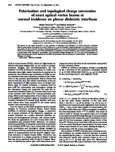
˚ X-ray structure of the b-domain of rat liver metallothionein-2 from PDB entry Figure 1. (a) The 1.6-A 4MT2.21 (b) The respective MD structure averaged over the last 9800 ps of the 10-ns CTPOLa simulation. (c) Comparison of the X-ray structure (in gray) of the CdZn2-Cys9 metal-binding site of rat liver metallothionein-2 (PDB entry 4MT2) with the respective average MD structure (in black) derived from the 10-ns CTPOLa simulation after a translational-rotational fit over the cysteine sulfur atoms. (d) The RMSDs of the protein Ca backbone (solid curve), Cd-Cys4 complex (gray curve), and average Zn-Cys4 complex (dotted curve) from the starting X-ray structure during the 10 ns simulation of the b-domain of rat liver metallothionein-2 using the CTPOLa force field. The RMSDs were calculated after a translation-rotational fit over all Ca atoms excluding the N-terminal Met-1 and Asp-2 residues as well as the last Lys-30 residue.
PEFs. The total energy and temperature remained constant (within 0.5% and 64 K, respectively) during each simulation.
Results and Discussion Convergence of the Metallothionein-2 b-Domain Simulations
To evaluate if the CTPOLa force field can be applied to proteins containing a polynuclear metal-binding site, this force field was used in simulations (totaling 19 ns) of the CdZn2-Cys9 b-domain ˚ of rat liver metallothionein-2 starting from the respective 1.6-A X-ray structure (PDB code 4MT2, see Calculations section). Subsequently, the RMSD of the Ca atoms in each simulation from the starting X-ray structure, omitting the N-terminal Met-1 and Asp-2 residues, which are only weakly defined in the X-ray
structure, as well as the last Lys-30 residue, was computed. The results show that the CTPOLa force field could maintain the structure of the overall protein and the 3-metal binding site: For each of the simulations, the Ca RMSD plateaued after 200 ps, indicating that the simulation has converged (see Fig. 1 and Supplementary Fig. 1). The Ca RMSD fluctuated around a mean of ˚ , which is significantly smaller than the (3 A ˚ RMSD 1.5–2.0-A of all the Ca atoms in the X-ray structure from those in the NMR structure of the Cd3 variant (PDB entry 2MRT).37 The RMSDs of the Cd-Cys4 complex (gray curve) and average ZnCys4 complex (dotted curve) from the starting X-ray structure in the 10-ns simulation fluctuated around a much lower mean of ˚ and 0.31 6 0.09-A ˚ , respectively, as compared to 0.42 6 0.08-A the Ca RMSD, indicating that the 3-metal-binding site is much more rigid than the rest of the protein.
Journal of Computational Chemistry
DOI 10.1002/jcc
Force Fields Including Charge Transfer and Local Polarization Effects
197
Table 3. Comparison Between the Experimental and Computed Average Metal Distances and Angles in the
b-Domain of Rat Liver Metallothionein-2. Method
Metala
˚) \Me2S[ (A
Exptb CTPOLac
Cd-66 Cd-66
CONVgd
Cd-66
2.52 2.47 (2.47 2.26
6 6 6 6
0.03 0.05 0.06) 0.02
2.49 2.43 (2.43 2.24
6 6 6 6
0.00 0.07 0.06) 0.02
2.54 2.50 (2.51 2.27
6 6 6 6
0.00 0.08 0.07) 0.02
111 106 (108 95
6 6 6 6
Exptb CTPOLac
Zn-67 Zn-67
CONVgd
Zn-67
2.41 2.36 (2.36 2.08
6 6 6 6
0.05 0.03 0.03) 0.03
2.36 2.34 (2.34 2.05
6 6 6 6
0.08 0.07 0.06) 0.02
2.45 2.38 (2.38 2.11
6 6 6 6
0.05 0.07 0.06) 0.02
102 101 (102 105
2.36 2.35 (2.35 2.07
6 6 6 6
0.01 0.03 0.04) 0.01
2.37 2.32 (2.32 2.05
6 6 6 6
0.00 0.04 0.04) 0.01
2.35 2.37 (2.38 2.08
6 6 6 6
0.03 0.09 0.04) 0.01
115 106 (106 105
b
Expt CTPOLac
Zn-68 Zn-68
CONVgd
Zn-68
˚) \Me2St[ (A
˚) \Me2Sb[ (A
\S2Me2S[ (8)
\St2Me2St[ (8)
\Sb2Me2Sb[ (8)
2 9 9) 2
113 106 6 9 (107 6 8) 96 6 2
109 106 6 6 (106 6 5) 93 6 2
6 6 6 6
3 9 9) 4
99 107 6 8 (105 6 7) 115 6 6
104 94 6 5 (97 6 5) 94 6 2
6 6 6 6
9 9 7) 3
106 108 6 9 (107 6 8) 112 6 3
123 103 6 7 (101 6 5) 98 6 2
a
The number after the metal ion corresponds to the numbering in the 4MT2 X-ray structure. ‘‘Expt’’ denotes average distances and angles in the 4MT2 structure. c ‘‘CTPOLa’’ denotes distances and angles averaged over each of the 19 simulations (excluding the first 200 ps of equilibration) using the CTPOL PEF [eq. (2)] and the MWa parameters; the numbers in brackets are the respective values derived from the single 10 ns simulation. d ‘‘CONVg’’ denotes distances and angles derived from a 10 ns simulation of the b-domain of rat liver metallothionein-2 using the conventional PEF [eq. (1)] with GROMOS96 Zn21 and estimated Cd21 vdW parameters.37 b
The CTPOLa Force Field Can Reproduce the Multi-Metal Binding-Site Geometry
This is evidenced from comparing the average Cd/Zn–S distance and S–Cd/Zn–S angle derived from the 10 simulations with the respective values obtained from the 4MT2 structure (Table 3). The respective distances and angles derived from the single 10ns simulation are also listed in Table 3 (numbers in parentheses) to compare with those from a 10-ns simulation of the b-domain of rat liver metallothionein-2 in previous work.37 In a polynuclear metal-binding site, two types of sulfur atoms can be distinguished: a terminal sulfur atom, St, coordinated to one metal ion and a bridging sulfur atom, Sb, coordinated to two metal ions. The mean distance from the metal ion (Me) to St and Sb as well as the St–Me–St, Sb–Me–Sb, and Me–Sb–Me, angles, were computed (Table 3). The computed and experimental overall \Me–S[ distances and \S–Me–S[ angles are in close agreement. In particular, the average distance from Cd-66 or Zn-67 to the terminal sulfur St is shorter than that to the bridging sulfur Sb in the CTPOLa simulations, as observed in the X-ray structure. However, the mean angle about the bridging sulfur, \Me–Sb–Me[, which is around 1048 6 28 in the X-ray structure, is significantly overestimated ((1318 6 148) indicating that the CHARMM vdW parameters for S(Cys2) in Table 2 may not be appropriate for the bridging sulfur atom. To ensure that the change in the charges of the coordinating S(Cys2) atoms and the metal ion does not significantly alter the respective torsion angles, we compared the average S–Cd/Zn–S– Cb dihedral angles derived from the mean MD structure with the respective values obtained from the 4MT2 X-ray and 2MRT NMR structures (see Supplementary Table 1). Because the NMR structure corresponds to the Cd3 variant, only the S–Cd66–S–Cb dihedral angles are common to all three structures. The com-
puted S–Cd66–S–Cb dihedral angles are generally closer to the respective X-ray values than the NMR numbers. Because the S– Cd66–S–Cb dihedral angles in the NMR structure of the Cd3 variant deviate by as large as 198 from the respective X-ray values, the average deviation of the computed S–Cd/Zn–S–Cb dihedral angles from the respective X-ray values of 118 seem acceptable. Thus, the CTPOLa force field can preserve the multimetal binding site geometry in the X-ray structure. In contrast to the CTPOLa results shown in Table 3, simulation of the b-domain of rat liver metallothionein-2 using the conventional PEF [eq. (1)] with GROMOS96 Zn21 (eZn 5 0.234 kcal/mol) and estimated Cd21 vdW parameters could not accurately reproduce the experimental structure of the multimetal ˚ ) and Zn–S binding site37: The average Cd-S (2.26 6 0.02 A ˚ ) distances are significantly shorter than the re(2.075 6 0.02 A ˚ and 0.31 A ˚ , respectively, spective X-ray values by about 0.26 A while the mean \S–Cd–S[ angle (958 6 28) is smaller than the X-ray value (1118 6 28).37 Comparison of these results with those from the 10-ns CTPOLa simulation suggest that including charge transfer and local polarization effects as well as appropriate metal vdW parameters are important in maintaining the structural integrity of the 3-metal binding site and overall protein structure. The CTPOLa Force Field Can Preserve the Observed Hydrogen-Bonding Pattern
To further validate the CTPOLa force field, the percentage occurrences of hydrogen bonds found in the 4MT2 X-ray structure of the rat liver metallothionein-2 b-domain during the simulations were computed. To compare with the respective results from previous work,37 we employed the same criteria for the existence of a hydrogen bond, i.e., the hydrogen-acceptor distance ˚ and the donor-hydrogen-acceptor angle is [1358. is \2.5-A
Journal of Computational Chemistry
DOI 10.1002/jcc
198
Sakharov and Lim • Vol. 30, No. 2 • Journal of Computational Chemistry Table 4. Hydrogen Bonds Present in the 4MT2 X-ray Structure and Their % Occurrence in Simulations Using the
CTPOLa/Conventional Force Fields.a Hydrogen bond Asn-4 HN) ) )Asp-2 Od Ser-12 HN) ) )Asp-10 Od Ala-16 HN) ) )Ser-28 Oc Ser-18 HN) ) )Cys-15 O Gln-23 HN) ) )Asn-4 O Lys-25 HZ) ) )Gln-23 O Lys-25 HZ) ) )Asp-2 Od Ser-28 Hc) ) )Cys-13 O Cys-29 HN) ) )Cys-26 O
X-Ray (%)b
NMR (%)c
100 100 100 100 100 100 100 100 100
– 100 – – – – – – –
CTPOLa (%)d 71 64 – 7 31 57 88 21 14
(89) (39) (–) (12) (84) (86) (91) (27) (7)
GROMOS (%)e 87 17 4 – 86 – 13 5 17
a
A dash denotes an absence of the hydrogen bond in the NMR/simulation structures. ‘‘X-ray’’ denotes hydrogen bonds in the original 4MT2 X-ray structure, which were computed by adding missing hydrogen atoms and minimizing them for 10 steps using steepest descent with the backbone and side chain heavy atoms fixed. c ’’NMR’’ denotes hydrogen bonds present in the 2MRT NMR structure. d Average percentage occurrence of the hydrogen bonds computed from each of the 19 simulations (excluding the first 200 ps of equilibration); the numbers in brackets are the respective values derived from the single 10 ns simulation. e Percentage occurrence from a 10-ns simulation reported by Berweger et al.37 b
According to these two criteria, nine hydrogen bonds are present in the 4MT2 X-ray crystal structure, while only the Ser-12 HN) ) )Asp-10 Od hydrogen bond is preserved in the corresponding 2MRT NMR solution structure of the Cd3 variant (Table 4). Simulations using the CTPOLa force field better preserve the hydrogen-bonding pattern seen in the X-ray structure than the respective simulation using the conventional force field. Only one X-ray hydrogen bond (Ala-16 HN-Ser-28 Oc) was lost and two occurred infrequently (\20%) during the CTPOLa simulations, whereas two hydrogen bonds were lost, and five occurred infrequently (\20%) during the corresponding conventional simulation.37 Importance of Charge Transfer and Local Polarization
The above structural analyses indicate that charge transfer effects are significant. Although the charges on the three metals are fixed at 12e using the conventional force field, the mean charge on Cd-66, Zn-67, and Zn-68 is 1.18e, 1.18e, and 1.16e, respectively during the CTPOLa simulations. The fluctuation in the charge transfer amount is relatively small, 60.04e, consistent with the small Me–S distance variation (Table 3). Although the mean distance from the metal to the bridging Sb is longer than that to the terminal St in the CTPOLa simulations, resulting in less charge transferred by Sb compared to St, the mean charge on Sb (20.41e) is less than that on St (20.58e), as Sb has to transfer charge to two metal ions. Despite the sizable magnitude of the charge transfer, including charge transfer effects alone would not be sufficient to reproduce the experimentally observed metal-binding site. This is because charge transfer reduces the absolute charges on the metal ion and S(Cys2), thus, attenuating their charge–charge interactions. To compensate for the decreased electrostatic interactions, the local polarization energy of Me21 and its ligands, which also depend on their partial charges, have to be included.
In fact, our previous simulations of a classical Cys2His2 Zn-finger domain showed that including charge transfer but omitting local polarization leads to a non-tetrahedral Zn–site with the experimental Zn–S(Cys2) and Zn–N(His0) distances overestimated (see Introduction).31 Charge Transfer and Local Polarization Cannot Yield a Linearly Coordinated Hg21
To evaluate if the CTPOLa force field can be applied to proteins containing heavy toxic metal ions such as Hg21, simulations of the Hg21–bound 18-residue MerP peptide were carried out with both the conventional and CTPOL PEFs [eqs. (1) and (2)] and the MWa vdW parameters for Hg21 in Table 2. Neither the conventional nor the CTPOL PEF with the MWa parameters could yield a linearly coordinated Hg21. Although the experimental (Cys)S–Hg–S(Cys) angle is 1628 in the NMR structure of the Hg21–bound 18-residue MerP peptide (1DVW) and 1728 in the X-ray structure of the Hg21–bound 72-residue MerP protein (1AFJ), the average (Cys)S–Hg–S(Cys) angle is 1278 in simulations with the conventional or CTPOL PEF and the MWa Hg21 vdW parameters (Table 5). Furthermore, the Hg21 is bound not only to the cysteines (Cys9 and Cys12) in the metal-binding loop, but it is also coordinated to three water molecules at a dis˚ so that its CN is 5. tance of 2.13 A Although NMR refinement of the MerP peptide employed the CHARMM force field,39 which does not include charge transfer and polarization effects, simulations excluding these effects yield ˚ ) that deviate from the experia Hg–S distance (2.53 6 0.09 A ˚ mental value (2.33 6 0.12 A), whereas simulations incorporating charge transfer and polarization effects yield a Hg–S ˚ ) within experimental error (Table 5). distance (2.44 6 0.07 A The \Hg–S[ distance in the CTPOLa simulation is shorter than that in the CONVa simulation due to polarization rather than charge transfer effects, as the average charge on Hg during the
Journal of Computational Chemistry
DOI 10.1002/jcc
Force Fields Including Charge Transfer and Local Polarization Effects
199
Table 5. The CNs and Average Metal Distances and Angles Derived from Simulations of the 18-Residue
MerP Peptide Bound to Hg21, Cd21, and Zn21 Using Various Force Fields. Metal
Method
s (ps)
CN
Hg21 Hg21 Hg21 Hg21 Cd21 Cd21 Zn21 Zn21
CONVaa CTPOLab CTPOL0 ac Exptd CONVaa CTPOLab CONVaa CTPOLab
400 1000 1000 2 400 1000 400 1000
5 5 2 2 5 5 5 4
˚) Ca RMSD of a.a. 6212 (A 2.14 6 2.12 6 1.03 6 2 2.13 6 2.08 6 2.18 6 1.93 6
0.32 0.47 0.19
˚) \Me2S[ (A 2.53 2.44 2.44 2.33 2.58 2.50 2.34 2.19
0.36 0.24 0.39 0.34
6 6 6 6 6 6 6 6
˚) \Me2O[ (A 2.13 6 2.13 6 3.05 6 2 2.17 6 2.18 6 1.98 6 1.98 6
0.09 0.07 0.09 0.12 0.06 0.05 0.08 0.04
0.19 0.15 0.46 0.05 0.06 0.06 0.05
\S2Me2S[ (0) 127 127 170 162 128 128 112 132
6 6 6 6 6 6 6 6
12 13 17 6 09 10 11 12
a
‘‘CONVa’’ denotes distances and angles averaged over the last 200-ps simulation of the MerP peptide using the conventional PEF [eq. (1)] and the MWa metal vdW parameters in Table 2. b ‘‘CTPOLa’’ denotes distances and angles averaged over the last 800-ps simulation of the MerP peptide using the CTPOL PEF [eq. (2)] and the MWa parameters. c ‘‘CTPOLa0 ’’ is the same as CTPOLa except that an angular restraint [eq. (11)] is added to the CTPOL PEF so that eq 10 is used instead of eq. (2). d ‘‘Expt’’ denotes average distances and angles in the 1DVW NMR structure of the Hg212bound MerP peptide.
CTPOLa simulation (0.89 6 0.04e) is almost half the fixed charge (62e) in the CONVa simulation, which would attenuate the Hg–S distance. The CTPOL0 PEF Yields a Linearly Coordinated Hg21
The missing physics in the empirical PEF that prevents a proper prediction of the Hg21 linear geometry is thought to be relativistic effects, which results in a large 6s–6p energy gap that limits effective hybrid orbital formation to linear sp.60 Thus, to reproduce the observed linear coordination of Hg21 to Cys9 and Cys12, an angular restraint [eq. (11)] was incorporated in the CTPOL PEF, as described in the Methods section. Using this modified CTPOL0 PEF [eqs. (10) and (11)] and MWa Hg21 vdW parameters (referred to as CTPOL0 a), Hg21 became linearly coordinated to Cys9 and Cys12 with an average (Cys)S–Hg–S(Cys) angle of 1708 in the 1 ns simulation. Despite changes in the charges of the coordinating atoms and Hg, the SCys12–Hg–SCys9–Cb and SCys9– Hg–SCys12–Cb dihedral angles in the mean MD structure (1698 and 2628) are close to the respective values averaged over nine NMR structures (1728 and 2528) (see Table 6). The CTPOL0 PEF yields not only the observed linear bicoordination of Hg21, but also a more faithful experimental metalbinding loop structure, as shown in Figure 2. Furthermore, the
RMSD of the Ca atoms of residues 6–12 in the CTPOL0 a simu˚ , which is less lation from the respective NMR structure is (1-A than half of that in the respective simulation with the conven˚ , Table 5). It also better preserves tional or CTPOL PEF ([2 A the two hydrogen bonds found in the NMR structure of the Hg21–bound MerP peptide (1DVW) than the conventional or CTPOL PEF, as shown in Table 7. CN of Hg21
Because rapidly exchanging water molecules would not be detectable by NMR, it is not clear if the Hg21 CN could be greater than 2. By averaging the Hg–O(water) distances of all water ˚ of the metal ion in the CTPOL0 a simulamolecules within 4 A ˚ from Hg21 tion, water oxygen atoms were found more than 3 A (see Table 5). This distance is significantly longer than the ˚ in aqueous solution,41 observed Hg–O(water) distance of 2.33 A indicating that the water molecules are not directly bound to Hg21. Interestingly, the measured Hg–S distance in the 18-resi˚ , 1DVW) or MerP protein due MerP peptide (2.33 6 0.12 A ˚ , LAFJ), where Hg21 is bound to two Cys2, is (2.33 6 0.12 A similar to that in the homologous CSD structure (MERSET01) ˚ ) and no other where Hg21 is bound to two ethylthiolates (2.34 A ligands such as water molecules. As the bond distances to the
Table 6. Comparison Between the Experimental And Computed Average Dihedral Angles About the Hg2S Bond in the 18-Residue MerP Peptide.
NMR1
NMR2
NMR3
NMR4
NMR5
NMR6
NMR7
NMR8
NMR9
NMR10
NMRa
MDb
SCys122Hg2SCys92Cb
167.9
172.6
170.5
173.3
174.2
176.5
168.5
175.4
170.9
223.0
169.3
SCys92Hg2SCys122Cb
244.7
251.8
245.8
250.9
256.0
259.8
249.0
258.5
249.9
285.2
152.7 172.2 255.2 51.8
Dihedral angle
a
The dihedral angle in regular font is obtained by averaging the angles in all 10 NMR structures in PDB entry 1DVW, whereas that in bold font is obtained by averaging the angles in all but the NMR10 structure. b Dihedral angles computed from the mean structure derived from the CTPOL0 a simulation.
Journal of Computational Chemistry
DOI 10.1002/jcc
261.9
200
Sakharov and Lim • Vol. 30, No. 2 • Journal of Computational Chemistry
Figure 2. (a) Comparison of the NMR structure (in gray) of the Hg21–bound 18-residue MerP peptide (PDB entry 1DVW) with the respective average MD structure (in black) derived from the CTPOL0 a simulation after a translational-rotational fit over the backbone heavy atoms, Hg21, and the Cys side chain heavy atoms. (b) Comparison of the NMR structure (in gray) of the Hg21–binding site with the respective average MD structure (in black) of the Hg21–binding site derived from the CTPOL0 a simulation after a translational-rotational fit over Hg21 and the Cys side chain heavy atoms.
metal ions are known to be sensitive to the coordination number,61 the Hg21 CN is predicted to be equal to two from the CTPOL0 a simulation. CN of Cd21 and Zn21
Because the CTPOLa force field can preserve the experimentally observed structure of (1) the classical Zn–Cys2His2 and Zn–Cys4 Zn-finger domains, as shown in our previous work,31 and (2) the CdZn2–Cys9 b-domain of rat liver metallothionein-2 (see above), it was used to predict the CNs of Zn21 and Cd21 bound to the 18-residue MerP peptide, as the respective NMR data cannot identify the number and type of additional metal ligands except for the two metal-bound cysteines comprising the MXCXXC motif.40 The CTPOLa simulations predict a CN of five for Cd21, but four for the smaller Zn21 ion. In the CTPOLa simulations, Cd21 is pentacoordinated to the Cys9 and Cys12 sulfur atoms and three water molecules at average distances of 2.50 ˚ , respectively, whereas Zn21 is tetracoordinated to and 2.18-A the same sulfur atoms and two water molecules at average dis˚ , respectively (see Fig. 3 and Table 5). tances of 2.19 and 1.98-A When charge transfer and local polarization were neglected in
the simulations, Cd21 remains pentacoordinated, whereas an extra water molecule becomes coordinated to Zn21 so that its CN is five (Table 5, CONVa). The latter is partly because excluding charge transfer from the two Cys2 ligands to Zn21 in simulations using the conventional PEF allows zinc to retain the full 12e charge, thus attracting an additional (third) water molecule to Zn21.
Conclusions The results for the CdZn2–Cys9 b-domain of rat liver metallothionein-2 (Fig. 1 and Table 3) show that the CTPOLa force field can be applied to simulations of proteins containing not only a single metal ion,31 but also to proteins containing polynuclear metal-binding sites in aqueous solution. Using a PEF that includes charge transfer and local polarization effects [eq. (2)] as well as metal vdW parameters that are consistent with the
Table 7. Hydrogen Bonds Present in the 1DVW NMR Structure and
Their % Occurrence in the Simulations Using the Conventional, CTPOLa, and CTPOL0 a Forcefields.
H-bond Cys-12 O) ) )Thr-15 HN Cys-12 O ) ) )Val-16 HN
NMR (%)
CONVa (%)
CTPOLa (%)
CTPOL0 a (%)
100 100
18 29
11 25
89 31
Figure 3. The average MD structures of the Zn21–binding site (left) and the Cd21–binding site (right) derived from CTPOLa simulations of the MerP peptide bound to Zn21 and Cd21, respectively.
Journal of Computational Chemistry
DOI 10.1002/jcc
Force Fields Including Charge Transfer and Local Polarization Effects
structural and thermodynamical properties of 22 dications,32 simulations of the b-domain of rat liver metallothionein-2 could maintain the overall protein structure and CdZn2–Cys9 complex geometry seen in the respective X-ray structure (PDB entry 4MT2). In contrast, using the conventional PEF, simulation of the same domain produced a too compact CdZn2–Cys9 metal cluster (i.e. too short Cd/Zn–S bonds) with a partially incorrect geometry. These results underline the importance of charge transfer and local polarization effects and appropriate metal vdW parameters in maintaining the structural integrity of polynuclear metal-binding sites. Although the CTPOLa force field can be applied to simulations of proteins containing Cd21 and/or Zn21, inclusion of charge transfer and local polarization effects does not suffice to describe the linear bicoordinate geometry of the heavy Hg21 often observed in mercury-binding proteins. Thus, we have introduced a new CTPOL0 PEF [eq. (10)], which includes an additional angular restraint term [eq. (11)], to correct the description of the angle distribution about Hg21. Using this new CTPOL0 PEF and the MWa parameters for Hg21, a one ns simulation of the 18-residue MerP peptide bound to Hg21 could reproduce the experimentally observed metal-binding loop structure and linear bicoordination of Hg21 (Fig. 2 and Table 5). It also predicts a CN of two for Hg21, while CTPOLa simulations of the Cd21 and Zn21–bound MerP peptide yield a predicted CN of five for Cd21 and four for Zn21 (see Fig. 3). This shows that the same metal-binding loop can bind metal ions with different CNs in different conformations.
References 1. 2. 3. 4. 5. 6. 7. 8. 9. 10. 11. 12. 13. 14. 15.
16. 17. 18. 19.
Sun, G.; Budde, R. J. A. Biochemistry 1999, 38, 5659. Vallee, B. L.; Auld, D. S. Proc Natl Acad Sci USA 1990, 87, 220. Christianson, D. W. Adv Protein Chem 1991, 42, 281. Coleman, J. E. Annu Rev Biochem 1992, 61, 897. Lippard, S. J.; Berg, J. M. Principles of Bioinorganic Chemistry; University Science Books: Mill Valley, California, 1994. Lipscomb, W. N.; Strater, N. Chem Rev 1996, 96, 2375. Dudev, T.; Lim, C. Chem Rev 2003, 103, 773, J Chin Chem Soc 2003, 50, 1093. Dudev, T.; Lim, C. Annu Rev Biophys 2008, 37, 97. Jin, G.; Inoue, S.; Urano, T.; Cho, S.; Ouchi, Y.; Cyong, J. Toxicol Appl Pharmacol 2002, 185, 98. Hess, E. V. Toxicology 2002, 181, 65. Asmuss, M.; Mullenders, L. H. F.; Hartwig, A. Toxicol Lett 2000, 112/113, 227. Razmiafshari, M.; Kao, J.; d’Avignon, A.; Zawia, N. H. Toxicol Appl Pharmacol 2001, 172, 1. Predki, P. F.; Sarkar, B. J Biol Chem 1992, 267, 5842. Hartwig, A. Antioxid Redox Signal 2001, 3, 625. Hartwig, A.; Asmuss, M.; Blessing, H.; Hoffmann, S.; Jahnke, G.; Khandelwal, S.; Polzer, A.; Burkle, A. Food Chem Toxicol 2002, 40, 1179. Tai, H.-C.; Lim, C. J Phys Chem A 2006, 110, 452. Moteki, S. A.; Huang, M.; Shaw, C. F., III; Petering, D. H. J Inorg Biochem 1999, 74 239. Huan, Y.; Chu, D. Y.; Tang, Y.; Cao, W. Acta Phys Chim Sinica 2000, 16, 764. Petering, D. H.; Huang, M.; Moteki, S.; Shaw, C. F., III. Mar Environ Res 2000, 50, 89.
201
20. Roesijadi, G. Cell Mol Biol 2000, 46, 393. 21. Robbins, A. H.; McRee, D. E.; Williamson, M.; Collett, S. A.; Xuong, N. H.; Furey, W. F.; Wang, B. C.; Stout, C. D. J Mol Biol 1991, 221, 1269. 22. Opella, S. J.; DeSilva, T. M.; Veglia, G. Curr Opin Chem Biol 2002, 6, 217. 23. Nascimento, A. M. A.; Chartone-Souza, E. Genet Mol Res 2003, 2, 92. 24. Powlowski, J.; Sahlman, L. J Biol Chem 1999, 274, 33320. 25. Steele, R. A.; Opella, S. J. Biochemistry 1997, 36, 6885. 26. Brooks, B. R.; Bruccoleri, R. E.; Olafson, B. D.; States, D. J.; Swaminathan, S.; Karplus, M. J Comput Chem 1983, 4, 187. 27. Cornell, W. D.; Cieplak, P.; Bush, B. L. Comput Phys Commun 1995, 91, 1. 28. van Gunsteren, W. F.; Billeter, S. R.; Eising, A. A.; Hunenberger, P. H.; Kruger, P.; Mark, A. E.; Scott, W. R. P.; Tironi, I. G. vdf Hochschulverlag AG an der ETH: Zurich, 1996. 29. Dudev, T.; Lim, C. J Phys Chem B 2001, 105, 4446. 30. Dudev, T.; Lim, C. J Am Chem Soc 2002, 124, 6759. 31. Sakharov, D.; Lim, C. J Am Chem Soc 2005, 127, 4923. 32. Babu, C. S.; Lim, C. J Phys Chem A 2006, 110, 691. 33. Liang, J.-H.; Lipscomb, W. N. Proc Natl Acad Sci USA 1990, 87, 3675. 34. Pang, Y.-P. J Mol Model 1999, 5, 196. 35. Pang, Y.-P.; Xu, K.; El Yazal, J.; Prendergast, F. G. Protein Sci 2001, 9, 1857. 36. Pang, Y.-P. Proteins: Struct Funct Genet 2001, 45, 183. 37. Berweger, C. D.; Thiel, W.; van Gunsteren, W. F. Proteins: Struct Funct Genet 2000, 41, 299. 38. Rappe, A. K.; Casewit, C. J.; Colwell, K. S.; Goddard, W. A., III; Skiff, W. M. J Am Chem Soc 1992, 114, 10024. 39. Veglia, G.; Porcelli, F.; De Silva, T. M.; Prantner, A. M.; Opella, S. J. J Am Chem Soc 2000, 122, 2389. 40. DeSilva, T. M.; Veglia, G.; Porcelli, F.; Prantner, A. M.; Opella, S. J. Biopolymers 2002, 64, 189. 41. Ohtaki, H.; Radnai, T. Chem Rev 1993, 93, 1157. 42. Allen, F. H. Acta Crystallogr Sect B 2002, 58, 380. 43. Frisch, M. J.; Trucks, G. W.; Schlegel, H. B.; Scuseria, G. E.; Robb, M. A.; Cheeseman, J. R.; Montgomery, J. A., Jr.; Vreven, T.; Kudin, K. N.; Burant, J. C.; Millam, J. M.; Iyengar, S. S.; Tomasi, J.; Barone, V.; Mennucci, B.; Cossi, M.; Scalmani, G.; Rega, N.; Peterson, G. A.; Nakatsuji, H.; Hada, M.; Ehara, M.; Toyota, K.; Fukuda, R.; Hasegawa, J.; Ishida, M.; Nakajima, T.; Honda, Y.; Kitao, O.; Nakai, H.; Klene, M.; Li, X.; Knox, J. E.; Hratchian, H. P.; Cross, J. B.; Adamo, C.; Jaramillo, J.; Gomperts, R.; Stratmann, R. E.; Yazyev, O.; Austin, A. J.; Cammi, R.; Pomelli, C.; Ochterski, J. W.; Ayala, P. Y.; Morokuma, K.; Voth, G. A.; Salvador, P.; Dannenberg, J. J.; Zakrzewski, V. G.; Dapprich, S.; Daniels, A. D.; Strain, M. C.; Farkas, O.; Malick, D. K.; Rabuck, A. D.; Raghavachari, K.; Foresman, J. B.; Ortiz, J. V.; Cui, Q.; Baboul, A. G.; Clifford, S.; Cioslovski, J.; Stefanov, B. B.; Liu, G.; Liasehenko, A.; Piskorz, P.; Komaromi, I.; Martin, R. L.; Fox, D. J.; Keith, T.; Al-Laham, M. A.; Peng, C. Y.; Nanayakkara, A.; Challacombe, M.; Gill, P. M. W.; Johnson, B.; Chen, W.; Wong, M. W.; Gonzalez, C.; Pople, J. A. Gaussian 03. Gaussian: Pittsburgh, PA, 2003. 44. Hohenberg, P.; Kohn, W. Phys Rev B 1964, 136, 864. 45. Kohn, W.; Sham, L. J. Phys Rev A 1965, 140, 1133. 46. Slater, J. C. Quantum Theory of Molecules and Solids, Vol. 4: The Self-Consistent Field for Molecules and Solids; McGraw-Hill: New York, 1974. 47. Vosko, S. H.; Wilk, L.; Nusair, M. Can J Phys 1980, 58, 1200. 48. Fuentealba, P.; Preuss, H.; Stoll, H.; Szentpaly, L. V. Chem Phys Lett 1989, 89, 418.
Journal of Computational Chemistry
DOI 10.1002/jcc
202
Sakharov and Lim • Vol. 30, No. 2 • Journal of Computational Chemistry
49. Szentpaly, L. V.; Fuentealba, P.; Preuss, H.; Stoll, H. Chem Phys Lett 1982, 93, 555. 50. Hariharan, P. C.; Pople, J. A. Theor Chim Acta 1973, 28, 213. 51. Clark, T.; Chandrasekhar, J.; Spitznagel, G. W.; Schleyer, P. V. R. J Comput Chem 1983, 4, 294. 52. Krishnan, R.; Binkley, J. S.; Seeger, R.; Pople, J. A. J Chem Phys 1980, 72, 650. 53. MacKerell, J. A. D.; Bashford, D.; Bellott, M.; Dunbrack, R.; Evanseck, J. D.; Field, M. J.; Fischer, S.; Gao, J.; Guo, H.; Ha, S.; Joseph-McCarthy, D.; Kuchnir, L.; Kuczera, K.; Lau, F. T. K.; Mattos, C.; Michnick, S.; Ngo, T.; Nguyen, D. T.; Prodhom, B.; Reiher, W. E. I.; Roux, B.; Schlenkrich, M.; Smith, J. C.; Stote, R.; Straub, J.; Watanabe, M.; Wiorkiewicz-Kuczera, J.; Yin, D.; Karplus, M. J Phys Chem B 1998, 102, 3586.
54. Johnson, W. R.; Kolb, D.; Huang, K. -N. At Mol Nucl Data Tables 1983, 28, 333. 55. Kaminski, G. A.; Jorgensen, W. L. J Chem Soc Perkin Trans2 1999, 2365. 56. Jorgensen, W. L.; Chandrasekhar, J.; Madura, J. D.; Impey, R. W.; Klein, M. L. J Chem Phys 1983, 79, 926. 57. Ryckaert, J. P.; Ciccotti, G.; Berendsen, H. J. C. J Comput Phys 1977, 23, 327. 58. Allen, M. P.; Tildesley, D. J. Computer Simulation of Liquids; Oxford University Press: NY, 1990. 59. Mezei, M. J Comp Chem 1997, 18, 812. 60. Richens, D. T. The Chemistry of Aqua Ions; Wiley: New York, 1997. 61. Dudev, T.; Lim, C. J Am Chem Soc 2000, 122, 11146.
Journal of Computational Chemistry
DOI 10.1002/jcc