POINT-OF-VIEW
POINT-OF-VIEW
Transcription 3:5, 1-5; September/October 2012; © 2012 Landes Bioscience
This manuscript has been published online, prior to printing. Once the issue is complete and page numbers have been assigned, the citation will change accordingly.
FoxO1 in embryonic development Anwarul Ferdous1,* and Joseph A. Hill1,2 Department of Internal Medicine (Cardiology); University of Texas Southwestern Medical Center; Dallas, TX USA; 2Department of Molecular Biology; University of Texas Southwestern Medical Center; Dallas, TX USA
1
E
stablishment of the maternal-fetal circulation during embryonic development is a fundamental process required for effective exchange of nutrients, waste products and signaling factors critical to all subsequent stages of fetal growth and development. Recent work has uncovered a previously unrecognized role of the transcription factor FoxO1 in the orchestration of molecular events underlying establishment of maternal-fetal circulatory interaction. These new data contribute to a larger body of literature implicating this protein in the governance of a wide array of processes during development and beyond.
genesis of specific tissues. The placenta is the first organ to form during mammalian embryogenesis, establishing the maternal-fetal circulatory system required for exchange of gases, nutrients, waste products and growth factors.4,5 Genesis of placental tissues commences with the chorion, a membrane formed by extra-embryonic ectoderm derived from the polar trophectoderm of the blastocyst around embryonic day 3.5–4.5 (E3.5-E4.5) in mice (Fig. 1A).4-6 At the same time, the inner cell mass (ICM) of the blastocyst gives rise to the primitive ectoderm and endoderm; the former contributes to the genesis of embryonic stem (ES) cells, tissues of the embryo proper (including the cardiovascular system), amnion and allantois, a mesodermal extra-embryonic sac that emerges at the posterior end of the embryo (Fig. 1B).4-6 Subtle perturbations in placental morphogenesis and function due to either genetic or environmental insult underlie many aspects of organ malformation, pregnancy complications and early pregnancy loss.4,5 Studies in mice have significantly enriched our understanding of the genetic control of placental development and identified a host of signaling and transcription factors that participate in placental morphogenesis.4,5,7
© 2012 Landes Bioscience. Do not distribute.
Keywords: FoxO, cardiovascular, development, allantois, maternal-fetal circulation, circulation, placenta, embryogenesis Submitted: 04/03/12 Revised: 06/06/12 Accepted: 06/07/12 http://dx.doi.org/ *Correspondence to: Anwarul Ferdous; Email:
[email protected]
www.landesbioscience.com
Congenital heart disease contributes significantly to morbidity and mortality in pediatric, and increasingly, adult populations around the world.1 Further, it is estimated that the incidence of congenital heart disease is more than 10-fold higher in non-viable embryos.2,3 More than half a million pregnancy losses occur annually due to miscarriage in the United States alone. Certainly, sad statistics such as these highlight the critical need to unveil mechanisms of embryonic development, including cardiovascular and placental morphogenesis, as important contributors to congenital heart disease pathogenesis. Fetal growth and viability rely critically on numerous complex processes of organ and organ system development. Indeed, mammalian organ morphogenesis entails multiple precisely orchestrated and overlapping molecular events that direct survival, growth and differentiation of progenitor/stem cells toward a specific cell fate, ultimately contributing to the
FoxO in Embryonic Development FoxO (Forkhead box-containing protein, O subfamily) factors are a subclass of the large family of Forkhead transcriptional regulators characterized by a conserved 110-amino acid DNA-binding motif (“forkhead box” or “winged helix” domain).8-10 Based on homologies within
Transcription 1
effects of vascular/cardiovascular and placental malformations. In striking contrast to the normal cardiovascular system observed in FoxO1null mice at early stages of development, these embryos manifest a profoundly swollen/hydropic allantois, which fails to attach with its counterpart, the chorion (Fig. 1B).19 Additional studies went on to suggest that the markedly perturbed allantoic phenotype and consequent failure to establish effective maternal-fetal circulatory exchange observed in FoxO1null embryos is not associated with, or a consequence of, vascular/cardiovascular malformation; rather, disruption of chorioallantoic attachment in mutant embryos triggered progenitor cell death, cardiovascular malformation and, ultimately, embryonic lethality.19 In aggregate, these studies point to FoxO1 as a new element in the growing list of diverse cellular factors involved in placental development during embryogenesis.4,5,7
© 2012 Landes Bioscience. Figure 1. Contributions of specific cell types within the blastocyst to the genesis of distinct tissues during embryogenesis (A) and proteins involved in chorioallantoic attachment (B). Note that the inner cell mass (ICM) of the blastocyst contributes to the genesis of both the allantois and tissues of the embryo proper, including the cardiovascular system. On the other hand, Vcam-1 expression in the allantois and α4 integrin expression in the chorion are critically involved in chorioallantoic attachment at E8.5, and inactivation of the FoxO1 gene disrupted that process due to attenuated expression of Vcam-1 within allantois.19
FoxO1 in Placental Morphogenesis
Do not distribute.
the forkhead box domain, 39 distinct members of the human Forkhead family are categorized into 19 subclasses (FoxAFoxS).10,11 The FoxO subgroup comprises four members (FoxO1, FoxO3, FoxO4 and FoxO6), which recognize a consensus DNA-binding element, 5'-RYAAAYA-3' (where R = A/G, Y = C/T), in the promoter and enhancer region of numerous genes. The end result is effects on a wide array of cellular functions, including cell differentiation, proliferation, survival, metabolism and homeostasis of stem/progenitor cells.12-15 Among these processes, it is clear that the Forkhead family of transcription factors plays essential roles in development.9,16-19 Among them, a specific role for the Forkhead protein Foxf1 in placental development20 suggests that other Forkhead factors might have similar roles in establishment of maternal-fetal circulatory interaction. Gene mutation strategies have uncovered unique roles of FoxO factors in development. For example, mice lacking FoxO3 and FoxO4 are viable, but FoxO3null female mice manifest age-dependent infertility.16,17 On the other hand, mice
2
lacking FoxO1, but not FoxO3 or FoxO4, die in utero at E10.5,13,17,18 highlighting a specific role of FoxO1 in development, which cannot be compensated by other gene family members. Although abnormal vascular/cardiovascular morphogenesis was thought to be the primary mechanism underlying the embryonic lethality of FoxO1-null embryos,13,17,18 recent work has described additional molecular events underlying these defects.19 We recently uncovered an essential and previously unrecognized role for FoxO1 in placental morphogenesis,19 demonstrating that, apart from its role in cardiovascular development, FoxO1 also participates in the development of other organs during embryogenesis, including the placenta. We, and others, have shown that FoxO1null embryos are indistinguishable from wild-type at early (E8.0-E9.0) stages of gestation; gross morphology, cardiovascular structures and vascular gene expression are indistinguishable from wild-type (WT) littermates.18,19 This observation suggests strongly that the embryonic lethality of FoxO1-null mice is due either to a placental defect or the combinatorial
Transcription
Chorioallantoic attachment is a critical aspect of placental morphogenesis. As such, defects in this process are among the most common causes of mid-gestation embryonic lethality.4,5,7 Precise mechanisms governing chorioallantoic attachment remain unknown. However, current thinking holds that interaction between allantoic Vcam-1 (vascular cell adhesion molecule-1) with its counterpart, α4 integrin, within the chorion is critically required (Fig. 1B).4,5,21-23 And consistent with a critical role for FoxO1 in orchestration of chorioallantoic interaction, gene expression analyses of numerous transcription factors and signaling molecules involved in placental morphogenesis revealed that Vcam1 gene expression was significantly attenuated in FoxO1null heart and allantois as compared with FoxO1-WT (Fig. 1B).19 These findings suggest that a FoxO1Vcam1 axis participates in both placental and cardiovascular morphogenesis at distinct developmental stages. This notion is corroborated by observations that chorioallantoic development in FoxO1null embryos phenocopies Vcam1-null
Volume 3 Issue 5
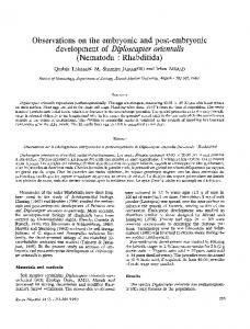
Figure 2. Analysis of the system-specific role(s) of FoxO1 during cardiovascular morphogenesis. (A) Aggregation strategies (S1, S2) of FoxO1-mutant ES cells/diploid embryo to rescue the placental phenotype and define the autonomous role of FoxO1 in cardiovascular morphogenesis. Blastocyst formation from aggregation of the eight-cell FoxO1 mutant diploid embryo with lacZ-expressing FoxO1-WT (FoxO1+/+) ES cells (S1) or FoxO1-null ES cells and tetraploid (4n) cells derived from FoxO1-WT diploid embryo (S2) is proposed. Blastocysts generated from each aggregation strategy and the relative rescue of cardiovascular and placental phenotypes will be evaluated in live FoxO1-null embryos harboring FoxO1-WT or heterozygous allantois at or beyond the original embryonic lethal stage.19 Note that cardiovascular malformation in FoxO1-null mice could be secondary to the placental phenotype (A, Option #1 and B) or FoxO1 could play an autonomous role in placental and cardiovascular morphogenesis at distinct developmental stages (A, Option #2/3 and C).
© 2012 Landes Bioscience. embryos.19,22 However, the fact that an abnormal cardiovascular phenotype in Vcam1 and several other mutant mouse models precedes placental malformation,21-25 confounds analysis of the autonomous roles of FoxO1 and Vcam-1 in cardiovascular development. Similarly, the relative contributions of FoxO1 gene silencing to embryonic cardiovascular malformation afforded by placental malformation as opposed to a direct effect on cardiovascular system development are yet to be defined (Fig. 2). Another intriguing observation is that lack of allantoic expression of the genes coding for α4 integrin (Itga4)23 or a co-chaperone protein (Mrj/ Dnajb6 )24 elicits phenotypic consequences similar to those observed in FoxO1- and Vcam1-deficient mice,19,22 indicating that the mechanism of chorioallantoic attachment is much more complex than anticipated. Further investigation will also be required to determine whether the placental phenotype of FoxO1-null mice is restricted to precursor of allantoic mesoderm or trophoblast populations. Apart from known positive and negative regulatory mechanisms governing FoxO1 activity by post-translational modifications,15 accumulating evidence suggests that protein-protein interactions between FoxO1 and a host of
transcriptional activators and co-activators can also regulate gene expression and govern diverse cellular functions.26 For example, interaction of FoxO1 with HNF-4 (hepatocyte nuclear factor-4)27 and STAT3 (signal transducer and activators of transcription-3)28 modulates glucose and lipid metabolism.26 However, it is presently unknown whether their cooperative activity is also involved in placental morphogenesis. During murine gastrulation, the visceral endoderm (VE) derives from the primitive endoderm and plays an essential role in differentiation of ectoderm to produce the three germ layer cells from which the fetus is derived (Fig. 1A). Targeted disruption of Hnf-4 gene in mice revealed abnormal gastrulation, indicating an essential role of HNF-4 in VE, which secrets several essential serum factors including transthyretin, a downstream target of HNF-4.29 On the other hand, STAT proteins are upstream transcriptional regulator of suppressor of cytokine signaling (SOCS) genes, which play an essential role in placental development. Mice lacking SOCS3 die at mid-gestation and revealed abnormalities in placental development and leukemia inhibitory factor receptor α signaling as well as an increased numbers of mature trophoblast
giant cells (Fig. 1A).30 Collectively, these studies suggest that fine-tuning of diverse transcriptional and signaling pathways in embryonic and extra-embryonic progenitor cells is critical for normal placental and fetal development during and following gastrulation. Therefore, it is conceivable that the absence of FoxO1 may lead to aberrant activation or repression of genes that are not FoxO1 downstream targets. A comprehensive genome-wide gene expression analysis would be essential to identify such target genes.
Do not distribute.
FoxO1 in Cardiovascular Development To unravel the mysteries of FoxO1 governance of cardiovascular development, engineering of chimeric mice by means of a combinatorial ES cell aggregation strategy generating diploid and tetraploid embryos will be an invaluable tool.6 Indeed, this approach has been utilized by numerous investigators to complement placental phenotypes in chimeric mice.25,29-36 For example, this strategy was employed to deconvolute the role of the basic helixloop-helix transcription factor Hand1 in placental and cardiovascular development at distinct developmental stages.25 Moving forward, complementation of the
www.landesbioscience.com Transcription
3
placental phenotype in FoxO1-null mice using FoxO1-mutant ES cell and diploid embryos will be critical to parse the relative contributions of FoxO1 to embryonic cardiovascular morphogenesis. Complementation of the placental phenotype in FoxO1-null mice can be achieved using blastocysts derived from aggregation of FoxO1-mutant diploid embryos and lacZ-expressing ES cells,34 which are wild-type at the FoxO1 locus (FoxO1+/+) (Fig. 2A and Strategy 1). Using this approach, the diploid mutant cells contribute to the genesis of both embryonic and extra-embryonic tissues, including heart and placenta (see Fig. 1A). On the other hand, the contribution of the ES cells will be restricted to tissues of the embryo proper and allantois; further, the relative contribution of ES cells vs. mutant cells to the embryo proper and allantois can be unambiguously defined by analyzing β-galactosidase activity (see Fig. 1A).34 This aggregation-based strategy will lead to the following possibilities: FoxO1null mice are alive at E10.5, and cardiovascular and placental phenotypes are absent (Option #1, Fig. 2A). Such an observation would suggest that cardiovascular malformations in FoxO1-null mice arise secondary to the placental defect (Fig. 2B) 13,17-19 and that the placental phenotype is restricted to the allantoic mesoderm. Alternatively, if mutant embryos manifest only cardiovascular malformation and die at later stages of development, an autonomous role for FoxO1 in cardiovascular morphogenesis would be suggested (Option #2, Fig. 2A and C). In that case, isolation of embryos at distinct developmental stages would allow for evaluation of the stage of lethality and the underlying cardiovascular phenotype. Finally, if null embryos manifest both cardiovascular and placental phenotypes, critical roles for FoxO1 in either/both placental and cardiovascular morphogenesis at distinct development stages would be suggested (Option #3, Fig. 2A and C). In that eventuality, the complementation assay could be repeated using blastocysts derived from aggregation of FoxO1-WT tetraploid cells (4n) and FoxO1-null ES cells (Fig. 2A and Strategy 2). In this approach, ES cells will not contribute to placental development,
and as such, any cardiovascular phenotype would imply a cell-autonomous role for FoxO1 in cardiovascular morphogenesis (Fig. 2C). Conclusion and Perspective Despite recent advances in elucidating anatomical defects associated with congenital heart disease, molecular mechanisms underlying these perturbations in the developing and adult heart are incompletely defined. Recent work has uncovered a previously unrecognized role for the transcription factor FoxO1 in placental and cardiovascular morphogenesis. Additional work is warranted within this fascinating biology to dissect underlying mechanisms in an effort to develop novel therapeutic insights with potential clinical relevance.
7. Cross JC, Baczyk D, Dobric N, Hemberger M, Hughes M, Simmons DG, et al. Genes, development and evolution of the placenta. Placenta 2003; 24:12330; PMID:12596737; http://dx.doi.org/10.1053/ plac.2002.0887. 8. Clark KL, Halay ED, Lai E, Burley SK. Co-crystal structure of the HNF-3/fork head DNArecognition motif resembles histone H5. Nature 1993; 364:412-20; PMID:8332212; http://dx.doi. org/10.1038/364412a0. 9. Carlsson P, Mahlapuu M. Forkhead transcription factors: key players in development and metabolism. Dev Biol 2002; 250:1-23; PMID:12297093; http:// dx.doi.org/10.1006/dbio.2002.0780. 10. Kaestner KH, Knochel W, Martinez DE. Unified nomenclature for the winged helix/forkhead transcription factors. Genes Dev 2000; 14:142-6; PMID:10702024. 11. Wijchers PJ, Burbach JP, Smidt MP. In control of biology: of mice, men and Foxes. Biochem J 2006; 397:233-46; PMID:16792526; http://dx.doi. org/10.1042/BJ20060387. 12. Accili D, Arden KC. FoxOs at the crossroads of cellular metabolism, differentiation and transformation. Cell 2004; 117:421-6; PMID:15137936; http:// dx.doi.org/10.1016/S0092-8674(04)00452-0. 13. Paik JH, Kollipara R, Chu G, Ji H, Xiao Y, Ding Z, et al. FoxOs are lineage-restricted redundant tumor suppressors and regulate endothelial cell homeostasis. Cell 2007; 128:309-23; PMID:17254969; http:// dx.doi.org/10.1016/j.cell.2006.12.029. 14. Paik JH, Ding Z, Narurkar R, Ramkissoon S, Muller F, Kamoun WS, et al. FoxOs cooperatively regulate diverse pathways governing neural stem cell homeostasis. Cell Stem Cell 2009; 5:54053; PMID:19896444; http://dx.doi.org/10.1016/j. stem.2009.09.013. 15. Ferdous A, Battiprolu PK, Ni YG, Rothermel BA, Hill JA. FoxO, autophagy and cardiac remodeling. J Cardiovasc Transl Res 2010; 3:355-64; PMID:20577843; http://dx.doi.org/10.1007/ s12265-010-9200-z. 16. Castrillon DH, Miao L, Kollipara R, Horner JW, DePinho RA. Suppression of ovarian follicle activation in mice by the transcription factor Foxo3a. Science 2003; 301:215-8; PMID:12855809; http:// dx.doi.org/10.1126/science.1086336. 17. Hosaka T, Biggs WH 3rd, Tieu D, Boyer AD, Varki NM, Cavenee WK, et al. Disruption of forkhead transcription factor (FOXO) family members in mice reveals their functional diversification. Proc Natl Acad Sci USA 2004; 101:2975-80; PMID:14978268; http://dx.doi.org/10.1073/pnas.0400093101. 18. Furuyama T, Kitayama K, Shimoda Y, Ogawa M, Sone K, Yoshida-Araki K, et al. Abnormal angiogenesis in Foxo1 (Fkhr)-deficient mice. J Biol Chem 2004; 279:34741-9; PMID:15184386; http://dx.doi. org/10.1074/jbc.M314214200. 19. Ferdous A, Morris J, Abedin MJ, Collins S, Richardson JA, Hill JA. Forkhead factor FoxO1 is essential for placental morphogenesis in the developing embryo. Proc Natl Acad Sci USA 2011; 108:16307-12; PMID:21930913; http://dx.doi. org/10.1073/pnas.1107341108. 20. Mahlapuu M, Ormestad M, Enerbäck S, Carlsson P. The forkhead transcription factor Foxf1 is required for differentiation of extra-embryonic and lateral plate mesoderm. Development 2001; 128:155-66; PMID:11124112. 21. Gurtner GC, Davis V, Li H, McCoy MJ, Sharpe A, Cybulsky MI. Targeted disruption of the murine VCAM1 gene: essential role of VCAM-1 in chorioallantoic fusion and placentation. Genes Dev 1995; 9:1-14; PMID:7530222; http://dx.doi.org/10.1101/ gad.9.1.1.
© 2012 Landes Bioscience. Acknowledgements
We thank Dr. Thomas Gillette for critical reading of the manuscript. We also thank the members of the Hill and Ferdous labs for their support. Funding for these studies was provided by the NIH (HL-075173; HL-080144; HL-090842; U01-HL100401–01; P30-HL101254–01), AHA (0640084N), ADA (7–08-MN-21ADA), the AHA-Jon Holden DeHaan Foundation (0970518N), and the March of Dimes (5-FY09–21).
Do not distribute.
4
References 1. Roger VL, Go AS, Lloyd-Jones DM, Benjamin EJ, Berry JD, Borden WB, et al.; American Heart Association Statistics Committee and Stroke Statistics Subcommittee. Heart disease and stroke statistics—2012 update: a report from the American Heart Association. Circulation 2012; 125:2-220; PMID :22179539; http://dx.doi.org/10.1161/ CIR.0b013e31823ac046. 2. Hoffman JI. Incidence of congenital heart disease: I. Postnatal incidence. Pediatr Cardiol 1995; 16:10313; PMID:7617503; http://dx.doi.org/10.1007/ BF00801907. 3. Hoffman JI. Incidence of congenital heart disease: II. Prenatal incidence. Pediatr Cardiol 1995; 16:155-65; PMID:7567659. 4. Rossant J, Cross JC. Placental development: lessons from mouse mutants. Nat Rev Genet 2001; 2:538-48; PMID:11433360 ; http://dx.doi. org/10.1038/35080570. 5. Watson ED, Cross JC. Development of structures and transport functions in the mouse placenta. Physiology (Bethesda) 2005; 20:180-93; PMID:15888575; http://dx.doi.org/10.1152/physiol.00001.2005. 6. Tanaka M, Hadjantonakis AK, Nagy A. Aggregation chimeras. Combining ES cells, diploid and tetraploid embryos. Methods Mol Biol 2001; 158:135-54; PMID:11236654.
Transcription
Volume 3 Issue 5
22. Kwee L, Baldwin HS, Shen HM, Stewart CL, Buck C, Buck CA, et al. Defective development of the embryonic and extraembryonic circulatory systems in vascular cell adhesion molecule (VCAM-1) deficient mice. Development 1995; 121:489-503; PMID:7539357. 23. Yang JT, Rayburn H, Hynes RO. Cell adhesion events mediated by alpha 4 integrins are essential in placental and cardiac development. Development 1995; 121:549-60; PMID:7539359. 24. Hunter PJ, Swanson BJ, Haendel MA, Lyons GE, Cross JC. Mrj encodes a DnaJ-related co-chaperone that is essential for murine placental development. Development 1999; 126:1247-58; PMID:10021343. 25. Riley P, Anson-Cartwright L, Cross JC. The Hand1 bHLH transcription factor is essential for placentation and cardiac morphogenesis. Nat Genet 1998; 18:271-5; PMID:9500551; http://dx.doi. org/10.1038/ng0398-271. 26. Barthel A, Schmoll D, Unterman TG. FoxO proteins in insulin action and metabolism. Trends Endocrinol Metab 2005; 16:183-9; PMID:15860415; http:// dx.doi.org/10.1016/j.tem.2005.03.010. 27. Hirota K, Daitoku H, Matsuzaki H, Araya N, Yamagata K, Asada S, et al. Hepatocyte nuclear factor-4 is a novel downstream target of insulin via FKHR as a signal-regulated transcriptional inhibitor. J Biol Chem 2003; 278:13056-60; PMID:12519792; http://dx.doi.org/10.1074/jbc.C200553200.
28. Kortylewski M, Feld F, Krüger KD, Bahrenberg G, Roth R A, Joost HG, et al. Akt modulates STAT3-mediated gene expression through a FKHR (FOXO1a)-dependent mechanism. J Biol Chem 2003; 278:5242-9; PMID:12456685; http://dx.doi. org/10.1074/jbc.M205403200. 29. Duncan SA, Nagy A, Chan W. Murine gastrulation requires HNF-4 regulated gene expression in the visceral endoderm: tetraploid rescue of Hnf4(-/-) embryos. Development 1997; 124:279-87; PMID:9053305. 30. Boyle K, Robb L. The role of SOCS3 in modulating leukaemia inhibitory factor signalling during murine placental development. J Reprod Immunol 2008; 77:1-6; PMID:17408753; http://dx.doi. org/10.1016/j.jri.2007.02.003. 31. Takahashi Y, Dominici M, Swift J, Nagy C, Ihle JN. Trophoblast stem cells rescue placental defect in SOCS3-deficient mice. J Biol Chem 2006; 281:11444-5; PMID:16517610; http://dx.doi. org/10.1074/jbc.C600015200. 32. Goto Y, Takagi N. Tetraploid embryos rescue embryonic lethality caused by an additional maternally inherited X chromosome in the mouse. Development 1998; 125:3353-63; PMID:9693139.
33. Bissonauth V, Roy S, Gravel M, Guillemette S, Charron J. Requirement for Map2k1 (Mek1) in extra-embryonic ectoderm during placentogenesis. Development 2006; 133:3429-40; PMID:16887817; http://dx.doi.org/10.1242/dev.02526. 34. Voss AK, Thomas T, Gruss P. Mice lacking HSP90β fail to develop a placental labyrinth. Development 2000; 127:1-11; PMID:10654595. 35. Sugimoto M, Karashima Y, Abe K, Tan SS, Takagi N. Tetraploid embryos rescue the early defects of tw5/tw5 mouse embryos. Genesis 2003; 37:16271; PMID:14666509; http://dx.doi.org/10.1002/ gene.10238. 36. de Bruin A, Wu L, Saavedra HI, Wilson P, Yang Y, Rosol TJ, et al. Rb function in extraembryonic lineages suppresses apoptosis in the CNS of Rb-deficient mice. Proc Natl Acad Sci USA 2003; 100:654651; PMID:12732721; http://dx.doi.org/10.1073/ pnas.1031853100.
© 2012 Landes Bioscience. Do not distribute.
www.landesbioscience.com Transcription
5