n
fo r
Dentine Regeneration: Key Roles for Stem Cells and Molecular Signalling
ot
Q ui
by N ht
pyrig No Co t fo rP ub lica tio n te ss e n c e
Anthony J. Smith, Minal Patel, Lee Graham, Alastair J. Sloan, Paul R. Cooper Oral Biology, School of Dentistry, University of Birmingham, Birmingham, UK
Summary: The exquisite regenerative potential of the dentine-pulp complex provides an important defence wound-healing role for the dental tissues after injury. However, the diversity of such regenerative responses questions the traditional paradigm that all such responses are specifically dentinogenic in nature. Possible variations in stem/progenitor cells and the molecular signalling processes directing these regenerative responses may contribute to a spectrum of responses ranging in nature from specifically dentinogenic to a less-specific mineralised tissue type of response. Understanding of the features of these diverse regenerative responses may allow their exploitation in the development of future novel clinical therapies. Key words: dentine, pulp, regeneration, stem cells, cell signalling Oral Biosci Med 2005; 2/3: 127-132
INTRODUCTION The exquisite regenerative potential of the dentine-pulp complex has long been recognised and one of the earliest reports was in 1771 by Hunter describing the tooth’s response to caries (Hunter, 1771). During the past six to seven decades, dentistry has been at the forefront of regenerative medicine in the use of pulp-capping agents, such as calcium hydroxide, for reparative dentinogenesis in the stimulation of dentine bridge formation at sites of pulpal exposure. This has led to an established paradigm that any tissue regeneration in the dentine-pulp complex represents reparative dentine. However, our increasing understanding of the biology of the dentine-pulp complex places this paradigm in question and highlights the need to carefully characterise the molecular and biological processes underpinning dentine regeneration if we are going to successfully exploit it for development of novel clinical therapies to extend and replace more traditional dental restorative procedures. Nevertheless, the clinical opportunities and benefits from development of such therapies are immense and could have major impact on the practice of restorative dentistry and endodontics.
Vol 2. No 2/3. 2005
DIVERSITY OF REGENERATIVE RESPONSES AND TERTIARY DENTINOGENESIS It is important to initially define what processes are taking place during regeneration in the dentine-pulp complex. The concepts of reactionary and reparative dentinogenesis (Smith et al, 1995) were proposed for this purpose. Following primary dentinogenesis, which provides the bulk of the dentine constituting the crown and root of the tooth, down-regulation of odontoblast secretory activity is seen and the quiescent odontoblasts continue to slowly secrete secondary dentine gradually occluding the pulp chamber (Ten Cate, 1998). Injury to the tooth, however, can lead to up-regulation of odontoblast activity and tertiary dentine secretion focally at the site of injury (Kuttler, 1959). The events leading to tertiary dentinogenesis, however, represent a spectrum of cellular responses dependent on the intensity of the injury and hence, the need to more accurately define the processes taking place. Reactionary dentinogenesis is defined by a tertiary dentine matrix secreted by surviving post-mitotic odontoblast cells in response to an appropriate stimulus. Clearly, this represents a specific dentinogenic response since the cells responsible for
127
n
ot
Q ui
fo r
secretion of the reactionary tertiary dentine are the primary odontoblasts, which differentiated during tooth development. Reparative dentinogenesis is defined by a tertiary dentine matrix secreted by a new generation of odontoblast-like cells in response to an appropriate stimulus, after the death of the original post-mitotic odontoblasts responsible for primary and physiological secondary dentine secretion. This represents a more complex sequence of events than reactionary dentinogenesis. The reparative response, which also includes dentine bridge formation after pulp-capping, may encompass both specific dentinogenic processes and also, possibly less specific mineralised tissue formation. Despite the diversity of responses seen, they are all generally termed as being reparative dentinogenesis in nature, which does not allow a mechanistic understanding of the biological events taking place. A variety of data point to a spectrum of dentinogenic processes during reparative dentinogenesis and hard tissue formation in the dentine-pulp complex including: • Variation in regularity of the tubular structure of the matrix (Baume, 1980). • Initial deposition of a fibro-dentine matrix often following pulp-capping (Baume, 1980). • Deposition of atubular matrix at sites of tissue regeneration in the pulp after application of bio-active molecules, such as growth factors (Rutherford et al, 1993). • Common occurrence of pulp stones and the heterogeneity in their form (Trowbridge et al, 2002). The biological processes involved in reactionary and reparative dentinogenesis differ in their complexity and highlight the opportunities for specificity in the regenerative processes (Fig 1). Reactionary dentinogenesis sim-
by N ht
Smith et al
pyrig No Co t fo rP ub lica ply represents an up-regulation of the synthetic / secreti tory activity of the existing odontoblasts, whilst te repara- on ss eornprotive dentinogenesis involves recruitment of stem ce genitor cells, induction of their differentiation into odontoblast-like cells and then, up-regulation of their synthetic/secretory activity. Thus, reparative dentinogenesis involves a more complex cascade of biological processes inherent in which there are opportunities for biological diversity. This biological diversity may well impact on the specificity of the processes taking place and their correlation to physiological dentinogenic events. Two aspects, which may importantly contribute to the possible diversity of events during reparative dentinogenesis, are the origin of the stem/progenitor cells participating in the reparative dentinogenesis and the molecular signals responsible for induction of their differentiation into odontoblast-like cells.
STEM/PROGENITOR CELLS AND DENTINE REGENERATION During tooth development, the primary odontoblasts are derived from cells which have migrated to the first branchial arch from the cranial neural crest and which, following a series of reciprocal epithelial-mesenchymal interactions in the tooth germ, give rise to the cells of the dental papilla and are induced by signals derived from the inner dental epithelium and mediated by the dental basement membrane to differentiate into odontoblasts (Ruch et al, 1995). As the dental pulp develops, it becomes increasingly populated by cells of non-neural crest origin, presumably derived from first branchial arch mesenchyme, (Chai et al, 2000) and the mature pulp comprises a heterogeneous population of cells. The derivation of the stem / progenitor cells during reparative dentino-
Up-regulation of o 1 Odontoblasts
REACTIONARY DENTINOGENESIS
Up-regulation of 1 Odontoblasts
REPARATIVE DENTINOGENESIS
Stem / progenitor cell recruitment
o
TERTIARY DENTINE
Induction of Odontoblast-like Cell Differentiation
Fig 1 Key processes involved in reactionary versus reparative dentinogenesis and their differing degrees of complexity.
128
Oral Biosciences & Medicine
Q ui
by N ht
pyrig No Co t fo Signalling Dentine Regeneration: Key Roles for Stem Cells and Molecular rP ub lica tio n te ss e n c e
ot
n
256
fo r
Events
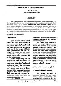
Fig 2 Flow cytometric analysis of low affinity NGF-R labelled rat pulp cells ( ) and their isotype control ( ) showing a population of specifically labelled stem / progenitor cells ( ), which can be isolated on the basis of this marker.
A
B
0 10
0
10
1
10
2
10
3
10
4
Relative fluorescence intensity
C
D
Fig 3 Expansion culture of pulp cells from primary culture (a, c) to passage 7 (b, d) favours the myofibroblast phenotype as indicated by sq-RTPCR detection of smooth muscle α-actin (a, b top versus normalised GAPDH control a, b bottom) and SEM (bar = 50 μm) morphological appearance (c, d).
genesis remains elusive, although a variety of derivations may go some way towards explaining the diversity of responses seen during regeneration in the dentine-pulp complex. Possible derivations suggested for these stem/ progenitor cells include the undifferentiated mesenchymal cells in the cell-rich zone of Höhl adjacent to the odontoblasts (Cotton, 1968), perivascular cells, undifferentiated mesenchymal cells and fibroblasts (Fitzgerald et al, 1990). Recently, Gronthos et al (2000, 2002) have reported on the presence of a unique population of postnatal dental pulp stem cells. These studies suggested a hierarchy of progenitors in adult dental pulp, including a minor population of self-renewing, highly proliferative, multi-potent stem cells within a larger compartment of perhaps more committed progenitors (Gronthos et al, 2002). Attempts to isolate stem cells from first branchial arch tissue, which are capable of differentiating into odontoblasts and might have potential for tissue engiVol 2. No 2/3. 2005
neering applications, has allowed identification of some appropriate markers for these cells (Deng et al, 2004) and low affinity nerve growth factor receptor has been targeted for magnetic-activated cell sorting of these cells (Deng et al, unpublished). Use of this cell surface marker to identify possible post-natal stem/progenitor cells from mature rodent dental pulp using flow cytometry has yielded a small population (less than 1% of the total) of cells (Fig 2), whose potentiality in terms of odontoblast differentiation is now being investigated. Interestingly, expansion by serial passage of primary cultures of the same rodent pulp cells tends to give rise to cells of myofibroblast appearance (Fig 3) and strong expression of actin. This may simply reflect stronger competitive growth by progenitors of the myofibroblasts present. However, it is possible that myofibroblasts may represent a form of “default” differentiation since the neural crest pheno129
ot
fo r
The molecular signalling of differentiation of odontoblast-like cells during reparative dentinogenesis is now becoming clearer and may recapitulate some of the events of physiological odontoblast differentiation during tooth development (Smith and Lesot, 2001). Presentation of inner dental epithelium derived growth factors, particularly of the TGF-β family, by the dental basement membrane leads to odontoblast differentiation during the bell stage of tooth development (Ruch et al, 1995). Expression of TGF-β isoforms by the odontoblasts (Sloan et al, 2000) leads to their sequestration in the dentine matrix (Cassidy et al, 1997) where they become “fossilised” unless released as a result of matrix breakdown during tissue injury. Whilst agents, such as EDTA, capable of dissolution of the dentine matrix can release these and other growth factors (Cassidy et al, 1997; Roberts-Clarke and Smith, 2000), we now have data demonstrating that both lactic acid, responsible for carious dissolution of the dental tissues, and calcium hydroxide, used in pulp-capping to stimulate reparative dentinogenesis, can also release TGF-β1 from dentine matrix (Fig 4). In vivo application of EDTA-soluble preparations of dentine matrix (Smith et al, 1990) and members of the TGF-β family, TGF-βs and BMPs, (Rutherford et al, 1993, 1994; Nakashima, 1994a,b; Nakashima et al, 1994; Tziafas et al, 1998; Hu et al, 1998) can stimulate reparative dentinogenesis in a pulp-capping situation and
n
MOLECULAR SIGNALLING AND DENTINE REGENERATION
Q ui
type has been suggested to be unstable with Schwann cells able to trans-differentiate into myofibroblasts, which is also strongly promoted by 1ng/ml TGF-β1 (Real et al, 2005).
by N ht
Smith et al
pyrig No Co t fo rP ub lica similar events have been reported in vitro in the absence ti of systemic influences (Sloan et al, 1999, te 2000). on However, the response seen during this sreparative se nc e dentinogenesis highlights the spectrum of specificity of the process with the morphology of the reparative dentine varying from being nearly indistinguishable from physiological primary dentine to an atubular matrix, sometimes incorporating the formative cells, and resembling bone. This may reflect divergence from the closely regulated temporo-spatial and dose-dependent signalling of odontoblast differentiation seen during physiological tooth development. Release of the cocktail of growth factors from dentine matrix may occur with a range of kinetics as well as concentrations of these molecules, which might be expected to give rise to a wide spectrum of signalling situations. Many of the growth factors exhibit a variety of cell signalling activities (e.g. proliferation, differentiation, apoptosis etc), which are dose-dependent and it is perhaps not surprising that the pulpal reactions to carious and other tissue injury give rise to such a spectrum of regenerative responses. Bjorndal (2001) has already elegantly demonstrated the correlation between clinical progression of carious lesions and the occurrence of regeneration in the form of tertiary dentinogenesis. These data have highlighted the absence of tertiary dentinogenesis beneath rapidly progressing more aggressive carious lesions. In such circumstances, the rate and level of dissolution of dentine matrix components would be expected to be significant. We have recently demonstrated that a dose-dependent Smad-mediated induction of apoptosis by TGF-β1 can occur in odontoblast-like cells (He et al, 2005) and similar responses appear to be apparent when odontoblastlike cells are cultured with higher concentrations of dentine matrix preparations (Table 1). Thus, at lower concentrations growth factors like TGF-β, and dentine
1.6 1.4 1.2 1 0.8 0.6 0.4 0.2 0 EDTA
130
Ca(OH)2
Lactic acid
Fig 4 Release of TGF-β1 (ng/mg of freeze-dried extract) from human dentine by treatment with 10% EDTA, pH 7, 0.02 M calcium hydroxide, pH 11.7 or 0.1 M lactic acid, pH 4.0.
Oral Biosciences & Medicine
ot
fo r
Vol 2. No 2/3. 2005
by N ht
It is thus clear that both the stem/progenitor cells giving rise to odontoblast-like cells for reparative dentinogenesis and the molecular signalling responsible for their differentiation may have a profound effect on the regenerative response and may account for the biological diversity observed in this response. The paradigm that any reparative tissue laid down in the dentine-pulp complex after injury is a form of dentine may be erroneous and an understanding of the origins of the stem/progenitor cells and the signalling of these cells is critical to identification of the nature of these responses. Where regenerative events clearly recapitulate embryonic development in terms of cell derivation and signalling, a true dentinogenic response is possible. However, the heterogeneity of the pulpal cell populations and the cell signalling events triggered post-injury, both as a result of local release of matrix-bound bio-active molecules and inflammatory or other defence processes, provide the opportunity for less specific cellular responses. Some of these responses may show very limited correlation to developmental events in the tooth resulting in deposition of reparative mineralised matrices with little resemblance in structure and/or composition to physiological dentine. Although such arguments may appear a little academic, they may be important in providing a practical clinical basis for development of new regenerative therapies. The tubular nature of dentine, in contrast to bone and other mineralised tissues, provides the property of permeability and the opportunity for greater cellular communication within the matrix through cell processes extending along the tubules. The ability to determine such properties for a reparative tissue, based perhaps both on the nature of the progenitor cells and the molecules responsible for signalling their differentiation,
n
CONCLUSIONS
Q ui
matrix preparations containing such growth factors, may be stimulatory in signalling reparative dentinogenesis and regeneration, but at higher concentrations these molecules may induce apoptosis in those cells potentially capable of giving rise to regeneration. Such observations concur well with clinical observations where carious injury of the greatest intensity may lead to an environment in the dentine-pulp complex, which does not favour regeneration. The complexity of the cell signalling events within the pulp during caries is emphasised by the changes in pulpal gene expression identified by microarray analysis (McLachlan et al, 2005). The observed up-regulation of inflammatory mediators (McLachlan et al, 2005) may impede regenerative events, as has also been reported after application of BMP-7 to inflamed ferret pulps (Rutherford and Gu, 2000).
pyrig No Co t fo Signalling Dentine Regeneration: Key Roles for Stem Cells and Molecular rP ub li Table 1 Cell survival (% of unexposed control) when immor-ca tio n talised MDPC-23 cells are exposed to differing concentrations te ss e nproof a preparation of EDTA-soluble human dentine matrix ce teins (DMPs) for five days DMP exposure concentration
Cell survival (%)
1 μg/ml DMPs
1000 μg/ml DMPs
98.9%
10.9%
may provide a powerful tool for the clinician to direct a regenerative response most appropriate to the needs of the application. Thus, restoration of tooth crown structure may seek to regenerate tubular dentine matrix resembling physiological dentine, whilst endodontic applications may seek a less permeable atubular matrix during regeneration. Greater focus should now be directed towards better characterising these two aspects of dentine regeneration to provide the basis for development of novel biologically based treatment modalities for future clinical exploitation.
REFERENCES Baume LJ. Biology of Pulp and Dentine. Basel, Switzerland: S Karger, 1980. Bjorndal L. Presence or absence of tertiary dentinogenesis in relation to caries progression. Adv Dent Res 2001;15:80-83. Cassidy N, Fahey M, Prime SS, Smith AJ. Comparative analysis of Transforming Growth Factor-Beta (TGF-β) isoforms 1-3 in human and rabbit dentine matrices. Arch Oral Biol 1997;42:219-223. Chai Y, Jiang X, Ito Y, Bringas P, Han J, Rowitch DH, et al. Fate of the mammalian cranial neural crest during tooth and mandibular morphogenesis. Development 2000;127:1671-1679. Cotton WR. Pulp response to cavity preparation as studied by the method of thymidine 3H autoradiography. In: Finn SB (ed) Biology of the Dental Pulp Organ. Tuscaloosa, AL: Univ Alabama Press 1968;69. Deng MJ, Jin Y, Shi JN, Lu HB, Liu Y, He DW, et al. Multi-Lineage differentiation of ecto-mesenchymal cells isolated from the first branchial arch. Tiss Eng 2004;10:1597-1606. Fitzgerald M, Chiego JD, Heys DR. Autoradiographic analysis of odontoblast replacement following pulp exposure in primate teeth. Arch Oral Biol 1990;35:707-715. Gronthos S, Mankani M, Brahim J, Robey PG, Shi S. Postnatal human dental pulp stem cells (DPSCs) in vitro and in vivo. Proc Natl Acad Sci USA 2000;97:13625-13630. Grontos S, Brahim J, Li W, Fisher LW, Cherman N, Boyde A, et al. Stem cell properties of human dental pulp stem cells. J Dent Res 2002;81:531-535. He W-X, Niu Z-Y, Zhao S, Smith AJ. Smad Protein Mediated Transforming Growth Factor beta1 Induction of Apoptosis in the MDPC-23 Odontoblast-like Cell Line. Arch Oral Biol 2005; in press (online).
131
ot
n
fo r
132
Q ui
Hu CC, Zhang C, Qian Q, Tatum NB. Reparative dentin formation in rat molars after direct pulp capping with growth factors. J Endodont 1998;24:744-751. Hunter J. The Natural History of the Human Teeth. London: Johnson, 1771. Kuttler Y. Classification of dentin into primary, secondary and tertiary. Oral Surg 1959;12:996-1001. McLaclan JL, Smith AJ, Bujalska IJ, Cooper PR. Gene expression profiling of pulpal tissue reveals the molecular complexity of dental caries. Biochim Biophys Acta 2005; in press (online). Nakashima M. Induction of dentin formation on canine amputated pulp by recombinant human bone morphogenetic proteins (BMP) -2 and -4. J Dent Res 1994;73:1515-1522. Nakashima M. Induction of dentine in amputated pulp of dogs by recombinant human bone morphogenetic proteins -2 and -4 with collagen matrix. Arch Oral Biol 1994;39:1085-1089. Nakashima M, Nagasawa H, Yamada Y, Reddi AH. Regulatory role of transforming growth factor-beta, bone morphogenetic protein-2 and protein-4 on gene expression of extracellular matrix proteins and differentiation of pulp cells. Dev Biol 1994;162:18-28. Real C, Glavieux-Pardanaud C, Vaigot P, Le-Douarin N, Dupin E. The instability of the neural crest phenotypes: Schwann cells can differentiate into myofibroblasts. Int J Dev Biol 2005; 49:151-159. Roberts-Clark D, Smith AJ. Angiogenic growth factors in human dentine matrix . Arch Oral Biol 2000;45:1013-1016. Ruch JV, Lesot H, Begue-Kirn C. Odontoblast differentiation. Int J Dev Biol 1995;39:51-68. Rutherford RB, Gu K. Treatment of inflamed ferret dental pulps with recombinant bone morphogenetic protein-7. Eur J Oral Sci 2000;108:202-206. Rutherford RB, Wahle J, Tucker M, Rueger D, Charette M. Induction of reparative dentine formation in monkeys by recombinant human osteogenic protein-1. Arch Oral Biol 1993;38:571-576. Rutherford RB, Spanberg L, Tucker M, Rueger D, Charette M. The time-course of the induction of reparative dentine formation in monkeys by recombinant human osteogenic protein-1. Arch Oral Biol 1994;39:833-838.
by N ht
Smith et al
pyrig No Co t fo rP ub Sloan AJ, Smith AJ. Stimulation of the dentine-pulp complex oflic rat a incisor teeth by transforming growth factor-‚ isoforms 1-3 in vitro. tio n te Arch Oral Biol 1999;44:149-156. e s s c en Sloan AJ, Rutherford RB, Smith AJ. Stimulation of the rat dentine-pulp complex by BMP7 in vitro. Arch Oral Biol 2000;45:173-177. Smith AJ, Tobias RS, Plant CG, Browne RM, Lesot H, Ruch JV. In vivo morphogenetic activity of dentine matrix proteins. J Biol Buccale 1990;18:123-129. Sloan AJ, Perry H, Matthews JB, Smith AJ. Transforming growth factor-B isoform expression in mature human molar teeth. Histochem J 2000;32:247-252. Smith AJ, Cassidy N, Perry H, Begue-Kirn C, Ruch JV, Lesot H. Reactionary dentinogenesis. Int J Dev Biol 1995;39:273-280. Smith AJ, Lesot H. Induction and regulation of crown dentinogenesis – embryonic events as a template for dental tissue repair. Crit Rev Oral Biol Med 2001;12:425-437. Ten Cate AR. Oral Histology – Development, Structure and Function. St Louis: Mosby 1998. Trowbridge H, Kim S, Suda H. Structure and functions of the dentin and pulp complex. In: Cohen S, Burns RC (eds). Pathways of the Pulp. St Louis: Mosby 2002. Tzaifas D, Alvanou A, Papadimitriou S, Gasic J, Komnenou A. Effects of recombinant fibroblast growth factor, insulin-like growth factor -II and transforming growth factor-β1 on dog dental pulp cells in vivo. Arch Oral Biol 1998;43:431-444. Reprint requests: Anthony J Smith, Professor Oral Biology School of Dentistry University of Birmingham St Chad’s Queensway Birmingham, B4 6NN UK E-mail:
[email protected]
Oral Biosciences & Medicine