against cardiac ganglionic structures and myenteric nervous plexuses appears strongly to depend on the parasite strain used to produce chagasic infection.
SAO PAULO
\L Dalmo de Souza Amorim, Jose Antonio Marin Neto
Functional alterations of the autonomic nervous system in Chagas' heart disease Division of Cardiology, Department of Internal Medicine, Faculdade de Medicina de Ribeiriio Pre to da Universidade de Siio Paulo - Siio Paulo, Brazil
Several independent pathological studies in experimental models and in human beings showed conspicuous autonomic denervation in Chagas' disease. In spite of the inherently complex structural organization of the autonomic nervous system, the parasympathetic and sympathetic divisions are involved, as shown by many functional studies. Hence, Chagas' disease represents a unique model of impairment of the autonomic control of the heart, in absence of the nonspecific effects of cardiac failure. An important limitation of the studies thus far carried out is the lack of a better knowledge of the molecular biology characteristics of different strains of T. cruzi. This could explain some geographical discrepancies found in the clinical behaviour of Chagas' disease, and contribute to a better understanding of its pathophysiology. UNITERMS: Chagas' disease. Autonomic nervous system.
INTRODUCTION
S
ince the primordial studies by Carlos Chagas and coworkers, the pathological involvement of the nervous system in the trypanosomiasis discovered by him has been the subject of research and speculation (14). The existence of a nervous form characterized by central manifestations of neuropathy, including psychiatric disorders, has indeed been proposed (75); however, this
Address for correspondence: Dalmo de Souza Amorim Faculdade de Medicina de Ribeirao Preto da Universidade de Sao Paulo Divisao de Cardiologia - Departamento de Clfnica Medica Ribeirao Preto - SP - Brasil - CEP 14049-900
772
form did not receive definitive clinical acceptance despite the fact that it was specifically investigated in studies using different methodological approaches. Nevertheless, not only it is possible. to detect anatomically polyfocal aggressions against the central nervous system, but there are studies demonstrating a certain degree of sensorimotor functional impairment detected by laboratory tests in patients with Chagas' disease (57,67). There is ample evidence that Chagas' disease as characterized by a marked preference for involvement of the muscle and nervous systems (35,36). In this respect, the physiopathology of several clinical syndromes associated with Chagas' disease, such as cardiopathy, esophagopathy, colopathy etc., should logically involve essentially muscular and nervous lesions in the corresponding organs. Thus, the involvement of the neurovegetative system seems to be the essential element in the digestive manifestations of the disease, with a
Sao Paulo Medical Journal/RPM
113(2) Marl Apr 1995
predominance of this mechanism over the concomitant direct myopathic process in the esophagus and colon (36,52). However, the mechanisms responsible for triggering and installation of the muscular and nervous lesions detected during the acute and chronic phases of the disease have not been sufficiently clarified. Furthermore, the physiopathological role of the nervous lesions documented in Chagas' disease continues to be controversial, especially with respect to Chagas' heart disease.
ANATOMICAL DEMONSTRATION OF AGGRESSION AGAINST THE PARASYMPATHETIC SYSTEM OF THE HEART
On the basis of the results obtained in autopsy studies at several independent centers, it is possible to conclude that lesions of the intracardiac nervous system are practically constant in human trypanosomiasis occurring in Brazil (35,36,54,55). These lesions diffusely involve the cardiac nervous structures, although they are characterized by their peculiar focal nature of irregular and unpredictable distribution. Systematic evaluations using light and electron microscopy have demonstrated degenerative lesions in subpepicardial ganglia, in Schwann and satellite cells, and in myelinic and amyel!nic nerve fibers deep inside the myocardium (54,72). In order to quantify the extent of parasympathetic denervation in the chagasic heart, K6berle and collaborators adopted, starting in the 1950's, techniques of neuronal counting based on exhaustive and standardized microscopic exploration of serial histological sections obtained from the so-called intercaval atrial band (8,36,55). This method permitted the definite detection of cardiac neuron depopulation in autopsied chagasics. Different quantitative methods exploring other anatomical regions of the heart adopted by independent researchers confirmed the indisputable reduction of atrial intramural neurons in individuals who had died during different phases, or with different forms of Chagas' disease (42,54,64,72). Since the beginning, extreme variability in individual neuronal counts was observ~d in autopsied chagasics. There were cases in which destruction appeared to be total (not a single intact neuron was found), side by side with cases in which the number of nervous cardiac cells was within the limits. usually detected in "normal" controls (54,72).
Sao Paulo Medical Journal/RPM
113(2) Mar/Apr 1995
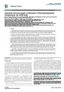
On the other hand, although this atrial (parasympathetic) neuronal depopulation cannot be considered an attribute of Chagas' disease alone, in all comparative studies with other diseases such as rheumatism (39,64), endomyocardial fibrosis (l 0), dilated cardiomyopathy (8,10) and hypertensive heart disease (39), the phenomenon was found to be much more conspicuous in Chagas' heart disease (Fig. 1). Another important feature of the parasympathetic neuronal depopulation of chagasic etiology is the absence of a correlation between the intensity of denervation and other cardiac anatomopathological characteristics (15), the type of death (sudden, during cardiac failure) (40) or the clinical phase (form) of the disease at the time of death (41). As an exception to this rule, only with respect to the concomitance of digestive involvement (megaesophagus and/or megacolon), a significant correlation with the intensity of cardiac parasympathetic depopulation has been recorded (2). Apparently, it was only in a systematic study of the cardiac parasympathetic nervous system carried out in
Right atrium gJnglia
.--------., 200
I I I I I j
•
I I
I
I I I I I I I I
•
I
... _------ .. I
I
•
~(5 :; 100 l1i
•• I
C
"0
o
Figure 1. Number of ganglion cells per mm of right atrial tissue. The full line is the average and the dashed lines are the standard deviations for five control hearts from adults aged 44.0 :t 6.0 years. The solid circles indicate individual values for seven patients with dilated cardiomyopathy, while the open circles reveal very low neuronal count in three chagasic hearts. (From Amorim and Olsen, 198213).
773
Venezuela (70), that the phenomenon of neuronal depopulation in autopsied chagasics was denied. According to that study, there may be a mild fiber degeneration without a significant reduction in number of neurons. It should be pointed out that, at least with respect to quantitative aspects, these investigators probably made the mistake of counting only ganglia and not actual neurons. Other possibilities that may eventually explain this discrepancy between the study in question and the others will be analyzed further on. The experim~ntal studies carried out on various animal models of laboratory infection with T. cruzi may be considered to confirm qualitatively and quantitatively the aggression of chagasic etiology against the intracardiac nervous structure (22,71). However, two additional relevant aspects emerge from these studies: a) electron microscopy and histochemical techniques demonstrate the generalized occurrence of pathological involvement of atrial cardiac nervous structure in terms of anatomical and functional damage in acutely infected mice (71); b) the potential for aggression against cardiac ganglionic structures and myenteric nervous plexuses appears strongly to depend on the parasite strain used to produce chagasic infection and on its particular interaction with the inoculated host (65). To illustrate, in studies on chronic infection in mice, the Y strain proved to be much more pathogenic for the colonic Auerbach plexus than the Colombia strain, and the neuron-damaging potential of the Berenice strain appeared to be greater than that of the ABC strain, whereas strains isolated in the region of the Brazilian Bahian "Reconcavo" seem to present low virulence against the nervous structures of the digestive tube (9). In another study, the Y and Bolivia strains showed greater neuron-destroying ability than the PF strain (65).
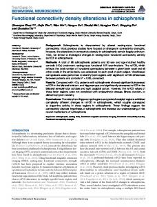
caused the paradoxical effect of reducing the number of auricular beats" (14). In 1955, A. Brasil again pointed out, as part of the so-called "autonomic sinoatrial blockade", the ineffectiveness of atropine in inducing tachycardia in chronic chagasic patients (12). Among these original studies were those on the participation of nonspecific mechanisms of depression of cardiac vagal control linked to cardiac failure of any etiology (17,29). Starting in the 1960's, an extensi ve series of studies was undertaken in the Laboratory of Cardiac Catheterization of the Faculty of Medicine of Ribeirao Preto. The studies were carried out on chagasic patients carefully selected on the basis of the absence of current or previous manifestations of heart failure and demonstrated that chronic chagasic patients show: a) an abnormal response to pharmacological inhibition of the efferent cardiac parasympathetic system (non-evoked sinus tachycardia, like in normal individuals, by intravenous atropine administration) (5,48) (Fig. 2). b) Abnormal response to reflex inhibition by physiological stimuli of the efferent cardiac parasympathetic system (deficient sinus tachycardiac response during the phase of hypotensive compression of the Valsalva maneuver (44), at low-intensity levels of
HEART RATE RESPONSES TO ATROPINE
-+60
130
FUNCTIONAL REPERCUSSIONS OF PARASYMPATHETIC CARDIAC DAMAGE
----
e120
~1l0 jlOO ~90
774
Q::
~
:>
80
~ 70 ~60 C
A
C
f-
I
•••
c:
'j;
3i40
.8 .....
I
The original papers by Carlos Chagas and collaborators mention a peculiar absence of the chronotropic response to the vagolytic agent atropine in chagasic patients: "On the basis of a large number of cases, we conclude that the dromotropic action of this drug is quite considerable and that its habitual chronotropic action is very small in many cases". "In the presence of complete blockade, the dromotropic action of the drug was absent. In the same cases, the chronotropic action was of low intensity and was often in the negative direction, i.e., it
IV
NORMALS CHAGASICS
CHAGASICS
NORMALS
0.04 mglkg
~-I••••
~+20
- I
Ii: ~O
-