MOLECULAR AND CELLULAR BIOLOGY, Nov. 2003, p. 8345–8351 0270-7306/03/$08.00⫹0 DOI: 10.1128/MCB.23.22.8345–8351.2003 Copyright © 2003, American Society for Microbiology. All Rights Reserved.
Vol. 23, No. 22
Functional Characterization of a Testis-Specific DNA Binding Activity at the H19/Igf2 Imprinting Control Region Aaron B. Bowman,† John M. Levorse, Robert S. Ingram, and Shirley M. Tilghman* Howard Hughes Medical Institute and Department of Molecular Biology, Princeton University, Princeton, New Jersey 08544 Received 13 May 2003/Returned for modification 10 July 2003/Accepted 7 August 2003
The DNA methylation state of the H19/Igf2 imprinting control region (ICR) is differentially set during gametogenesis. To identify factors responsible for the paternally specific DNA methylation of the ICR, germ line and somatic extracts were screened for proteins that bind to the ICR in a germ line-specific manner. A specific DNA binding activity that was restricted to the male germ line and enriched in neonatal testis was identified. Its three binding sites within the ICR are very similar to the consensus sequence for nuclear receptor extended half sites. To determine if these binding sites are required for establishment of the paternal epigenetic state, a mouse strain in which the three sites were mutated was generated. The mutated ICR was able to establish a male-specific epigenetic state in sperm that was indistinguishable from that established by the wild-type ICR, indicating that these sequences are either redundant or have no function. An analysis of the methylated state of the mutant ICR in the soma revealed no differences from the wild-type ICR but did uncover in both mutant and wild-type chromosomes a significant relaxation in the stringency of the methylated state of the paternal allele and the unmethylated state of the maternal allele in neonatal and adult tissues. The reciprocal imprinting of the maternally expressed H19 and paternally expressed Igf2 genes in mammals depends on a 2-kb differentially methylated imprinting control region (ICR) upstream of the H19 gene (21, 27). The ICR is a bifunctional genetic element, acting on the unmethylated maternal chromosome as a chromatin boundary to insulate enhancers downstream of H19 from the Igf2 promoter without hindering expression of the H19 promoter (1, 11, 16, 17). In sperm the ICR is methylated, and that methylation spreads in the embryo to the H19 promoter to silence the gene (9). The ICR methylation also inactivates the chromatin boundary, thereby permitting the enhancers to activate the Igf2 promoter. It has been shown recently that the activity of the chromatin boundary requires the binding of the transcription factor CTCF and that methylation inhibits that binding (1, 11, 14). Conversely CTCF binding to the ICR is required to maintain the unmethylated state of the maternal ICR in the soma (25, 26). Thus, DNA methylation and CTCF binding are mutually exclusive at the ICR. This has led to the proposal that the differential DNA methylation of the ICR that is set in the germ line is directly responsible for dictating the functional state of the ICR. A key unanswered question is the mechanism whereby the methylated states are set differently in the two germ lines. One plausible candidate has been ruled out by Schoenherr et al. (26), who recently demonstrated that CTCF itself is not required for proper establishment of the methylation of the ICR in sperm or the absence of methylation in oocytes. The differential epigenetic states (or imprints) are inherited in the fertilized egg and survive genome-wide demethylation
and remethylation events early in development (3). The exception to this rule is the embryonic germ line. Shortly after primordial germ cells are set aside and prior to sexual dimorphism the majority of the imprints on the parental chromosomes are erased. At the H19/Igf2 locus the ICR begins to show loss of methylation as early as 11.5 days postcoitem (dpc) and that loss continues until 12.5 to 13.5 dpc. Methylation of the ICR begins in prospermatogonia, around 14.5 dpc, and is almost complete by the end of the type A spermatogonial stage, as measured in cells derived from neonates at 8 days postpartum (dpp) (6, 29). The remaining small fraction of hypomethylated chromosomes reach full methylation by the spermatid stage. Interestingly it has been observed that the paternally inherited ICR acquires its methylation earlier than the maternally inherited ICR, suggesting that a full erasure of the paternal epigenetic state may not occur in the male germ line (5, 6). The acquisition of maternal methylation at imprinted loci is thought to occur during the oocyte growth phase (20, 23). One DNA methyltransferase family member, Dnmt3L, has been implicated in the establishment of maternal methylation imprints, but its role in male methylation could not be assessed because the homozygous males are sterile (2, 13). The mechanism by which de novo methyltransferases are specifically targeted to the correct ICRs in the appropriate germ line is unknown. Models for directing methylation establishment in a germ line-specific fashion fall into two classes: one in which a protein is needed to protect the ICR from methylation and one in which a protein is required to target methylation to the ICR. While these models are not mutually exclusive, they require at least one germ line-specific DNA binding protein at the time of imprint establishment. For this reason we examined germ line and somatic protein extracts for a germ line-specific DNA binding activity capable of binding to the H19/Igf2 ICR at the time of imprint establishment.
* Corresponding author. Mailing address: Howard Hughes Medical Institute and Department of Molecular Biology, Princeton University, Princeton, NJ 08544. Phone: (609) 258-6100. Fax: (609) 258-1615. E-mail:
[email protected]. † Present address: Howard Hughes Medical Institute, Baylor College of Medicine, Houston, TX 77030. 8345
8346
BOWMAN ET AL.
MOL. CELL. BIOL. TABLE 1. Sequences of oligonucleotide probes Sequencea
Probe
AB-1 ............................................................................................GACTCGGACTCCCAAATCAACAAGGTCGGCTTACTCTCTGCAAAGAATC AB-1bsp ......................................................................................GACTCGGACTCCCAAATCAACCTGCGGCTTACTCTCTGCAAAGAATC AB-1stu .......................................................................................GACTCGGACTCCCAAATCAACAGGCCTCGGCTTACTCTCTGCAAAGAATC AB-2 ............................................................................................GCAATATCCCAGGGTCACCCAAATAGGGATTCATAGGGGTGGTAAGATG AB-2avr .......................................................................................GCAATATCCCTAGGCACCCAAATAGGGATTCATAGGGGTGGTAAGATG AB-2bsr .......................................................................................GCAATATCCCTGTACACCCAAATAGGGATTCATAGGGGTGGTAAGATG AB-2A .........................................................................................CGTTAATCCCAGGGTCACCCAAATAGGGATTCATAGGGGTGGTAAGATG AB-2B .........................................................................................GCAATTAGGGAGGGTCACCCAAATAGGGATTCATAGGGGTGGTAAGATG AB-2C .........................................................................................GCAATATCCCTCCCACACCCAAATAGGGATTCATAGGGGTGGTAAGATG AB-2D .........................................................................................GCAATATCCCAGGGTGTGGGAAATAGGGATTCATAGGGGTGGTAAGATG AB-2E .........................................................................................GCAATATCCCAGGGTCACCCTTTATGGGATTCATAGGGGTGGTAAGATG AB-2F..........................................................................................GCAATATCCCAGGGTCACCCAAATACCCTATCATAGGGGTGGTAAGATG AB-3 ............................................................................................GGGTAGCTCCTTCAGTCTTGCGCCCTTCACGATCGATCGGTTCACTCTC AB-3spe.......................................................................................GGGTAGCTCCTTCAGTCTTGCGCCCTTACTAGTCGATCGGTTCACTCTC Nonspecific .................................................................................GGTTGGTGAGAAAATAGAGATTCTATTTTCATGTCCGGGGGATGAGCGT a
Mutated bases are underlined.
MATERIALS AND METHODS Protein extracts. Tissues were frozen in liquid nitrogen and stored at ⫺80°C until use. For neonatal testis and ovary at least 40 pairs of gonads were pooled. Tissues were homogenized with a Dounce homogenizer in a mixture containing 20 mM Tris-HCl (pH 7.9), 250 mM sucrose, 450 mM NaCl, 2 mM MgCl2, 2 mM CaCl2, 0.1% Triton X-100, and 1⫻ EDTA-free complete protease inhibitor cocktail (Roche). Nuclei were allowed to lyse for at least 15 min on ice. Supernatants were collected following centrifugation at 12,000 and 180,000 relative centrifugal force units, frozen in liquid nitrogen, and stored at ⫺80°C until use. EMSAs. A series of partially overlapping double-stranded 49-bp oligonucleotide probes that cover the H19 ICR were synthesized and used for the electrophoretic mobility shift assay (EMSA) screen (all sequences available upon request). The top strands of the probes described in this paper, written 5⬘ to 3⬘ relative to the direction of H19 transcription, are listed in Table 1. Probes were labeled with T4 polynucleotide kinase individually, annealed, and purified with a G50 column according to standard protocols. Gel shift reactions were performed in a mixture containing 20 mM HEPES (pH 7.9), 150 mM KCl, 5 mM MgCl2, 2 mM fresh dithiothreitol, 1 to 2 g of poly(dI:dC), 5% glycerol, 0.5% Triton X-100, probe at 20,000 cpm, 1 l of protein extract, and various concentrations of competitor probe. Reaction mixtures were incubated for 30 min and analyzed on a 5% acrylamide gel in 0.25⫻ Tris-borate-EDTA. Generation of mutant binding sites in mice. Mutations in the three AB binding sites were introduced into a 4.7-kb XbaI genomic DNA fragment (from ⫺5.5 to ⫺0.8 kb relative to the H19 transcription start site). The mutations correspond to AB-1stu, AB-2bsr, and AB-3spe in Table 1. The modified fragment was substituted for the XbaI fragment in the targeting construct described by Schoenherr et al. (26). The vector is a modified pACN derivative that utilizes a neomycin resistance gene (neo) and the cre recombinase gene under the control of a testis-specific promoter, both flanked by loxP sites (4). The final construct was sequenced over all cloning junctions and PCR-derived fragments to confirm its identity. CJ7 embryonic stem (ES) cells were targeted and chimeric animals were generated as previously described (26). Targeting at the endogenous locus was confirmed by Southern blot analysis with both external and internal probes. Excision of the neo gene was confirmed by Southern and PCR analyses. Mice were genotyped by PCR, and the results were confirmed by Southern blot analysis for the first two generations. PCR genotyping was performed on tail DNA with the primers 5⬘-TATTCTTGGACGTCTGCTGAATC and 5⬘-TTGA GAACGTTTTATCAAGGACTAGC. The product, which covers mutant site AB-1stu, was digested with StuI to detect the presence of the mutant allele. PCR genotyping to detect excision of the neo gene was performed with three primers: a single forward primer, 5⬘-CCATCTAGAACAATTCGGCGC, and two reverse primers, 5⬘-TACTATCCCACGGAGTATGAAC (which amplifies only the excised allele) and 5⬘-CGGCGAGGATCTCGTCGTGAC (which amplifies only the unexcised allele). Bisulfite sequencing. Five micrograms of genomic DNA was denatured in 20 mM NaOH for 15 min at 37°C. Denatured DNA was deaminated with sodium bisulfite (3.3 M) and hydroquinone (0.4 mM) under mineral oil, cycling between 240 min at 55°C and 5 min at 95°C for 16 h. DNA was purified by Qiagen PCR purification and desulfonated with 0.3 M NaOH for 15 min at 37°C, followed by neutralization with 3 M ammonium acetate, pH 7.0, and ethanol precipitation.
The treated DNA was resuspended in 50 l of water and stored in the dark at ⫺20°C until use. Fully nested PCR was performed as described previously with primers 5⬘-CCTCATAAAACCCATAACTAT and 5⬘-GTAAGGAGATTATGT TTTATTTTTGG (23, 28). PCR products were cloned, and colonies were picked randomly, irrespective of colony size or color (except dark blue). More than 99% of non-CpG cytosines were converted in all clones sequenced. Mutant and wild-type chromosomes were distinguished by differences in sequence over the mutation at AB-1.
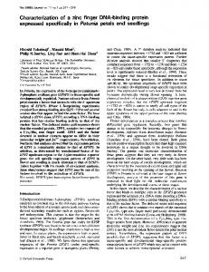
RESULTS Screen for germ line-specific binding proteins. To identify factors responsible for establishing the epigenetic state at the H19/Igf2 ICR, protein extracts were generated from neonatal mouse gonads at a time when imprints were being established (6). Mouse testes from 1- to 4-dpp mice and ovaries from 7- to 10-dpp mice were employed, along with young adult mouse liver as a somatic tissue control. Although neonatal ovaries are enriched for germ cells compared to adult ovaries, the neonatal ovary extracts contain significantly less germ cell-derived proteins than the neonatal testis extracts. Fifty-nine partially overlapping 49-bp oligonucleotides spaced every 21 bases and covering both regions of nuclease hypersensitivity in the mouse ICR were synthesized (12). This design allowed for every possible hexamer-sized binding site in the region to be tested with at least an 11-bp overhang on either side. The extracts were tested for protein binding by EMSA with all 59 probes. A male germ line-specific DNA binding activity. The survey uncovered a number of novel and previously known (e.g., CTCF) protein binding activities. One of these novel activities was testis specific and bound at three sites within the ICR. These sites were designated sites AB-1, AB-2, and AB-3 (Fig. 1A to C). The three sites are located between the pairs of CTCF sites that define hypersensitive regions I and II in the mouse ICR (11) (Fig. 1A). As shown for AB-2 in Fig. 1B, the AB binding activity is restricted to testis extracts and is enriched in neonatal testes compared to adult testes. Imprint establishment occurs primarily in spermatogonial cells, which are highly enriched in neonatal testes (⬃100% of germ cells) compared to adult testes, where they represent less than 1% of germ cells. Furthermore the activity is not detected in ovary, brain, liver, or 11- or 16-dpc embryo extracts (Fig. 1B and data not shown).
VOL. 23, 2003
NEONATAL TESTIS-SPECIFIC DNA BINDING SITE IN H19 ICR
8347
FIG. 1. Characterization of the AB binding sites by EMSA. (A) Illustration of the relative locations of AB and CTCF binding sites within the ICR. (B to E) Positions of AB binding activity (●). (B) AB-2 binding activity in protein extracts from various tissues. NT, neonatal testis; L, adult liver; NO, neonatal ovary; B, adult brain; T, adult testis; E, 16-dpc embryo. (C) Binding of AB-1, AB-2, and AB-3 in neonatal testis extract in the absence (⫺) or presence of AB-2 or a nonspecific probe. (D and E) AB binding activity with wild-type (WT) and mutant probes (see Table 1).
8348
BOWMAN ET AL.
MOL. CELL. BIOL.
TABLE 2. Sequence alignment of AB binding sites with consensus nuclear receptor extended half-site binding site Site
NR ext. halfc AB-1 AB-2 AB-3 AB-1stu AB-2bsr AB-3spe AB-2A AB-2E
Sequence alignment (5⬘–3⬘)a
TCAAGGTCA CCAAATCAACAAGGTCGGCTTACTC GCAATATCCCAGGGTCACCCAAATA TGCGCCCTTCACGATCGATCGGTTC CCAAATCAACAGGCCTCGGCTTACC GCAATATCCCTGTACACCCAAATAG TGCGCCCTTACTAGTCGATCGGTTC CGTTAATCCCAGGGTCACCCAAATA GCAATATCCCAGGGTCACCCTTTAT
Bindingb
⫹⫹ ⫹⫹⫹ ⫹ ⫺ ⫺ ⫺ ⫹⫹⫹ ⫹⫹⫹
a Identity to consensus sequence is indicated by boldface. Mutated bases are underlined. b Relative strength of EMSA bandshift. ⫺, none; ⫹, weak; ⫹⫹, strong; ⫹⫹⫹, strongest. c Nuclear receptor extended half site binding domain consensus sequence.
To determine if binding was sequence specific and to establish whether the three sites bound the same factor, we tested whether the three sites competed for binding. Figure 1C shows that AB-2 competes effectively with both AB-1 and AB-3 for binding. In contrast a nonspecific fragment was unable to act as a competitor. Similar competition experiments in different combinations established that AB-2 binds with the strongest affinity, whereas AB-3 has the weakest affinity. Characterization of the AB binding site. Sequence comparison of the three binding sites revealed a single shared sequence motif, CANG(A/G)TC, with homology to the orphan nuclear receptor family binding site (TCAAGGTCA) called the extended half-site or SF-1 binding site (Table 2). Furthermore there is a strong correlation between the relative affinity of the binding site and its similarity to the SF-1 consensus. To determine if this region of homology was required for AB binding activity, new probes with mutations in the core region of homology that altered 5 or fewer bases without changing any CpG residues were synthesized (Table 1). In every case, the mutations eliminated binding to the probe (Fig. 1D and Table 2), establishing that the region of homology is required for AB binding activity. To determine the extent of the binding site a series of probes with mutations across the length of the AB-2 were synthesized and the mutant probes were tested for binding (AB-2A to -2F in Table 1). Three mutant probes, covering 15 bases centered on the region of homology, failed to bind protein, while probes with mutations peripheral to these 15 bases had no effect on binding (Fig. 1E and Table 2). This further localized the binding site to no more than 15 bases, any 5 of which are required for binding activity. Mutating the AB binding sites in vivo. We set out to determine the potential role of the binding activity in paternal imprint establishment by generating mutations at all three sites within the ICR by homologous recombination in mouse ES cells (Fig. 2A). The mutations (AB-1stu, AB-2bsr, and AB3spe) eliminate AB binding activity as determined by EMSA (Fig. 1D and Table 2). The targeting construct included a 4.7-kb genomic DNA fragment containing the ICR, into which the three mutations had been incorporated, as well as genes encoding neomycin resistance and Cre recombinase under the control of a testis-specific promoter (Fig. 2C). The genes were surrounded by loxP sites, so that they could be excised in the germ line of male chimeras (4, 26).
Two targeted ES cell lines containing the mutated AB binding sites were identified by Southern analysis, and their structures were confirmed by hybridization with probes that lay outside both sides of the targeting vector (Fig. 2B and data not shown). The presence of all three mutations was confirmed by PCR analysis, as illustrated in Fig. 2D, using primers that amplified the region containing the StuI mutation at AB-1. F1 intercrosses yielded the expected frequency of wild type, heterozygote, and homozygote progeny (Fig. 2D). Establishment of imprinting in AB binding site mutant animals. If the AB binding sites are required to establish the methylation imprint on the paternal chromosome, then we would expect that male mutant mice would transmit an unmethylated ICR. Initial assessment of the imprinting status of the paternal inherited grandpaternal mutant chromosome (progeny of male F1 mice derived from a male chimera) by Southern blotting revealed wild-type levels of methylation for a paternal chromosome (data not shown). Furthermore, the mutant progeny were indistinguishable in size from wild-type littermates (data not shown). This suggested that at a gross level the mutations did not abolish the establishment of the paternal imprint on a grandpaternal chromosome. To ask whether the AB binding sites are required to establish a paternal imprint on an unmethylated chromosome that had been inherited from a mother, we examined 6-dpp progeny of a male F2 heterozygote that had inherited the mutation from his mother. To look for moresubtle changes in DNA methylation patterns, we used bisulfite DNA methylation analysis over hypersensitive region 1 (HS1) of the ICR that contains the AB-1 binding site, CTCF sites 1 and 2, and 16 CpG dinucleotides. The majority of the paternal grandmaternal mutant chromosomes were heavily methylated and indistinguishable from wild-type paternal chromosomes (Fig. 3A). However, we were surprised to see that 3 of 27 chromosomes had 63% or less CpGs methylated and that one chromosome was only 50% methylated. This level of undermethylation on the paternal chromosome had not been previously observed in studies employing embryonic or germ line tissues (24, 28, 30). To determine if this degree of undermethylation was due to the mutant sites, we sequenced individual chromosomes from a 6-dpp wild-type littermate. While we could not distinguish the maternal and paternal chromosomes by sequence, of the 18 chromosomes that we infer to be paternal (any chromosome with ⬎50% methylation), the overall level of methylation was similar to that of the mutant chromosomes (Fig. 3B). For example one wild-type chromosome had only 10 of 16 CpGs methylated. To allow us to definitively characterize the methylation status of paternally inherited wild-type chromosomes, we sequenced chromosomes from the progeny of a female F1 heterozygote. The level of methylation seen on these wild-type paternal chromosomes is indistinguishable from that on the mutant paternal chromosomes (Fig. 3D). Thus the variation in methylation observed on paternal mutant chromosomes cannot be attributed to the mutations, but rather represents the normal degree of variation observed at the locus. Establishment of the paternal imprint occurs before meiosis when germ cells are diploid. To determine if the wild-type chromosome can rescue a mutant phenotype in trans we sequenced F3 chromosomes from homozygous fathers. The level
VOL. 23, 2003
NEONATAL TESTIS-SPECIFIC DNA BINDING SITE IN H19 ICR
8349
FIG. 2. Mutating the AB binding sites in ES cells and mice. (A) Illustration of the targeting strategy, with (from top to bottom) the structure of the wild type chromosome (WT), the targeting construct, the modified chromosome in ES cells, and the vector-excised chromosome (F1). The location of the external probe used in panel B is indicated, along with the SpeI sites. Rectangles, H19 structural gene and ICR. (B) Southern analysis of SpeI-digested DNA isolated from ES cell colonies. T, correctly targeted ES cells used to generate chimeric animals; ⴱ, incorrectly targeted ES cells. (C) PCR genotyping of F1 offspring. PCR discriminates between the animals in which the selectable marker has not been excised (flox’ed allele) and those that have undergone its deletion (⌬ Neo 䡠 Cre). (D) PCR genotyping of the mutant allele in the progeny of an F1 intercross. The mutant product was digested with SpeI.
of methylation on these chromosomes is indistinguishable from those on other mutant and wild-type paternally inherited chromosomes (Fig. 3C). Although there is no detectable AB binding activity in the female germ line, we determined the methylation state of maternally inherited mutant chromosomes. The overall methylation states of maternally inherited mutant chromosomes and maternally inherited wild-type chromosomes in heterozygous
animals were indistinguishable (Fig. 4A and B). The majority of maternal chromosomes are completely devoid of methylation. However, a minority of chromosomes have a significant level of methylation. We detected 8 of 68 definitive wild-type maternal chromosomes with 25% or more of CpGs methylated (Fig. 4B). One chromosome had 12 of 16 methylated CpGs, a level of methylation that has not been previously reported. To determine if the relaxation in the parent-specific meth-
8350
BOWMAN ET AL.
FIG. 3. Methylation analysis of neonatal AB binding site mutant animals. Shown are bisulfite methylation sequence data obtained from 6-dpp liver DNA of mutant alleles (PM and MM, reflecting whether the chromosome was inherited from a father or mother, respectively) or wild-type alleles (PWT and MWT). The region contains 16 CpG residues (open [unmethylated] and solid [methylated] circles). Numbers indicate the numbers of individual chromosomes (when more than one) with identical CpG methylation patterns. Vertical line between CpG 9 and 10, position of the AB-1 mutation. CpG residues 4 and 5 are within CTCF site 1; CpG residues 11 to 13 are within CTCF site 2. (A) Paternal grandmaternal mutant chromosomes. (B) Inferred paternal wildtype chromosomes, based on ⬎50% methylation. (C) Paternal mutant chromosomes of progeny from a paternal homozygote (PM/M). (D) Paternal wild-type chromosomes of animals inheriting a maternal mutation.
MOL. CELL. BIOL.
were mutated so that they no longer bound the protein activity, we found that these mutant sites had no effect on either imprint establishment or maintenance. Either these binding sites have no function in imprinting or they are redundant with other factors that bind the ICR. While no other DNA binding proteins that were unique to neonatal testis were identified in the screen, we detected 10 sites that bound a factor in neonatal testis that was more abundant in adult testis and brain but undetectable in neonatal ovary and adult liver (data not shown). The sequences of the core AB binding sites and yeast onehybrid analysis (data not shown) suggest the possibility that the sites bind a member of the nuclear receptor family of proteins. Nuclear receptors have been implicated in changes in epigenetic states in other systems based on their ability to recruit DNA methyltransferases and histone deacetylases to establish repressive chromatin epigenetic states (8, 10, 31). Such an activity is consistent with a factor acting in the male germ line to establish the repressed H19/Igf2 paternal epigenetic state. The paternal and maternal H19/Igf2 ICR is inherited in either a fully methylated or fully unmethylated state, respectively, and it had been thought that this epigenetic state was maintained throughout the life of the animal. Our DNA methylation analysis revealed low but readily detectable hypomethylated paternal chromosomes and hypermethylated maternal chromosomes. For example, we identified wild-type maternal chromosomes in which all CpG residues within CTCF binding sites 1 and 2 are fully methylated. While such relaxation of the differential methylation at the H19/Igf2 ICR has been reported in cells exposed to in vitro culture (7, 19, 22), it has not been previously reported in vivo, perhaps because the majority of bisulfite sequence analyses on somatic cell chromosomes have been performed on embryonic tissues (24, 28, 30). In addition
ylation differences that we observed in neonates become more pronounced on the mutant chromosome with age, which would suggest a role in maintaining the epigenetic state, we performed bisulfite sequence analysis on 35-dpp liver DNA of paternally inherited mutant and wild-type chromosomes. The level of methylation was indistinguishable from that of 6-dpp neonatal liver, and no difference between wild-type and mutant chromosomes was seen (Fig. 4C and D). Undermethylated paternal chromosomes were observed at approximately the same frequency as in younger animals, and maternal chromosomes showed similar levels of methylation (data not shown). Thus the AB sites have no detectable role in imprint establishment or maintenance. DISCUSSION The mechanisms by which genomic imprints are differentially established in the male and female germ line are unknown. In an effort to understand this important issue in epigenetics, we set out to identify germ cell-specific factors that bind to the ICR of the H19/Igf2 locus at the time of imprint establishment. Utilizing protein extracts prepared from both ovaries and testes we identified a binding activity that appears to be unique to the male germ line that bound to three sites within the ICR. However, when we tested the in vivo function of these sites by generating a mouse strain in which the sites
FIG. 4. Bisulfite methylation analysis of adult AB binding site mutant animals. Bisulfite sequencing was performed on 6-dpp (A and B) or 35-dpp (C and D) liver DNA. (A) Maternal grandmaternal mutant chromosomes. (B) Maternal wild-type chromosomes from animals inheriting mutant paternal chromosomes. (C) Paternal grandmaternal mutant chromosomes. (D) Inferred paternal chromosomes of wildtype littermates of animals in panel C. All nomenclature is described in the Fig. 3 legend.
NEONATAL TESTIS-SPECIFIC DNA BINDING SITE IN H19 ICR
VOL. 23, 2003
assessments of adult levels of methylation have predominantly used methods that would not detect the low fraction of chromosomes with aberrant methylation. A notable exception comes from a study in which the methylation state of the human H19 ICR in normal adult lymphocytes was examined as a control: 6% of all chromosomes sequenced after bisulfite treatment were found to have 40 to 50% levels of methylation, consistent with our measured level of ⬃5% (18). This suggests that relaxation in the stringency of differential methylation is not exclusively associated with the mouse ICR. Though we find that these hypomethylated and hypermethylated chromosomes are rare, their existence illustrates that the maintenance of differential methylation on maternal and paternal chromosomes is not absolute. There does not appear to be an intrinsic bias in favor of either methylation or demethylation of the ICR in the soma, but rather an apparent stochastic inability to maintain a fixed state. CTCF binding sites have been shown in vivo to be necessary to maintain the unmethylated state (25, 26) but may not be sufficient. For example Hori et al. (15) have identified a single conserved dyad oct-binding protein motif within the mouse ICR that is required for the demethylation of transgenes containing a low level of methylation after transfection into embryonal carcinoma cells but no effect on highly methylated transgenes. No protein has been implicated to date in maintaining the methylated state of the paternal ICR. ACKNOWLEDGMENTS We thank M. Cleary, S. Cole, D. Katz, D. Mancini-DiNardo, C. Schoenherr, E. Semenova, S. Steele, and E. Wieschaus for helpful discussions of this work and Georgia Guan for technical assistance. A.B.B. is a Life Sciences Research Foundation Fellow sponsored by the Howard Hughes Medical Institute. S.M.T. was an investigator of the Howard Hughes Medical Institute. This work was supported by a grant from the National Institute for General Medical Sciences. REFERENCES 1. Bell, A. C., and G. Felsenfeld. 2000. Methylation of a CTCF-dependent boundary controls imprinted expression of the Igf2 gene. Nature 405:482– 485. 2. Bourc’his, D., G. L. Xu, C. S. Lin, B. Bollman, and T. H. Bestor. 2001. Dnmt3L and the establishment of maternal genomic imprints. Science 294: 2536–2539. 3. Brandeis, M., T. Kafri, M. Ariel, J. R. Chaillet, J. McCarrey, A. Razin, and H. Cedar. 1993. The ontogeny of allele-specific methylation associated with imprinted genes in the mouse. EMBO J. 12:3669–3677. 4. Bunting, M., K. E. Bernstein, J. M. Greer, M. R. Capecchi, and K. R. Thomas. 1999. Targeting genes for self-excision in the germ line. Genes Dev. 13:1524–1528. 5. Davis, T. L., J. M. Trasler, S. B. Moss, G. J. Yang, and M. S. Bartolomei. 1999. Acquisition of the H19 methylation imprint occurs differentially on the parental alleles during spermatogenesis. Genomics 58:18–28. 6. Davis, T. L., G. J. Yang, J. R. McCarrey, and M. S. Bartolomei. 2000. The H19 methylation imprint is erased and re-established differentially on the parental alleles during male germ cell development. Hum. Mol. Genet. 9:2885–2894. 7. Dean, W., L. Bowden, A. Aitchison, J. Klose, T. Moore, J. J. Meneses, W. Reik, and R. Feil. 1998. Altered imprinted gene methylation and expression in completely ES cell-derived mouse fetuses: association with aberrant phenotypes. Development 125:2273–2282. 8. Di Croce, L., V. A. Raker, M. Corsaro, F. Fazi, M. Fanelli, M. Faretta, F. Fuks, F. Lo Coco, T. Kouzarides, C. Nervi, S. Minucci, and P. G. Pelicci. 2002. Methyltransferase recruitment and DNA hypermethylation of target promoters by an oncogenic transcription factor. Science 295:1079–1082.
8351
9. Drewell, R. A., C. J. Goddard, J. O. Thomas, and M. A. Surani. 2002. Methylation-dependent silencing at the H19 imprinting control region by MeCP2. Nucleic Acids Res. 30:1139–1144. 10. Fuks, F., W. A. Burgers, N. Godin, M. Kasai, and T. Kouzarides. 2001. Dnmt3a binds deacetylases and is recruited by a sequence-specific repressor to silence transcription. EMBO J. 20:2536–2544. 11. Hark, A. T., C. J. Schoenherr, D. J. Katz, R. S. Ingram, J. M. Levorse, and S. M. Tilghman. 2000. CTCF mediates methylation-sensitive enhancerblocking activity at the H19/Igf2 locus. Nature 405:486–489. 12. Hark, A. T., and S. M. Tilghman. 1998. Chromatin conformation of the H19 epigenetic mark. Hum. Mol. Genet. 7:1979–1985. 13. Hata, K., M. Okano, H. Lei, and E. Li. 2002. Dnmt3L cooperates with the Dnmt3 family of de novo DNA methyltransferases to establish maternal imprints in mice. Development 129:1983–1993. 14. Holmgren, C., C. Kanduri, G. Dell, A. Ward, R. Mukhopadhya, M. Kanduri, V. Lobanenkov, and R. Ohlsson. 2001. CpG methylation regulates the Igf2/ H19 insulator. Curr. Biol. 11:1128–1130. 15. Hori, N., H. Nakano, T. Takeuchi, H. Kato, S. Hamaguchi, M. Oshimura, and K. Sato. 2002. A dyad oct-binding sequence functions as a maintenance sequence for the unmethylated state within the H19/Igf2-imprinted control region. J. Biol. Chem. 277:27960–27967. 16. Kaffer, C. R., M. Srivastava, K. Y. Park, E. Ives, S. Hsieh, J. Batlle, A. Grinberg, S. P. Huang, and K. Pfeifer. 2000. A transcriptional insulator at the imprinted H19/Igf2 locus. Genes Dev. 14:1908–1919. 17. Kanduri, C., V. Pant, D. Loukinov, E. Pugacheva, C. F. Qi, A. Wolffe, R. Ohlsson, and V. V. Lobanenkov. 2000. Functional association of CTCF with the insulator upstream of the H19 gene is parent of origin-specific and methylation-sensitive. Curr. Biol. 10:853–856. 18. Kerjean, A., J. M. Dupont, C. Vasseur, D. Le Tessier, L. Cuisset, A. Paldi, P. Jouannet, and M. Jeanpierre. 2000. Establishment of the paternal methylation imprint of the human H19 and MEST/PEG1 genes during spermatogenesis. Hum. Mol. Genet. 9:2183–2187. 19. Khosla, S., W. Dean, D. Brown, W. Reik, and R. Feil. 2001. Culture of preimplantation mouse embryos affects fetal development and the expression of imprinted genes. Biol. Reprod. 64:918–926. 20. Kono, T., Y. Obata, T. Yoshimzu, T. Nakahara, and J. Carroll. 1996. Epigenetic modifications during oocyte growth correlates with extended parthenogenetic development in the mouse. Nat. Genet. 13:91–94. 21. Leighton, P. A., R. S. Ingram, J. Eggenschwiler, A. Efstratiadis, and S. M. Tilghman. 1995. Disruption of imprinting caused by deletion of the H19 gene region in mice. Nature 375:34–39. 22. Liang, L., C. Kanduri, M. Pilartz, K. Svensson, J. H. Song, P. Wentzel, U. Eriksson, and R. Ohlsson. 2000. Dynamic readjustment of parental methylation patterns of the 5⬘-flank of the mouse H19 gene during in vitro organogenesis. Int. J. Dev. Biol. 44:785–790. 23. Lucifero, D., C. Mertineit, H. J. Clarke, T. H. Bestor, and J. M. Trasler. 2002. Methylation dynamics of imprinted genes in mouse germ cells. Genomics 79:530–538. 24. Olek, A., and J. Walter. 1997. The pre-implantation ontogeny of the H19 methylation imprint. Nat. Genet. 17:275–276. 25. Pant, V., P. Mariano, C. Kanduri, A. Mattsson, V. Lobanenkov, R. Heuchel, and R. Ohlsson. 2003. The nucleotides responsible for the direct physical contact between the chromatin insulator protein CTCF and the H19 imprinting control region manifest parent of origin-specific long-distance insulation and methylation-free domains. Genes Dev. 17:586–590. 26. Schoenherr, C. J., J. M. Levorse, and S. M. Tilghman. 2003. CTCF maintains differential methylation at the Igf2/H19 locus. Nat. Genet. 33:66–69. 27. Thorvaldsen, J. L., K. L. Duran, and M. S. Bartolomei. 1998. Deletion of the H19 differentially methylated domain results in loss of imprinted expression of H19 and Igf2. Genes Dev. 12:3693–3702. 28. Tremblay, K. D., K. L. Duran, and M. S. Bartolomei. 1997. A 5⬘ 2-kilobasepair region of the imprinted mouse H19 gene exhibits exclusive paternal methylation throughout development. Mol. Cell. Biol. 17:4322–4329. 29. Ueda, T., K. Abe, A. Miura, M. Yuzuriha, M. Zubair, M. Noguchi, K. Niwa, Y. Kawase, T. Kono, Y. Matsuda, H. Fujimoto, H. Shibata, Y. Hayashizaki, and H. Sasaki. 2000. The paternal methylation imprint of the mouse H19 locus is acquired in the gonocyte stage during foetal testis development. Genes Cells 5:649–659. 30. Warnecke, P. M., J. R. Mann, M. Frommer, and S. J. Clark. 1998. Bisulfite sequencing in preimplantation embryos: DNA methylation profile of the upstream region of the mouse imprinted H19 gene. Genomics 51:182–190. 31. Wei, L. N., X. Hu, D. Chandra, E. Seto, and M. Farooqui. 2000. Receptorinteracting protein 140 directly recruits histone deacetylases for gene silencing. J. Biol. Chem. 275:40782–40787.