Article
https://doi.org/10.1038/s41586-018-0027-0
Functional circuit architecture underlying parental behaviour
Johannes Kohl1, Benedicte M. Babayan2, Nimrod D. Rubinstein1, Anita E. Autry1, Brenda Marin-Rodriguez1, Vikrant Kapoor2, Kazunari Miyamishi3, Larry S. Zweifel4,5, Liqun Luo3, Naoshige Uchida2 & Catherine Dulac1*
Parenting is essential for the survival and wellbeing of mammalian offspring. However, we lack a circuit-level understanding of how distinct components of this behaviour are coordinated. Here we investigate how galanin-expressing neurons in the medial preoptic area (MPOAGal) of the hypothalamus coordinate motor, motivational, hormonal and social aspects of parenting in mice. These neurons integrate inputs from a large number of brain areas and the activation of these inputs depends on the animal’s sex and reproductive state. Subsets of MPOAGal neurons form discrete pools that are defined by their projection sites. While the MPOAGal population is active during all episodes of parental behaviour, individual pools are tuned to characteristic aspects of parenting. Optogenetic manipulation of MPOAGal projections mirrors this specificity, affecting discrete parenting components. This functional organization, reminiscent of the control of motor sequences by pools of spinal cord neurons, provides a new model for how discrete elements of a social behaviour are generated at the circuit level.
Although essential for survival at a multigenerational time scale, parental care entails sacrifices without immediate benefits for the caregiver, suggesting that this behaviour is driven by evolutionarily shaped, hard-wired neural circuits1,2. Parenting, similar to other naturalistic behaviours, comprises multiple coordinated components, such as specific motor patterns, an enhanced motivation to interact with infants, distinct hormonal states and often the suppression of other social activities such as mating. We aimed to exploit the recent identification of MPOAGal neurons as a key node in the control of parenting in mice3 to uncover organizational principles of associated neural circuits. We hypothesized that the function of MPOAGal neurons in parental behaviour requires integration of external signals, such as stimuli from pups and other environmental sources, and internal hormonal and metabolic information, as well as the ability to coordinate the motor, motivational, hormonal and social components of parenting.
Identity and activity of MPOAGal inputs
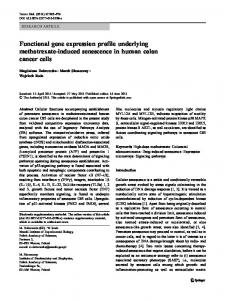
To determine brain-wide inputs into MPOAGal neurons, we used rabies virus-mediated retrograde trans-synaptic tracing4 (Fig. 1a), and found that MPOAGal neurons receive direct inputs from more than 20 areas in both male and female mice (Fig. 1b, c, Extended Data Fig. 1a and Extended Data Table 1). Presynaptic neurons within the MPOA itself provided the highest fractional input (approximately 20%), and hypothalamic inputs accounted for about 60% of the presynaptic neurons, suggesting that extensive local processing occurs (Fig. 1c). MPOAGal neurons also receive inputs from monoaminergic and neuropeptidergic modulatory areas, the mesolimbic reward system, pathways associated with pheromone-processing, and hypothalamic as well as septal areas involved in emotional states (Fig. 1c and Extended Data Fig. 1a). Inputs from the paraventricular hypothalamic nucleus (PVN), a key area for homeostatic and neuroendocrine control, were particularly abundant. Notably, MPOAGal neurons did not receive direct inputs from oxytocin (OXT)-secreting PVN (PVNOXT) neurons, which are implicated in parturition, lactation and maternal behaviour1,2,5, but instead received inputs from vasopressin-expressing PVN (PVNAVP) neurons, which are associated with the modulation of many social behaviours6 and nest
building7 (Fig. 1d). MPOAGal neurons also received inputs from AVP+, but not OXT+, neurons of the supraoptic nucleus (Extended Data Fig. 1d). Input fractions were similar in males and females, with a few exceptions (Fig. 1e, f and Extended Data Fig. 1a). Therefore, MPOAGal neurons appear to be anatomically well-positioned to integrate external (sensory) as well as internal (modulatory) signals that are relevant to parenting in both sexes. Next, we investigated MPOAGal input activation during parenting according to the animal’s sex and reproductive state. In laboratory mice, virgin females and sexually experienced males and females show parental behaviours, whereas virgin males typically attack and kill pups3,8,9. We combined rabies tracing with immunostaining for the activity marker Fos after parenting in primiparous females (mothers), virgin females and fathers (Fig. 1g) and compared the Fos+ fraction of input neurons between parental animals and non-pup-exposed controls (Fig. 1h–j). Local MPOA inputs were specifically activated during parenting in all groups (Fig. 1h–j), whereas the activation of other inputs was dependent on sex and reproductive state: in parents, but not virgin females, a subset of reward-associated and modulatory inputs were activated (Fig. 1h–j). Presynaptic neurons in pheromone-processing pathways (the medial amygdala (MeA) and bed nucleus of the stria terminalis (BNST)) were selectively activated in fathers and virgin females, but not in mothers (Fig. 1h–j). Because pup-directed aggression in virgin mice is pheromone-dependent3,8, the MeA–BNST pathway might remain partially active in sexually experienced males and parental virgin females, whereas it is fully silenced only in mothers. Intriguingly, the largest number of inputs was activated in fathers (Fig. 1j), and non-overlapping subsets of inputs were activated in mothers and virgin females (Fig. 1h, i). These results suggest that MPOAGal neurons perform different computations of inputs according to the animal’s sex and reproductive state.
Input–output logic of the MPOAGal circuit
To identify MPOAGal projections and synaptic targets, we infected MPOAGal neurons with adeno-associated viruses (AAVs) encoding the fluorophore tdTomato as well as the presynaptic marker
1 Howard Hughes Medical Institute, Department of Molecular and Cellular Biology, Center for Brain Science, Harvard University, Cambridge, MA, USA. 2Department of Molecular and Cellular Biology, Center for Brain Science, Harvard University, Cambridge, MA, USA. 3Howard Hughes Medical Institute, Department of Biology, Stanford University, Stanford, CA, USA. 4Department of Pharmacology, University of Washington, Seattle, WA, USA. 5Department of Psychiatry and Behavioral Sciences, University of Washington, Seattle, WA, USA. *e-mail:
[email protected]
N A t U r e | www.nature.com/nature
© 2018 Macmillan Publishers Limited, part of Springer Nature. All rights reserved.
RESEARCH Article a
c
b
Gal::cre
V
d
PVN
e
AVPe
Rabies OXT
Rabies TH
f
AHPM
h
Rabies
Brain region
MPOA
Rabies Fos
*
MPOA SNpc AVPe PVN MeA BNST AH VMH RM LS PVT DM MS VMPO PMV AHPM MnPO NAsh SON Arc NAc VOLT
i *** **
Parenting Control
0
5
10
Activated fraction (%)
15
VTA
MnPO
AHPM
MPOA
BMA
MeA VMPO SON
Fos
AAV-FLEx-TVA-mCherry AAV-FLEx-RG + EnvA-ΔG-rabies
PVN AH
VOLT
Rabies AVP
g
BNST MS
AVPe
AAV-FLEx-TVA-mCherry AAV-FLEx-RG + 14 days EnvA-ΔG-rabies
LC
LS
NAsh NAc
MPOA SNpc AVPe PVN MeA BNST AH VMH RM LS PVT DM MS VMPO PMV AHPM MnPO NAsh SON Arc NAc VOLT
***
Arc
j
***
V 0
>16 14 12 10 8 6 4 2