Jonathan ReizerS, Sarah L. Sutrina, Long-Fei Wus, Josef Deutscherll, Prasad Reddy 11, ...... of this protein (Kapadia et al., 1991; Liao et al., 1991). The. Bacillus ...
Vol. 267, No. 13, Issue of May 5, pp. 91.%%9169.1992 Printed in U.S.A.
THEJOURNAL OF BIOLOGICAL CHEMISTRY 0 1992 by The American Society for Biochemistry and Molecular Biology, Inc.
Functional Interactions between Proteins of the Phosphoeno1pyruvate:Sugar Phosphotransferase Systems of Bacillus subtilis and Escherichia coli* (Received for publication, June 5, 1991)
Jonathan ReizerS, Sarah L. Sutrina, Long-Fei Wus, Josef Deutscherll, Prasad Reddy 11, and Milton H. Saier, Jr. From the Department of Biology, University of California at SanDiego, La Jolla, California 92093-0116 and the IICenter for Advanced Research in Biotechnology, National Institute of Standards andTechnology, Rockville,Maryland 20850
Proteins of the phosphoeno1pyruvate:sugarphosphotransferase system (PTS) of Bacillus subtilis were overexpressed,purifiedtonear homogeneity, and characterized. The proteinsisolated include Enzyme I, HPr, the glucose-specific IIA domain of the glucosespecific Enzyme I1 (IIAg"), and the mannitol-specific IIA protein, IIAmt'. Site specific mutant proteins of IIAgk and HPr were also overexpressed and purified, and their properties were compared with those of the wild type proteins. These proteinsand their phosphorylated derivatives were characterized with respect totheir immunologicalcross-reactivities employing the Western blot technique and in terms of their migratory behavior during sodium dodecyl sulfate-gel electrophoresis, nondenaturing gel electrophoresis, and isoelectric focusing. The interactions between homologous and heterologous Enzymes I and HPrs, between homologous and heterologous HPrs and theIIAgl' proteins, and between homologous and heterologous IIAgl' proteins and IIBC"' of B. subtilis as well as IICBg"of Escherichia coli were defined and compared kinetically. The mutant HPrs andIIAg" proteins were also characterized kinetically as PTS phosphocarrier proteins and/oras inhibitors of the phosphotransferase reactions of the PTS. These studies revealedthat comof HPr in plexation of IIAgl' with the mutant form which serine 46 wasreplaced by aspartate (S46D) did not increase therate of phosphoryl transfer from phospho Enzyme I to S46DHPr more than when IIA"" was complexed to S46D HPr. These findings do not support a role for HPr(Ser-P) in the preferential utilization of one PTS carbohydrate relative to another. Functional analyses in E. coli established that IIAg" of B. subtilis can replace IIA"' of E. coli with respect both to sugar transport and to regulation of non-PTS permeases, catabolic enzymes, and adenylatecyclase. Site-specific mutations in histidyl residues 68 and 83 (H68A and H83A) inactivated IIAg" of B. subtilis with respect to phosphoryl transfer and its various regulatory roles.
* This work was supported by National Institute of Allergy and Infectious Diseases Grants 5R01 AI 21702 and 2R01 AI 14176. The costs of publication of this article were defrayed in part by the payment of page charges. This articlemust therefore be hereby marked "advertisement" in accordance with 18 U.S.C. Section 1734 solely to indicate this fact. $ T o whom correspondence and reprint requests should be addressed. Tel.: 619-534-4084;Fax: 619-534-7108. Present address: Laboratory of Microbiology, Bat. 406, INSA, 20 Ave. A. Einstein, 69621 Villeurbanne cedex, France. 11 Present address: Max-Planck Institute for System Physiology, Rheinlanddamm 201, D-4600, Dortmund 1, Germany.
The bacterial phosphoeno1pyruvate:sugarphosphotransferase system (PTS)l in enteric bacteria and all well characterized Gram-positive bacteria consists of two general energycoupling proteins, Enzyme I and HPr, as well as the sugarspecific permeases, the Enzymes I1 which consist of three functional domains, the IIA, IIB, and IIC domains (Saier, 1985;Reizer et al., 198813; Meadow et al., 1990; Saier and Reizer, 1990). In each permease, the IIA domain or protein, also sometimes referred to as an Enzyme 111, bears the first phosphorylation site, the IIB domain or protein bears the second phosphorylation site, and the IIC domain bears the transmembrane channel and sugar binding site (Saier and Reizer, 1990). The glucose phosphotransferase proteins of Escherichia coli have been characterized extensively both genetically and biochemically. Thus, Enzyme I, HPr, and the glucose-specific Enzyme IIA (IIAgl")comprise an operon on the E. coli chromosome, while the glucose-specific Enzyme IICB (IICBg") maps to a distinct chromosomal location. All four genes have been sequenced, and the encoded proteins have been purified to near homogeneity (for cited references see Meadow et al., 1990). In Bacillus subtilis, the situation is quite different. Enzyme IIglC,Enzyme I, and HPr are encoded by contiguous cistrons, present in this order, and the membranal IIgLc protein covalently incorporates the IIAgl"domain a t its C terminus (Gonzy Trkboul et al., 1989; Sutrina et al., 1990).Thus, theprotein is correctly designated IICBAg'". The covalently bound IIAg" domain of the B. subtilis IIgl"plays a dual role in the uptake of glucose and sucrose (Sutrina et al., 1990) and may play an additional role in the regulation of transcriptional antitermination of the sac regulon (Steinmetz and Aymerich, 1990; Crutz et al., 1990; Le Coq et al., 1990) and of glycerol kinase activity (Reizer et al., 1984b; Romano et al., 1990). In contrast to enteric bacteria, Gram-positive bacteria possess an HPr(Ser) kinase (Reizer, 1989; Reizer and Peterkofsky, 1987; Reizer et al., 1988a, 1988b) which has been suggested to function in the regulation of PTS transport (Deutscher et al., 1984; for recent reviews see Reizer, 1989; Reizer and Peterkofsky, 1987). These proposed regulatory roles, however, are not yet established (Reizer, 1989a, Reizer et al., 1989a, 1989b;Sutrina et al., 1990). Further, allbiochemical and molecular genetic studies performed to date to characterize these regulatory interactions have utilized heterologous systems with proteins derived from more than one Gram-
' The abbreviations used are: PTS, phosphoeno1pyruvate:sugar phosphotransferase system; SDS, sodium dodecyl sulfate; PAGE, polyacrylamide gel electrophoresis; DTT, dithiothreitol; PEP, phosphoenolpyruvate; TMG, thiomethyl-j3-galactoside;HPr, histidinecontaining phosphocarrier protein of the phosphotransferase system.
9158
Proteins of the B. subtilis PTS positive genus (Deutscher et al., 1984; Reizer et al., 1989b). The use of heterologous systems in the studies described previously was necessitated by the lack of availability of the complete complement of Gram-positive PTS proteins from any one source. In the present paper, we describe the overexpression and purification of all of the soluble proteins of the B. subtilis glucose and mannitol PTS including the IIAglC domain of IIgIc,an accomplishmentwhich allowsdefinition of the protein-protein interactions with a single homologous system. We further carry out the requisite kinetic analyses to define the protein-protein interactions, both homologous and heterologous, between components of the B. subtilis and E. coli PTS constituents and show that IIAg'" of B. subtilis can substitute for IIAg'"of E. coli with respect to all of its known regulatory functions. These studies suggest that contrary to a previous proposal, complexation ofIIAglC with HPr(Ser-P) does not allow preferential glucose phosphorylation relative to mannitol phosphorylation when IIAm" is complexed with HPr(Ser-P). Consequently, it is likely that HPr(Ser) phosphorylation and preferential complexation of IIAglc with the HPr(Ser-P) derivative is not responsible for the existence of a hierarchy of sugar utilization with glucose at thetop of the hierarchy.
9159
1mM EDTA, 0.2 mM DTT, and 0.1 M KC1 but to elute as a dimer when phosphorylated and run at room temperature under buffer conditions similar to those used in the present study (Waygood and Steeves, 1980). Temperature-dependent, as well as Mg2+ and PEP-dependent, association has been reported similarly for Enzyme I of S. typhimurium (Kukuruzinska et al., 1982, 1984).
Purification of IIA" of B. subtilis
The wild-type IIAg"-domain of the B. subtilis IIgl' and the site-directed mutant forms H68A and H83A of this protein were purified from E. coli MZ1 bearing the overexpression vector pRE33, pRE33H6, or pRE33H8, respectively. Purification of the wild-type IIAglc-domain abythree-stepprocedure (see "Experimental Procedures") is shown in Fig. 1and Table I. Following chromatography on DEAE-Sephacel, Sephadex G-50, and hydroxylapatite, the 5.6-fold purified IIAg'"-domain migrated on SDS-PAGE as a single band corresponding to a molecular weight of about 21,000. The final yield ofIIAglC activity from the crude extract was approximately 55%) and most of the loss occurred during the final hydroxylapatite chromatography step. Since the 5.6-fold purification of wildtype IIA"' yielded nearly homogeneous protein, we can conclude that approximately 18%of the total cellular protein in EXPERIMENTAL PROCEDURES~ crude extracts of E. coli MZ1 bearing the overexpression vector pRE33 represented IIAgIc. RESULTS In addition to analysis by SDS-PAGE (Figs. lA and 2 A ) and nondenaturing PAGE (Figs. 1B and 2B), the purified, Purification of Enzyme I wild-type IIA& was subjected to N-terminal aminoacyl seEnzyme I of B. subtilis was purified from E. coli MZ1 quencing. The N-terminal aminoacyl sequence was found to carrying the overexpression vector pREll using ion exchange be identical with that predicted from the DNAsequence chromatography on DEAE-Sephacelfollowed by gelfiltration (Sutrina et al., 1990), i.e. (M)IAEPLQNEI (50% of the Non Sephacryl S-200 and chromatography on hydroxylapatite. terminal methionine was cleaved off), and the analysis indiThis three-step procedure yielded 170 mg of Enzyme I from cated that the protein was at least 98% pure. Isoelectric 6 liters of a logarithmic culture. The final product was esti- focusing analyses yielded PI values of 4.7 and 3.8 for IIAfi!:. mated to be >90% pure based on SDS and nondenaturing andits phosphorylated derivative, respectively (data not PAGE analyses. The isoelectric point of Enzyme I, as deter- shown). The unphosphorylated IIA& consistently focused as mined by isoelectric focusing, was 4.2 & 0.1. The purified a diffuse band. The estimated PI of the E. coli IIAglc wasfound Enzyme I of E. coli focused at a slightly higher pH (4.5 f 0.1) to be4.5 in agreement with the value reported for the S. in agreement with the reported isoelectric point of Enzyme I typhimurium IIAgl' (Meadow and Roseman, 1982). The two of Salmonella typhimurium (Weigelet al., 1982).The apparent mutant proteins H83A and H68A behaved similarly to wild molecular weight of the B. subtilis Enzyme I as determined type IIAglC of B.subtilis as they also focused as diffuse bands by SDS-PAGEwas 66,000(see Fig. 2 A ) . In order to determine at a position correspondingto a PIof 4.7.Followingincubation the H68A mutant IIAglc the molecular weight of the native protein, gel filtration on with Enzyme I, HPr,andPEP, calibrated columns of Sephacryl S-200 (1.6 X 210 cm) was focused as a sharp band at pH 3.8 while the H83A mutant performed. When 1 mg of Enzyme I in 4 ml of column buffer protein remained unaltered, focusing at pH 4.7 (data not (50 mM potassium phosphate buffer, pH 6.5, 10 mM MgClZ, shown). These results indicate that the H68A mutant IIAglC 0.2 mM DTT, 1 mM EDTA) was loaded onto the column and is capable of accepting the phosphoryl groupfrom HPr chromatography was carried out at 4 "C, Enzyme I activity A B eluted at a position corresponding to a molecular weight of 1 2 3 4 5 6 66,000. A similar molecularweight(72,000)was obtained 9,,4 - ~. ". ... when chromatography was performed at room temperature. 68... In a thirdexperiment, PEP (5 mM) was added to theEnzyme 43u I preparation, and the mixture was incubated for 5 min at 25.7 37 'C in order to phosphorylate the Enzyme I before loading !!! it onto the same column at room temperature. The main peak 18.4 14.3 of activity eluted at a position (Mr = 78,000) closer to that 12.3 expected fora monomer than for a dimer, but a small peak of 6.2 activity eluted at higher molecular weight (-128,000). Enzyme I of E. coli has been reported to migrate as a monomer on gel FIG. 1. Polyacrylamide gels showing the protein profile of filtration columns at 4 "C in 10 mM KP04,pH 7.5, containing the various stepsof IIAR'.:. purification and the purity of this " "
.
Y
8 3
-
phosphocarrier protein.A, SDS-polyacrylamide gel(15%)containProcedures," parting the following amounts of protein of the indicated purification of "Results," and Figs.4-9) are presented in miniprint at the end of steps: crude extract, 111 pg, lune I; DEAE-Sephacel pool,27 pg, lune 2; Sephadex G-50pool, 49 pg, lune 3; hydroxylapatite pool,10 pg, 20 this paper.Miniprint is easily read with the aid of astandard magnifying glass. Full size photocopies are included in the microfilm pg, and 30 pg, lunes 4, 5, and 6, respectively. B, nondenaturing gel (15%)containing 30 pg of IIAg:. from the hydroxylapatite pool. edition of the Journal that is available from WaverlyPress.
* Portionsof this paper (including"Experimental
Proteins of the B. subtilis PTS
9160
TABLE I Purification of IIA% Total IIA"' activity
Total protein Specific
nmol a-MG-Plmin
17.3 84.1
Crude extract 950 DEAE-Sephacel 381 Sephadex199 G-50 Hydroxylapatite 94
mg
16,440 16,800 16,730 9,120
activity
nmol a-MG-Plminjmg protein
Purification factor -fold
1
97
5.6
Yield
%
100 55
Molecular Weight Determinationand the Effect of Phosphorylation on the Electrophoretic Mobility ofZZAR"of B. subtilis
slightly lower than that reported for the E. coli counterpart (18,099; Saffen et al., 1987; De Reuse and Danchin, 19881, a significantly greater mobility was observed for the E. coli phosphocarrier protein than for that of B. subtilis. Interestingly, similar results were obtained with the purified HPr proteins of these organisms: a higher mobility was observed with HPr derived from E. coli compared to thatderived from B. subtilis although the calculated MI of the E. coli protein (9,109; De Reuse and Danchin, 1988; De Reuse et al., 1985; Saffen et al., 1987) is similar to the calculated MI of the B. subtilis HPr (9,121; Gonzy-TrGboul et al., 1989; Reizer et al., 1988b; compare lanes 2-5 with lanes 7-10 in Fig. 2 A ) . Conceivably, these results are due to thedifferences in the amino acid compositions of these phosphocarrier proteins since previous studies have established that even a single aminoacyl substitution can affect protein migration on SDS gels, presumably by affecting the ratio of SDS bound per unit of size (Noel et al., 1979). Lanes 3 and 8 show the migratory behavior of the purified IIAgl' proteins of B. subtilis and E. coli after exposure to PEP, Enzyme I, and HPr. The phosphorylated form of the E. coli IIAg" exhibited a slightly lower electrophoretic mobility than thenonphosphorylated protein, (compare lane 8 with lanes 6 and 7). A lower migratory behavior on SDS-PAGE of the phosphorylated form of IIAgl' of enteric bacteria has been reported (Nelson et al., 1986; Dorschung et al., 1984). The migratory behavior ofIIAgICof B. subtilis was not affected upon phosphorylation (compare lane 3 with lanes1 and 2 in Fig. 2 A ) . Fig. 2B shows the electrophoretic mobilities of IIAgtCon a nondenaturing polyacrylamide gel. It can be seen that IIAgIc of B. subtilis (lane3 ) and thatof E. coli (lane 8 ) both exhibited slightly higher mobilities upon phosphorylation compared to the nonphosphorylated proteins. It is noteworthy that the shift inmobility upon phosphorylation of either theB. subtilis HPr (compare lane 4 with lanes 2 and 5; see also Reizer et al., 1984a) or theE. coli HPr (compare lane 7 with lane 9; see also Reizer et al., 1984a) was substantially greater than that obtained upon phosphorylation of the corresponding IIAgl' proteins. No significant difference in electrophoretic mobility upon phosphorylation of Enzyme I of B. subtilis or E. coli was observed either in nondenaturingor SDS-polyacrylamide gels (Fig. 2, A and B).
The apparent molecular weight of IIAg" of B. subtilis was determined by SDS-PAGE (see Figs. IA and 2 A ) and by gel filtration of the native protein on a calibrated column (1.6 x 93 cm) of Sephadex G-75 superfine. The molecular weight determined by both methods gave similar results, 21,000 & 1,000, somewhat higher than theMI of 17,381calculated from the deduced aminoacyl sequence of the protein (Sutrina et al., 1990). The relative electrophoretic mobilities of the purified IIAg" proteins of E. coli and B. subtilis on SDS andnondenaturing PAGE are shown in Fig. 2A. Lanes 1 and 6 in Fig. 2A show the mobilities of these proteins inthe absence of phosphorylation. Although the calculated M, of IIAg" of B. subtilis is
Zmmunodetection of ZZAR" Fig. 3 shows Western blot analyses of the IIAR"proteins of E. coli and B. subtilis as well as of the H68A and H83A mutant IIAgl' proteins of the latter organism. Although the IIAplcdomain of B. subtilis exhibits 43% identity with IIARlc ofE. coli, only slight immunological cross-reactivity between antibodies raised against each of these two proteins was observed. Thus, antibodies directed against IIAptCof E. coli exhibited much stronger immunoreactivity with this protein than with the IIAP1'-domainof B. subtilis (compare lane 1 to lane 2 in Fig. 3A). Similarly, antibodies raised against IIAR1'of B. subtilis exhibited strong immunoreactivity with this protein
A
97.4 68 43 25.7
6. subtilis E.coli
" 1 2 3 4 56 7 8 9 1 0
---
-
Enzyme I
-
J
18.4 14.3 + 12.3 + 6.2
-.- + + + +
Enzyme1
HPr
-+++ +
IIA& IIA":.
HPr (B.s.) HPr (E.c.)
-++++ -++++
IIAg''+++--
PEP
----
Y
--++B.subtilis
+++--
--+++
E.coli
" 1 2 3 4 5 6 7 8 9
Enzyme I HPr (B.s.) IIAt;,
4
-
Enzyme1 HPr IIAg"
PEP
+
HPr (E.c.)
+
IIAf:.
-+ + + + - + + +
-+ +++ -+++ + + +-- + + +-- + + - -- + +
FIG.2. Electrophoretic analysesof the freeand phosphorylated forms of Enzyme I, HPr, and IIAgl' isolated from B. subtilis and E. coli. Samples loaded on SDS-polyacrylamide gel (1576,panel A ) or nondenaturing gel (15%, panel B ) contained the following amounts of PTS proteins: 2 pg of E. coli Enzyme I; 3 pg of R. subtilis Enzyme I; 4 pg of E. coli HPr; 10 pg of B. subtilis HPr; 15 pg of IIAf,':. and 10 pg of IIAg;., as indicated at thetop of the gels and below the corresponding lanes. The phosphorylation reactions (at 37 "C for 15 min) contained 50 mM Tris-HCI buffer, pH 7.2, 2 mM dithiothreitol, 5 mM MgClz, 12.5 mM PEP, and the indicated PTS protein species as shown below the corresponding lanes.
(His-P), whereas the H83A mutant IIAg" cannot serve as a phosphoacceptor protein.
Proteins of the B. subtilis PTS A
B
9161
in HPr on the interaction of this phosphocarrier protein with Enzyme I, the phosphorylation of a radiolabeled sugar was 1 2 1 2 1 2 3 4 studied as a function of HPr concentration with Enzyme I present in limiting amounts while PEP, the sugar substrate, and the Enzyme I1were present in excess. The results of FIG.3. Western blot analysis of IIAg'.:.,IIAK!:., and the site- these experiments using the B. subtilis glucose permease and directed mutants H68A and H83A of IIAgZ.. Duplicate samples [14C]methyla-glucoside (a-MG) aresummarized in Table 11. (10-15 pg) of the purified HA&. (lane I ) and IIA& (lanes 2 ) were Replacement of serine residue 46 with alanine (S46A), threseparated by SDS-PAGE, transferred electrophoretically to a nitro- onine (S46T)) or tyrosine (S46Y) had.little effect on the cellulose membrane, and detectedimmunochemically with antibodies interaction of HPr with Enzyme I. By contrast, replacement raised against HAC. (panel A ) or HAP,, (panel B ) . A similar procedure with antibodies raised against the purified IIAP,. protein was of serine 46 with anegatively charged aspartyl residue resulted used for immunodetection of HA$:. (lune I ) , IIAfk, (lune 21, as well in a dramatic change in the kinetic parameters that characas the site-directed mutants H68A (lune 3) and H83A (lane 4 ) of terize the interaction of this phosphocarrier protein with HA& (panel C). Enzyme I. Thus,the maximal velocity andthe apparent affinity were reduced 5- and 10-fold, respectively, compared but barely reacted with IIAR1' ofE. coli (compare lanes 1 and to those determined for the wild-type HPr or the mutant phosphocarrier proteins with neutral substitutions at residue 2 in Fig. 3 B ) . Because the purified H68A and H83A mutants of IIAR1' of 46. Phosphorylation of serine 46 in the wild-type HPr resulted B. subtilis were not active in PEP-dependent sugar phos- in a similar but more dramatic reduction of both the apparent phorylation assays (see below), it was essential to establish affinity and the V,,, values (Fig. 4 and Table 11). These that they were indeed mutant forms of IIAK'".The datain Fig. results are in qualitative agreement with our previous data 3C show that both mutant proteins readily reacted with the which were obtained with a heterologous system consisting of antibody preparationdirected against the wild-type B. subtilis Enzyme I of Enterococcus faecalis, the wild-type and the mutant HPrproteins of B. subtilis, the Enzyme IICB1""/IIA'~C IIAR'"-domain. Two phosphocarrier proteins of Gram-positive bacteria, pair of S. aureus, and [I4C]TMGas thesugar substrate (Reizer IIAXY1 of Lactobacillus caseiand IIA'" of Staphylococcusaureus, et al., 1989b). To investigate the potential effect(s) of different sugarhave been shown to undergo a substantial conformational change upon phosphorylation (London and Hausman, 1983; specific IIA proteins on the interactions between Enzyme I Deutscher et al., 1982). This change in state results in con- and HPr of B. subtilis (Deutscher et al., 1984), a similar version of the hydrophilic IIPUKa'protein to a hydrophobic, experiment was conducted in which Enzyme IICBK1'of phosphorylated molecule and leads to loss of immunological B. subtilis and a-[14C]MG were replaced with the Enzyme reactivity with antibodies directed against the unmodified IICB1"'/IIA'"" pair of S. aureus and [14C]TMG, respectively. phosphocarrier protein. We did not detect such a dramatic As shown in Table11, the apparentK,,,values of the B. subtilis change upon phosphorylation ofIIAK1'of B. subtilis since Enzyme I for wild-type B. subtilis HPr, the S46D mutant Western blot analysis, following separation of P-IIApICfrom protein, and HPr(Ser-P) were in the same range (within a the unphosphorylated form on an isoelectric focusing gel factor of 2) as those found with the homologousglucose (Pharmacia LKBBiotechnology Inc., Phast system), revealed system, and a similar decrease in V,,, was observed for the that thephosphorylated protein readily reacted with antibod- phosphorylated or mutated derivative compared to the wildies directed against the unphosphorylated IIAR" (data not type HPr. Thus,a similar reduction in thephosphoryl accepshown). tor activity of HPr upon introduction of a negative charge at position 46was observed when Enzyme I from either B. Thermostability of IIA8" subtilis or E.faecalis was used and when two different IIAUKar The activity of the purified IIAplCin sugar phosphorylation proteins served as phosphoryl acceptors. We established previously that replacement of the funcassays was remarkably thermostable. The activity was completely stable following a 10-min incubation a t temperatures tional histidine residue in HPr with alanine (H15A) resulted up to85 "C, and a 10-min incubation a t 95 "C resulted in only in a phosphocarrier protein that is inactive in the Enzyme Ia 25% loss of activity. The residual phosphoryl transfer activ- catalyzed phosphorylation reaction (Reizer et al., 1989b). The ity of IIAg'' following a 10-min exposure to 121 "C (at 15 p.s.i.) data in Table I1 and Fig. 5 show that the H15A mutant was 20%. Similar remarkable thermostability was previously protein strongly inhibits the Enzyme I-catalyzed phosphorylreported for the IIAg'" ofS. typhimurium (Meadow and Rose- ation of the wild-type HPr. In fact, the apparent affinity (measured as Ki) for H15A was approximately 8-fold higher man, 1982). than for the wild-type phosphocarrier protein(Table 11). Kinetic Studies Interestingly, a higher rate of phosphorylation by the ATPThe availability of PTS constituents, i.e. Enzyme I, IIAgl', dependent kinase was observed with the H15A mutant HPr HPr, HPr(Ser-P), and site-directed mutants of HPr, all de- as compared with its wild-type counterpart (Reizer et al., rived from B. subtilis, allowed us to determine, for the first 1989b). Since the affinities of Enzyme I for the S46D mutant HPr time, the effect of HPr(Ser) phosphorylation on the kinetic and for HPr(Ser-P) appeared to be rather low,wewere parameters of homologous PTS proteins in PTS-mediated sugar phosphorylation reactions. Furthermore, availability of interested in determining whetherHPr(Ser-P) and theS46D the purified IIARICand the general energy-coupling proteins, HPr were effective inhibitors of the interaction of Enzyme I Enzyme I and HPr of E. coli, enabled us to determine the with the wild-type phosphocarrier protein. In contrast to the compatibility of heterologous PTS constituents, i.e. those H15A mutant, which clearly inhibited at concentrations as , S46D mutant HPr had little effect on the derived from E. coli with those derived from B. subtilis and low as 10 p ~ the phosphorylation of the wild-type HPr when present at 20 or vice versa. Interaction between Enzyme I and HPr-To determine the even 50 p~ (the wild-type HPr concentration range was 1. behaved similarly. Thus, themain physeffect of serine phosphorylation and amino acid substitutions 128 p ~ )HPr(Ser-P) yr
b
I)-
0
Proteins of the B. subtilis PTS
9162
HPr
TABLEI1 Kinetic parameters characterizing the interactions between Enzyme Z of B. subtilis or E. coli and various HPrproteins B. subtilis Enzyme I glucose-PTS B. subtilis Enzyme I lactose-PTS E. coli Enzyme I glucose-PTS' (B.S.)~ (Sa.)* Vma,(relative VmaX(relative Vmax(relative K," K" values) K" values) values) PM
PM
B. subtilis S46 H15, 23 f 10 52 1.0 f 16 77 1.0 0.06 S46A 29 f 10 1.5 NDd ND ND ND 0.65 ND NDND ND S46T 23 f 10 ND f 10 1.16 ND ND ND S46Y f 107237 0.20 f 132 0.19 ND ND 256 S46D HPr(Ser-P) 203 0.16 345 f 25 ND 0.25 f 60 ND (=K,) NA' ND ND ND ND H15A E. coli ND ND 0.05 f 14 2.7 f 0.6 1.0 S46 H15, a Assay mixtures (100 pl) contained limiting amounts of B. subtilis Enzyme I (8 ng) and excess IIAglc(4 pg of the B. subtilis or 1.6 pg of the E. coli protein), a-[14C]MG (50 p ~ ) PEP , (5 mM), and IICB"" (5-15 pg of protein of washed membranes from E. coli JLV-86 lacking IIAgl"activity or from B. subtilis JH642). Sugar phosphorylation was followed as a function of HPr concentration over a range of 1-64 p~ for the wild-type E. subtilis HPr (H15, S46), and the mutantproteins S46A, S46T, and S46Y; 4-256 p~ for the B. subtilis S46D and HPr(Ser-P); and4128 pM for E. coli HPr. IIAglc wasalways homologous to the HPrbeing tested. With B. subtilis HPr, results were similar if IICBg" of E. coli or IICBAgl' of B. subtilis was present in excess. K , values represent the average of several determinations, and the variability (in standard deviations) is provided. The number of independent determinations was as follows: E. subtilis H15, S46 (7); S46A, S46Y,and E. coli HPr (3); S46T, S46D, and HPr(SerP ) (2). Assay mixtures (100 pl) contained limiting amounts of B. subtilis Enzyme I (14 ng) and excess S. aureus IIA"" , (5 mM), and IICBK. (washed membranes from a ptsH mutant of S. aurew). (10 pg), ["CITMG (100 p ~ ) PEP Sugar phosphorylation was followed as a function of HPr concentration over a range of 2-128 p~ for the wildtype B. subtilis HPr (H15, S46) and 8-256 p~ for S46D and HPr(Ser-P). K , values represent the average of two independent determinations. e Assay conditions were similar to those of ( a ) except that assay mixtures contained E. coli Enzyme I (12 ng) rather than E. subtilis Enzyme I. IIAgl' was homologousto HPr, andwashed membranes from E. coli JLV-86 were the source of IICBgl'. Sugar phosphorylation was followed as a function of HPr concentration over a range of 0.516 p~ for E. coli HPr and 8-200 p~ for B. subtilis HPr. The K, value for E. coli HPr is the average of two independent determinations. ND, not determined. e NA, not applicable.
iological consequence of conversion of a fractionof the cellular HPr pool to HPr(Ser-P)would be to decrease the concentrareplace a fraction of it with tion of active HPr rather than to a competitive inhibitor of the Enzyme I-catalyzed phosphorylation reaction, in agreement with previously documented in uiuo results (Reizer et al., 1989b). To study the heterologous interaction between B. subtilis Enzyme I and E. coli HPr, conditions were established as before except that both constituents of the E. coli glucose permease (Enzyme IICBgicand IIAg") were present in excess, replacing their B. subtilis counterparts. Although the apparent affinities of the B. subtilis Enzyme I for the homologous and heterologous HPrs were similar, a significantly lower Vma, value was observed with HPr of E. coli as compared to that of B. subtilis (Table I1 and Fig. 6B). The kinetic parameters obtained for the reciprocal interaction, i.e. that of E. coli Enzyme I with E. coli or B. subtilis HPr are also presented (Table I1 and Fig. 6A). The maximal reaction rate of the heterologous system was only 6% of that of the homologous system, and the E. coli Enzyme I also exhibited much lower apparent affinity for HPr of B. subtilis as compared to its homologous phosphocarrier protein ( K , difference of 29-fold). The apparent K , value of the E. coli Enzyme I for the E. coli HPr obtained in the present study (2.7 f 0.6 p ~ is) in fairly good agreement with the value reported previously (9 f 3; Waygood and Steeves, 1980) and with the value reported for the corresponding proteins of S. typhirnuriurn (5.4 p ~ Weigel ; et al., 1982).
Interactions betweenIIAg" and H P r Proteins-To study the interaction of IIAglcwith HPr, conditions were established such that thepurified IIAg1"-domainof B. subtilis was present in limiting amounts, whereas Enzyme I (B. subtilis or E. coli, homologous to the HPrused), Enzyme IICBg'' (washed membranes from an E. coli crr nugE strain), and [14C]methylaglucoside werepresent in excess. Production of a-[14C]MG-P was studied as a function of HPr concentration,and the kinetic parameters were calculated from the corresponding Lineweaver-Burk plots; results areshown in Table 111. Similar VmaXvalues were obtained with IIAg" of B. subtilis and all the HPrs tested including the wild-type HPr protein of B. subtilis and its mutant forms, i.e. S46A, S46T, S46Y, and S46D, as well as HPr(Ser-P) and the E. coli HPr. No significant differences were observed in the apparent K,,, values of IIA"" for the B. subtilis wild-type HPr and its three mutantforms with a neutral substitution at position 46, i.e. S46A, S46T, and S46Y. By contrast, the presence of a negative charge at position 46 of HPr resulted in 4- to 10-fold increases in the apparent K,,, values (Table I11 and Fig. 7). Interestingly, the apparent affinity of the IIAglc-domainof B. subtilis for HPr of E. coli was &fold higher than for the homologous HPr. The data in Table I11 also present the kinetic parameters of the reciprocal interaction, i.e. between IIAglC ofE. coli and HPr of E. coli or B. subtilis. Although the maximal reaction rates were similar for the homologous and the heterologous systems, a considerably lower K,,, of IIA& was obtained for the homologous HPr thanfor HPr of B. subtilis ( K , difference was 300-fold).
Proteins of the B. subtilis PTS
9163
TABLE 111 Kinetic parameters characterizing the interactions between IIA*k of E. subtilis or E. coli and various HPr proteins
amounts, whereas purified Enzyme I and HPr (of E. coli or B. subtilis, both homologous to the IIAglcused) and LU-['~C] MG were present in excess. Sugar phosphorylation was studIIAX1'(B.s.)" IIA"' ( E . c . ) ~ ied asafunction of the IIAglc concentration,and kinetic HPr Vmax(relative parameters were derived as before. Maximal reaction rates Vma. (relative K, K, values) were similar for the purified IIAg'" of E. coli and B. subtilis values) (Table IV and Fig. 8). Nevertheless, the apparent affinity of BM IICBg" from E. coli for the homologousIIAgl"was 15-fold B. subtilis higher than that for the B. subtilis IIAgL(Table IV and Fig. 1.0 100 1.2 0.5 f 0.2 Wild-type 1.0 ND' ND S46A 0.4 f 0.1 8). The apparent K , value (1.7 p ~ of) IIA&. for the homoloND 1.1 ND S46T 0.6 f 0.1 gous IICBgl' is comparable to the value (3.4 p ~ reported ) for 1.0 ND ND S46Y 0.5 f 0.2 the corresponding proteins from S. typhimurium (Meadow 1.1 ND ND S46D 2.0 f 1.0 and Roseman, 1982) or E. coli (5 p ~ Grenier ; et al., 1986). 1.0 0.1 HPr(Ser-P)ND 5.3 fND We have recently demonstrated that the IIAg'" domain of E. coli the glucose permease of B. subtilis plays a dual functional role Wild-type 0.1 f 0.1 1.0 0.3 1.0 since it energizes both glucose and sucrose uptakes via the a Assay mixtures (100 pl) contained limiting amounts of E. subtilis IIA"' (200 ng) and excess Enzyme I (0.8 pg for the wild-type B. subtilis glucose-specific IICB and the sucrose-specific IIBC, respecHPr, and the mutant proteins S46A, S46T, and S46Y; 8 pg for S46D tively (Sutrina etal., 1990). Using an approach similar to that and HPr(Ser-P); and2 pg for E. coli HPr); PEP (5 mM); a-[14C]MG described above, we have determined the kinetic parameters (50 p ~ ) and ; Enzyme IICBK"(washed membranes from E. coli JLV- characterizing the interaction between 1IBCF.i.(washed mem86 lacking IIA"' activity; 10-20 pg of protein). Sugar phosphorylation branes from B. subtilis strain 3 s grown on sucrose) and the was followed as a function of HPr concentration over the range 0.053.2 p~ for the wild-type B. subtilis HPr, and the mutant proteins purified IIAglcproteins of E. coli and B. subtilis (Table IV and S46A, S46T, and S46Y, 0.1-6.4 pM for S46D, 0.1-12.8 p M for Fig. 9). Although the apparent K , values of 1IBCG.i. for the HPr(Ser-P), and0.005-0.8 p~ for E. coli HPr. The K, values repre- two purified IIAg'"proteins were similar, the maximal reaction sent the average of several determinations, and the variability in rate with IIAgl' ofE. coli was approximately 4-fold higher than standard deviations is provided for the following number of inde- that with the IIAgl'-domain of B. subtilis.
pendent determinations: E. subtilis wild-type HPr (5); S46D (4); HPr(Ser-P) andE. coli HPr (3); S46A, S46T, and S46Y (2). Lack of Interaction of the Site-specific IIAgk Mutant Proteins 'Assay conditions were similar to those in ( a ) except that assay of B. subtilis with HPr and E. coli Enzyme IICBgk rather than B. subtilis IIAK1'. mixtures contained E. coli HAKiC (24 ng) Purified Enzyme I of E. subtilis (0.8 pg) or E. coli (2 pg)was The two mutant IIAg'"proteins H83A and H68A werefound homologous to the HPrused. Sugar phosphorylation was followedas to have negligible activity (0.2 and 0.5% of wild type, respeca function of HPr concentration over the range 0.05-6.4 p~ for E. tively) in standard complementation assays. The H83A mucoli HPr and 1-128 p~ for the E. subtilis HPr. tant also did not appear to become phosphorylated in the ND, not determined.
presence of PEP, Enzyme I, and HPr (see above) which is consistent with the notion that His-83 serves as the phosInteraction between IIA'"' and HPr-Conditions were estab- phorylation site. By contrast, the H68A mutant protein, allished such that purified IIA'"'of S. aureus was present in though inactive as aphosphocarrier protein, was phosphoryllimitingamounts, whereas Enzyme IICB'"" (washed mem- ated in the presence of PEP, Enzyme I, and HPr. This mutant branes from S. aureus strain S797A (lacR ptsH)), purified protein thus accepts a phosphoryl group from HPr but cannot Enzyme I from E. coli or B. subtilis (depending on the HPr transfer it to Enzyme IICBg'". We also found that, like the E. used), aswell as [14C]TMG wereall present in excess, and the coli counterparts, H75Q and H90Q (Presper et al., 1989), the production of TMG-P was studied asa function of HPr B. subtilis IIAglcproteins H68A and H83A did not complement concentration. The apparent K , ofIIA'" for the wild-type each other. HPr ofB. subtilis was 0.7 f 0.3 p ~ a ,value similar to our In order to determine whether either of these mutant propreviously determined apparent K , (2 p ~Reizer ; et al., 1989b) and to the apparent K,,, of the B. subtilis IIAg'" for the same TABLEIV phosphocarrier protein (0.5 f 0.2 p ~ Table ; 111).By contrast, Interaction of IIAuk with Enzyme IZC&c. and Enzyme IIEG:, E. coli HPr functioned so poorly with IIAIac that kinetic Enzyme IICBg:. Enzyme IIBCI:. parameters could not be determined (13 p~ E. coli HPr gave IIAsl' barely detectable activity). VmaX Vmax Kmb (relative)b Km" (relative)" Interaction of IIA"'" with HPr Proteins-Conditions were established such that IIAmtlwas present in limiting amounts BM BM 25 18 f 6 1.0 1.0 (140 ng/100 plassay) while IICBmtl(-15 pg of protein; washed IIAk 17 1.7 1.0 3.9 IIA&. membranes from wild-type B. subtilis grown on mannitol), Enzyme 1 (6 pg), PEP (5 mM), and ['4C]mannitol (50 p ~ ) a Assay mixtures (100 pl) contained limiting amounts of IICB& were present in excess. The production of mannitol-P was (washed membranes from E. coli strain JLV86; 0.4 Fg of protein) and , I (1 pg), and HPr then followed as a function of the concentration of HPr over excess PEP (5 mM), a-[14C]MG(50 p ~ )Enzyme the range 0.05-6.4 pM. The K, values of IIAmt'for wild-type (1 pg). Enzyme I and HPr were homologous to IIAK"(E. coli or E. HPr and the S46D HPr were shown to be 0.62 p~ and 1.2 subtilis). Sugar phosphorylation wasfollowed as a function of the IIAK1'concentration over a range of 0.04-1.3 p~ for E. coli IIAK1' or p M , respectively. These values fall into the same range as 0.05-12 pM for E. subtilis IIAK1'. those of IIAfk. for the wild type andS46D HPr phosphocarrier Assay conditions were similar to those in Footnote a except that proteins (Table 111).There was no difference in V,,, for wild- limiting amounts IIBCfB'f,,(membranes from sucrose-grown E. subtilis protein) and excess["C]sucrose (50 p M ) type and mutant HPr proteins as was also observed with 3 s strain; 15-36pgof replaced IICBgl'and a-[I4C]MG.Sugar phosphorylation was followed IIA& (Table 111). Interaction of HAgk with IICBg?",.and IIBCg2,"Conditions as a function of IIAK"concentration over a range of 0.5-19 p~ for E. IIAgl' or 0.4-12.7 p~ for the E. coli protein. The K , value for were established such that IICB&. (washed membranes from subtilis E. subtilis IIAKi'represents an average of three independent determiE. coli strain JLV86 (crr, nagE)) was present in rate-limiting nations.
Proteins of the B. subtilis PTS
9164
teins could inhibit phosphorylation of the wild-type IIAgl', several experiments were carried out. In one set of experiments, IICBg:. was made rate-limiting while Enzyme I, HPr, and a-MG were present in excess, as described in Footnote a of Table IV. Sugar phosphorylation was followedas afunction of wild-type IIA&. concentration in the presence or absence offixed amounts of one of the B. subtilis IIAglc mutant proteins. In another set of experiments, HPr was made ratelimiting, with Enzyme I, IICBg:., and a-MG present inexcess, and sugar phosphorylation was followedas afunction of wildtype IIAfk, concentration in the absence or presence of fixed amounts of H68A or H83A IIAglC. Alternatively, increasing amounts of the mutant proteins were included in assay mixtures containing a fixed, subsaturatingconcentration of wild-type IIAglc. No inhibition by either of the mutants was observed under any of the conditions tested.
I n Vivo Evaluationof the Functional and Regulatory Properties of the E. subtilis IIAgkin E. coli The kinetic studies described abovesuggested that the purified IIAgLC domain of B. subtilis can assume the functional and regulatory roles of the E. coli IIAglC.To assess these roles under in vivo conditions, we transformed the E. coli strains JLV-86 (crr nagE m a d ) and LJ288 (Apts) with plasmids pBS33EC, pBS33, pBS33H6, and pBS33H8 bearing the crr gene of E . coli, the wild-type B. subtilis gene encoding IIAgl', and the mutant H68A or H83A gene of IIA&,, respectively. The activities of the various IIAgl' proteins were then evaluated by the fermentation responses of the transformants on MacConkey agar platescontaining various carbohydrates. The results shown in Table V clearly demonstrate that the wild-type IIAgl"of B. subtilis and the IIAg'" phosphocarrier protein of E. coli readily restored glucose utilization by E . coli JLV-86 (crr nagE md). By contrast, glucose was not utilized by the same strain that was transformed with a control Bluescript plasmid (pBS) or plasmids encoding the H68A or the H83A mutant forms of IIA&.. Thus, the phosphocarrier functions of IIA&. were impaired in uivo by substitution of the conserved residue His-83 or His-68 with the nonphosphorylatable amino acid, alanine. Similar results were documented previously for the E. coli crr gene product (Presper et al., 1989). The results summarized in Table V further demonstrate that theIIAgl'domain of B. subtilis can assume the regulatory roles of the unphosphorylated IIAgl'in E . coli. This conclusion
was derived from the fermentation patternof non-PTS sugars by E. coli LJ288 (Apts) that was transformed with plasmids bearing the wild-type IIAg" of either E . coli or B. subtilis or one of the mutant forms of the latterphosphocarrier protein. While introduction of a plasmid encoding one of the mutant forms of the E . subtilis IIAgl"(H83A or H68A) into the E. coli Apts mutant did not affect the fermentation of glycerol, lactose, maltose, or melibose, complete inhibition of fermentation of these non-PTS sugars was observed when the same strain was transformed with a plasmid encoding the wild-type IIAglcprotein of either E. coli or B. subtilis. In accordance with previous data (Saier andRoseman, 1976; Mitchell et al., 1987), galactose fermentation was not affected by IIAgl' of either E. coli or B. subtilis. The results clearly demonstrate that IIAglc of B. subtilis, similarly to its E. coli counterpart, can effectively mediate regulation of non-PTS sugar utilization most likely by allosteric interaction with glycerol kinase and the corresponding sugar permeases.
IIAgk of B. subtilis CanReplace IIAgk of E. coli i n Regulating P-Galactosidase Synthesis Table VI presents results summarizing the effects of plasmids encoding various IIAgl' proteins on P-galactosidase synthesis by two distinct strains of E. coli: JLV-86 which lacks IIAgl"as well as IInag and II""", and LJ288which is deleted for the entire pts operon encoding HPr, Enzyme I, and IIAgl'. In the former strain, wild-type IIAg'"of either E. coli or B. subtilis enhanced expression of @-galactosidaseand restored glucose repression. The repressed level of P-galactosidase expression wasbelow that observed in the control strain (Table VI). Either IIAgl' mayexert anegative effect or glucose metabolism might do so by a IIAgl'-independent mechanism (Saier, 1985). The two IIAg"mutant proteinsof B. subtilis, H68A and H83A, did not restore regulation. In the strain deleted for the pts operon, none of the plasmids altered the regulating response as expected assuming that IIAglc-Pis an activator, but IIAgl' is not an effector (Saier, 1989). These results indicate that wild-type B. subtilis IIAglccan substitutefor the E. coli protein with respect to regulation of p-galactosidase synthesis presumably by regulating the cyclic AMP-synthesizing enzyme, adenylate cyclase.
Kinetic Eualuation of the Proposed Role of HPr(Ser) Phosphorylation i n Regulation of t h e P T S
A previous study has suggested that differential complexation of HPr(Ser-P) with various IIA""ga'-specificproteins can
TABLE V Fermentation of sugars by E. coli JLV-86 (crr nagE, m a d ) and W 2 8 8 (Apts) bearing plasmids encoding the wild-type HAgkproteins of B. subtilis or E. coli Plasmid and encoded protein Strain
JLV-86 (crr nagE m a d )
Sugar pBS33H8 pBS33H6 PBSpBS33 (IIAK.) (none)
Fructose Glucose
+
-
pBS33EC
+ +
(IIAb':a.)
+ +
(IIA&, H68A)
(IIAb'YS.,H83A)
+
+-
-
U-288"(AptS)
Fructose Glucose + + Lactose + Maltose Glycerol + + Melibiose + Galactose Inclusion of methyl a-glucoside (0.1%)in the fermentationmedium did not alter the fermentation response of this strain. Similar fermentation responses were recorded for E. coli U288 bearing pBS33 on MacConkey agar plates containing glycerol, lactose, and maltose (1%each) and inmedium containing each of these sugars (at 1%) separately.
+
+ + +
Proteins of the B. subtilis PTS
9165
TABLEVI Synthesis of 0-galactosidase by E. coli JLV-86 (err nagE m a d )or LJ288 (Apts) bearing plasmids encoding various IIAgkproteins Plasmid
IIAg" encoded
u288 ( A p t s )
JLV-86 (crr nagE m a d ) IPTG"
IPTG
+ elucose
IPTG IPTG
+ elucose
A120/15rninfl0 Klett units of cell density
0.13 None Control (pBS) 0.23 IIAk pBS33EC 0.24 IIAK':. pBS33 0.14 IIAK':. (H68A) pBS33H6 0.14 IIAgCs. (H83A) pBS33H8 a IPTG, isopropyl 1-thio-0-D-galactopyranoside.
determine the hierarchical utilization of PTS sugars in Grampositive bacteria (Deutscher et al., 1984; for recent reviews see Reizer and Peterkofsky, 1987; Reizer et al., 1989a). According to thisproposal, the inhibition exerted by the negative charge at serine 46of HPr on the phosphorylation of this phosphocarrier protein by Enzyme I is minimized or abolished by thepresence of a IIAsUga'protein that mediates uptake of a preferred carbohydrate but not by the presence of a IIAsUga' protein that catalyzes translocation of a less favored carbohydrate (Deutscher et al., 1984). Since the experimental protocol which led to this conclusion utilized heterologous PTS proteins, i.e. IIA'" of S. aureus, IIA@ of E. fuecalis,HPr of S. lactis, and Enzyme I of E. faecalis or S. lactis, no concrete conclusion could be drawn regarding the physiological significance of this experiment. To eliminate the complication resulting from the use of a heterologous system, we purified the Enzyme IIA"'"of B. subtilis to apparent homogeneity and performed an experiment designed to investigate the type of interactions described by Deutscher et al., 1984, using only B. subtilis proteins. Kinetic parameters for the interaction between Enzyme I and eitherwild-type HPr or the S46D mutant were determined using the glucose system with IICBg:. and a-[14C]MG present in excess, or the mannitol system with IIAE,~,/IICBF.~. and [14C]mannitolpresent in excess. Results are presented in Table VII. With either the glucose or the mannitol system, the S46D mutant HPr proved to be a poor substrate of Enzyme I, with both a low affinity (K,= 8-10fold higher thanthat of wild-type HPr) anda low V,,, (approximately 30% of that obtained with wild type HPr). Nevertheless, although the phosphorylation by Enzyme I of wild-type HPr or the S46D mutant protein occurred at a higher rate in the presence of IIA& relative to IIAF.:., the ratio of the rates of the phosphorylation of wild-type HPr versus the S46D mutant protein was similar, approximately 4.0, regardless of the use of glucose- or mannitol-specific PTS proteins in these assays (see Table VII). Thus, we see no evidence for the suggestion that phosphorylation of HPr which bears a negative charge at serine 46 is preferentially stimulated in the presence of a IIAsUgarthat catalyzes the translocation of a preferred carbohydrate, i.e. glucose, relative to the phosphorylation of this protein in the presence of a IIAsUga'that mediates the translocation of a less favored carbohydrate, i.e. mannitol. For the glucose system, the results in Table VI1 are consistent with those of Table 11. DISCUSSION
The present study extends the work reported by Sutrina et al. (1990) in which the B. subtilis IIAglCwas shown to be covalently associated with IICBgl' but nevertheless to energize both glucose and sucrose uptake via their sugar-specific permeases. In the present paper, we describe the physical, biochemical, kinetic, and, inE. coli, regulatory properties of IIAgl"
0.16 0.07 0.07 0.14 0.18
0.18 0.22 0.19 0.16 0.18
0.2 0.17 0.17 0.17 0.15
of B. subtilis. This thermostable protein consists primarily of p structure, based on circular dichroism ~ t u d i e son , ~ collaborative two-dimensional and three-dimensional NMR studies (Fairbrother et al., 1991) and on x-ray crystallographic studies of this protein (Kapadia et al., 1991; Liao et al., 1991). The Bacillus protein is sufficiently similar to the E. coli IIAglcso as toallow it to substitute for the latterphosphocarrier protein in the regulation of E. coli permeases, catabolic enzymes, and adenylate cyclase. Thus,the fermentationdata shown in Table V reveal that the unphosphorylated B. subtilis IIAgl' can inhibit glycerol kinase of E. coli, as well as thepermeases specific for lactose, melibiose, and maltose. This conclusion has recently been substantiated for the maltose permease in a reconstitutedsystem (Dean et al., 1990).That theB. subtilis IIAgl' could replace the E. coli protein was surprising because the former protein lacks 8 N-terminal residues found in the E. coli and S. typhimurium IIAgl', and removal of the Nterminal 8 residues of the S. typhimurium and E. coli IIAglc has been reported to prevent it from functioning in sugar phosphorylation and reduce its ability to regulate the lactose permease (Meadow and Roseman, 1982; Meadow et al., 1986; Misko et al., 1987). The behavior of the E. coli and B. subtilis site-specific mutant proteins in which the phosphorylatable histidyl residue was replaced with glutamine (histidine residue 90 in the E. coli IIAglc;Presper et al., 1989) or alanine (histidineresidue 83 in the B. subtilis IIAgl'; this study) gave consistent results. The substitutions inactivated the proteins with respect to both phosphoryl transfer and regulation. While the E. coli H90Q IIAg" and the corresponding B. subtilis H83AIIAgIC could not be phosphorylated by phospho-HPr, the E. coli H75Q IIAglc and the B. subtilis H68A IIAg" could be phosphorylated by phospho-HPrbutnot dephosphorylated by IICBglc(Presper et al., 1989).3Further, thetwo mutants, H75Q and H90Q (Presper et al., 1989), or H68A and H83A (this study), did not complement each other, and the mutant proteins did not inhibit the activity of wild-type IIAg'". We have proposed that thephosphorylated form of IIAg'"activates the adenylate cyclase (Saier and Feucht, 1975; Saier, 1989). Our current analyses of adenylate cyclase activation by the B. subtilis IIAgl' proteins revealed that neither the H83A nor the H68A mutant protein could activate adenylate cyclase. This observation was surprising since the latter protein should be permanently phosphorylated in uiuo and therefore should activate. We do not yet have an explanation for this finding, but itis interesting to note that both the solution and crystal structures of the B. subtilis IIAglchave revealed that H68 and H83 are very close to each other in the three-dimensional structure (Fairbrother et al., 1991; Liao et al., 1991). Similar J. Reizer, S. L. Sutrina, L.-F. Wu, J. Deutscher, P. Reddy, and M. H. Saier, Jr., unpublished data.
4.0
Proteins of the B. subtilis PTS
9166
TABLE VI1 Kinetic parameters characterizingthe interaction between the B. subtilis Enzyme I and wild-type HPr or the S46D mutant protein inthe presence of glucose-specific or mannitol-specific PTS proteins Glucose PTS" HPr
K,
Mannitol PTSd
VmaX
(relative)bS d v S 4 6 D ) '
(VHW
IrM
K,
V,, (relative)b
(vH15,W/vS4ED)c
PM
H15, S46 20 1 4.5 0.3 S46D 167 0.08 48 0.3 4.3 "Assay mixtures (100 pl) contained a limiting amount of Enzyme I (24 ng) and excess of IICBA&. (-15 pg of , PEP (5 mM). Sugar protein of washed membranes from glucose-grown B. subtilis), a-["CIMG (50 p ~ ) and phosphorylation was followed as a function of HPr concentration over a range of 1-64 p M for wild-type HPr (H15, S46) or 4-512 p M for the S46D mutant protein. * Vmaxvalues are relative to thatobtained with wild type HPr (H15, S46) in the glucose-PTS assay. Values represent ratio of the rates calculated for the phosphorylation of wild type HPr (H15, S46) uersus that of the S46D mutant protein. Rates were calculated using the kinetic parameters presented in this table and 500 p~ as thecellular concentration of HPr. Assay mixtures (100 p1) contained a limiting amount of Enzyme I (24 ng) and excess of 1IAE.t. (2 pg), 15 pg of protein of washed membranes from mannitol-grown B. subtilis, ["Clmannitol (50 pM), and PEP (5 mM). Sugar phosphorylation was followed as a function of HPr concentration over the ranges given in Footnote a.
findings were recently reported for the secondary structure of IIAk. as determined by NMR spectroscopy (Pelton et al., 1991).Thus, a functional interaction between these 2 residues may be required for regulation. Alternatively, we cannot exclude the possibility that PTS-mediated phosphorylation of adenylate cyclasevia the phosphorylated intermediate of IIAgl'functions in part in the activation of adenylate cyclase. According to this interpretation, the inability of the H68A mutant IIAgl' protein to activate adenylate cyclase may be due to its inability to function as a phosphoryl donor. In an early elegant study, the interactions between the PTS constituents of S. aureus and E. coli were investigated (Simoni et al., 1973). Using [32P]phosphoenolpyruvate-labeling experiments, both Enzymes I were shown to phosphorylate the heterologous HPrs but markedly at reduced rates as compared to the homologous phosphocarrier proteins. Further, data obtained from complementation assays showed poor phosphoryl transfer activity from E. coli HPr(His-P) to S. aureus IIAlncor from S. aureus HPr(His-P) to E. coli IIAg". The data reported in the present study confirm and extend these observations. We have characterized the interactions between the E. coli and B. subtilis protein constituents of the PTS, and our data lead to theconclusion that poor cross-reactivity between systems occurs at more than one site. Results with the general energy coupling proteins and the glucose-specific PTS constituents of E. coli and B. subtilis are summarized in Fig. 10. The first heterologous phosphoryltransfer reaction, i.e. phosphorylation of the E. coli HPr by the B. subtilis Enzyme I or of the B. subtilis HPr by the E. coli Enzyme I had a low V,, value (about 5% relative to the homologous interactions). The E. coli Enzyme I also appeared to have a low affinity for the B. subtilis HPr relative the theE. coli HPr (29-fold difference in the apparent K,,,) while the B. subtilis Enzyme I exhibited similar affinity for both HPrs. In the second phosphoryltransfer reaction shown in Fig. 10, the homologous and heterologous interactions between IIAglcand HPr had similar Vmaxvalues, and the K, values were all in the same range except for the apparentK,,, of E. coli IIAgICfor the B. subtilis HPr(His-P) which wasapproximately 300-fold higher than the others. Thus, the B. subtilis IIAglCappeared to have high affinity for the E . coli HPr(His-P) while the E. coli IIAglChad low apparent affinity for the B. subtilis HPr(His-P). In a similar experiment, the apparentK, of the S. aureus IIA'" for B. subtilis HPr(His-P) was shown to be in the same range as that of B. subtilis IIAgl' for B. subtilis HPr(His-P), but E. coli HPr-P interacted so poorly with
sH15A
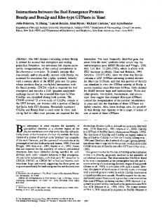
FIG. 10. Homologous and heterologous interactions of the
E. coli and B. subtilie glucose and sucrose PTS proteins. The homologous or heterologous interactionsare denoted by solid or dashed lines, respectively. Relative Vmaxvalues for the interactions of Enzyme I or IIAglc with the various HPrs or for the interactions of IICB& or IIBC;;. with IIA& or IIA& are shown in parentheses and brackets. In each case, the V,,, value for the homologous, wild-type, unmodified substrate has been set at 1.0, and values for the heterologous, phosphorylated, or mutant protein substrates are relative to this. Values for B. subtilis enzymes (Enzyme I, IIA&., and IIBCE;.) are given in parentheses, whereas Vmaxvalues for E. coli enzymes (Enzyme I, IIA&., and IICB&,) are given in brackets.
IIA'"' that kinetic parameters could not be determined; this result may bedue to a very high apparent K,,,, a very low V,.,, orboth. Finally, in the third phosphoryltransfer reaction shown in Fig. 10, phosphorylation of E. coli IICBg'" byeither E. coli or B. subtilis IIAg"-P showed similar Vmaxvalues, and the E. coli IICBg'" appeared to have a lower apparent affinity (apparent K,,, difference of 15-fold) for the heterologous IIAgl'-P. The reciprocal experiment could not be carried out due to unavailability of a B. subtilis IICBglc (Sutrina et al., 1990). Interestingly, the B. subtilis IIBC""' exhibited similar apparent affinity for IIAgl"-Pof either E. coli or B. subtilis, but theheterologous system showed a 4-fold higher V,,, value. It should be noted that for the glucose and sucrose systems the only phosphoryltransfer reaction showing depressed VmaX values for the heterologous systems relative to thehomologous system is the first reaction, phosphorylation of HPr by Enzyme I. The B. subtilis IIAglCcan be readily phosphorylated by HPr(His-P) of E. coli and can transfer its phosphoryl group to the E. coli IICBglcwith V,,, values which are comparable
Proteins of the B. subtilis PTS to those of the homologous systems. Interestingly, the apparent affinity of B. subtilis IIAgl' for E. coli HPr is high, but its apparent affinity for the IICBg'' of E. coli is low (15-fold lower than the affinity of the E. coliIIAg" for its homologous IICBg'"). These results suggest that the B. subtilis IIAg" can substitute for the E. coli IIAg'"in crr mutants providing it is expressed to high enough levels. The capability of the B. subtilis IIAg'"to complement a crr mutantof E. coli was indeed demonstrated (Table V). Employing proteins exclusively from B. subtilis, introduction of a negative charge at position 46of HPr, either by phosphorylation of serine 46 or by site-directed substitution of this residue with aspartate, strongly decreased both the apparent affinity of Enzyme I for HPr and the maximal reaction rate (Fig. 10). Replacement of serine 46 in the B. subtilis HPr with a neutral aminoacid, i.e. alanine, threonine, or tyrosine, did not affect significantly the kinetic parameters with the homologous interactive proteins, Enzyme I or IIAg". Theseresultsarein agreement with our previous kinetic analyses employing the E. faecalis-B. subtilis heterologous system (Reizer et al., 198913). The data presented in Table VI1 reveal that Enzyme Icatalyzed phosphorylation of HPr, or of the S46D mutant protein, occurs at a higher rate in thepresence of the glucosespecific PTS proteins than in the presence of the mannitolspecific PTS constituents. These data suggest that preferential translocation of one PTS sugar relative to the othercan be modulated at the level of HPr phosphorylation due to differential interaction and complex formation of the general energy-coupling protein(s) with sugar-specific PTS permeases. Studies are currently in progress to extend this observation and to confirm the significance of this modeof regulation. Previous studies have suggested that ( a ) ATP-dependent phosphorylation of HPr cancontrolPTS-mediated sugar uptake and ( b ) differential complexation of HPr(Ser-P) with various IIAsUm'proteins can determine the hierarchy of preferences of PTS sugar utilization in Gram-positive bacteria (Deutscher et al., 1984; for recent reviews see Reizer, 1989; Reizer et al., 1989a; Reizerand Peterkofsky, 1987). Since this proposal stems from in vitro studies that were performed with heterologous PTS proteins, we have re-examined this proposal using PTS proteins that were derived from a single organism. The data presented in this study (Table VII) do not support a role for HPr(Ser-P) in thepreferential utilization of one PTS carbohydrate relative to another. Thus, the rate of Enzyme I-catalyzed phosphorylation of the S46D mutant HPrwas inhibited to a similar extent (approximately 4-f0ld), relative to wild-type HPr, in the presence of PTS proteins that catalyze the translocation of a preferred carbohydrate, i.e. glucose,and in thepresence of PTS proteins that mediate the translocation of a less favored sugar, i.e. mannitol (see Table VII). In addition, the data shown in Table VI1 do not support the proposal (Deutscher et al., 1984) that phosphorylation of HPr(Ser-P) by Enzyme I occurs at a rate comparable to the phosphorylation of free HPr in thepresence of a IIA""gar ofthe preferred carbohydrate, i.e. glucose,but not in the presence of I1Asugar of a less favored carbohydrate, i.e. mannitol. Finally, we have recently demonstrated that the hierarchical utilization of glucose over mannitol in B. subtilis is unrelated to phosphorylation of HPr at serine residue 46 since ( a ) a B. subtilis mutant (SA003) that bears a chromosomal S46A mutant ptsH gene shows the preferential utilization of glucose over mannitol, and ( b ) no significant differences were observed in the rate of uptake of mannitol by the
9167
mutant SA003 and the wild-type B. subtilis ~ t r a i n s Alto.~ gether, the data presented in this communication and in our previously published studies (Reizer et al., 1989b; Sutrina et al., 1990; Reizer, 1989) clearly demonstrate that the ATPdependent phosphorylation of HPr isnot responsible for preferential utilization of one PTS sugar relative to another under the conditions employed in our studies. We cannot exclude the possibility that under other conditions, or for other bacterial species, the proposed regulatory mechanism is significant. The recent discovery that HPr(Ser) phosphorylation apparently regulates transport of non-PTS sugars and expression of genes concerned with non-PTS carbohydrate utilization4 suggests that the HPr(Ser) kinase can function in the regulation of non-PTS sugar utilization (see also Reizer et al., 1988a). Further studieswill be required to establish this postulate. Acknowledgments-We thank Drs. W. Epstein and J. W. Lengeler for bacterial strains used in the study, J. M. Tomich for determining the N-terminal aminoacyl sequence of IIA&., and Aiala Reizer for expert assistance inthe preparation of this manuscript.
REFERENCES Crutz, A. M., Steinmetz, M., Aymerich, S., Richter, R., and Le Coq, D. (1990)J. Bacteriol. 172, 1043-1050 De Reuse, H., and Danchin, A. (1988)J.Bacteriol. 170,3827-3837 De Reuse, H., Roy, A., and Danchin, A. (1985)Gene (Amst.) 35,199207 Dean, D. A., Reizer, J., Nikaido, H., and Saier, M. H., Jr. (1990)J. Biol. Chem. 265,21005-21010 Deutscher, J., Beyreuther, K., Sobek, H. M., Stuber, K., and Hengstenberg, W. (1982)Biochemistry 21,4867-4873 Deutscher, J., Kessler, U., Alpert, C. A., and Hengstenberg, W. (1984) Biochemistry 23,4455-4460 Dorschug, M., Frank, R., Kalbitzer, H. R., Hengstenberg, W., and Deutscher, J. (1984)Eur. J. Biochem. 144, 113-119 Fairbrother, W. J., Cavanagh, J., Dyson, H. J., Palmer, A.G. 111, Sutrina, S. L., Reizer, J., Saier, M. H., Jr., and Wright, P. E. (1991) Biochemistry 30,6896-6907 Gonzy-Treboul, G., and Steinmetz, M. (1987)J.Bacterwl. 169,22872290 Gonzy-TrGboul, G., Zagorec, M., Rain-Guion, M. C., and Steinmetz, M. (1989)Mol. Microbiol. 3, 103-112 Grenier, F. C., Waygood, E. B., and Saier, M. H., Jr. (1986)J. Cell. Bwchem. 31,97-105 Hengstenberg, W., Penberthy, W. K., Hill, K. L., and Morse, M. L. (1969)J. Bacteriol. 99,383-388 Kapadia, G., Chen, C. C. H., Reddy, P., Saier, M. H., Jr., Reizer, J., and Herzberg, 0.(1991)J. Mol. Biol. 221, 1079-1080 Kukuruzinska, M. A., Harrington, W. F., and Roseman, S. (1982)J. Bwl. Chem. 257,14470-14476 Kukuruzinska, M. A., Turner, B. W., Ackers, G. K., and Roseman, S. (1984)J. Biol. Chem. 259,11679-11681 Kundig, W., and Roseman, S. (1971)J. Biol. Chem. 246, 1393-1406 Le Coq, D., Crutz, A. M., Richter, R., Aymerich, S., Gonzy-Treboul, G., Zagorec, M., Rain-Guion, M.C., and Steinmetz, M. (1990)in Genetic Transformation and Expression (Buttler, L. O., Harwood, C., and Moseley, B. E. B., eds) pp. 447-456,Intercept, Andover,
UK Liao, D.-I., Kapadia, G., Reddy, P., Saier, M. H., Jr., Reizer, J., and Herzberg, 0.(1991)Biochemistry 30, 9583-9594 London, J., and Hausman, S. Z. (1983)J. Bacteriol. 156,611-619 Lowry, 0.H., Rosebrough, N. J., Farr, A. L., and Randall, R. J. (1951) J.Biol. Chem. 193,265-275 Meadow, N. D., and Roseman, S. (1982)J. Biol. Chem. 257,1452614537 Meadow, N. D., Coyle, P., Komoryia, A., Anfinsen, C. B., and Roseman, S. (1986)J. Biol. Chem. 261, 13504-13509 Meadow, N.D., Fox, D. K., and Roseman, S. (1990)Annu. Reu. Biochem. 59,497-542 Misko, T. P., Mitchell, W. J., Meadow, N. D., and Roseman, S. (1987) J.Biol. Chem. 262,16261-16266 J. Deutscher et al., unpublished data.
Proteins of the B. subtilis PTS
9168
Mitchell, W. J., Saffen, D. W., and Roseman, S. (1987)J. Bid. Chem. 262,16254-16260 Nelson, S. O., Schuitema, A. R. J., and Postma, P. W. (1986) Eur. J. Biochem. 154,337-341 Noel, D., Nikaido, K., and Ames, G. F.-L. (1979) Biochemistry 18, 4159-4165 Pelton, J. G., Torchia, D.A., Meadow, N.D., Wong,C.-Y., and Roseman, S. (1991) Proc. Natl. Acad. Sci. U. S. A. 88,3479-3483 Perego, M., Cole, S. P., Burbulys, D., Trach, K., and Hoch, J . A. (1989) J. Bacteriol. 1 7 1 , 6187-6196 Presper, K. A., Wong, C.-Y., Liu, L., Meadow, N. D., and Roseman, S. (1989) Proc. Natl. Acad. Sci. U. 5'. A. 86, 4052-4055 Reizer, J. (1989) FEMS Microbiol. Reu. 63, 149-156 Reizer, J., andPeterkofsky, A. (1987) in Sugar Transport and Metabolism in Gram-positive Bacteria (Reizer, J., and Peterkofsky, A., eds) pp. 333-364, Ellis Horwood, Chichester, UK Reizer, J., Novotny, M. J., Hengstenberg, W., and Saier, M. H., Jr. (1984a)J. Bacteriol. 160, 333-340 Reizer, J., Novotny, M. J., Stuiver, I., and Saier, M. H., Jr. (1984b) J. Bacteriol. 159, 243-250 Reizer, J., Peterkofsky, A., and Romano, A. H.(1988a) Proc. Natl. Acad. Sci. U. S. A. 8 5 , 2041-2045 Reizer, J., Saier, M. H., Jr., Deutscher, J., Grenier, F., Thompson, J., and Hengstenberg, W. (1988b) CRC Crit. Reu. Microbiol. 15,297338 Reizer, J., Deutscher, J., and Saier, M. H., Jr. (1989a) Biochimie 7 1 , 989-991 Reizer, J., Sutrina, S. L., Saier, M. H.,Jr., Stewart, G. C.,Peterkofsky, A., and Reddy, P. (1989b) EMBO J.8, 2111-2120
Romano, A. H., Saier, M. H., Jr., Harriott, 0. T., and Reizer, J. (1990) J. Bacteriol. 172,6741-6748 Saffen, D. W., Presper, K. A., Doering, T. L., and Roseman, S. (1987) J. Biol. Chem. 262,16241-16253 Saier, M. H., Jr. (1985) Mechanisms and Regulation of Carbohydrate Transport in Bacteria, Academic Press, New York Saier, M. H., Jr. (1989) Microbiol. Reu. 53, 109-120 Saier, M. H., Jr., and Feucht, B. U. (1975)J. Biol. Chem. 250,70787080 Saier, M. H., Jr., and Reizer, J. (1990) Res.Microbiol. 141, 10331038 Saier, M. H., Jr., and Roseman, S. (1976) J. Biol. Chem. 251,66066615 Sambrook, J., Fritsch, E. F., and Maniatis, T. (1989) Molecular Cloning: a Laboratory Manual, 2nd Ed, Cold Spring Harbor Laboratory Press, Cold Spring Harbor, NY Simoni, R. D., Nakazawa, T., Hays, J. B., and Roseman, S. (1973)J. Bid. Chem. 248,932-940 Steinmetz, M., and Aymerich, S. (1990) in Genetics and Biotechnology of Bacilli (Zukowski, M. M., Ganesan, A. T., and Hoch, J. A., eds) Vol. 3, pp. 303-311, Academic Press, New York Sutrina. S. L.. Reddv. P.. Saier. M. H.. . Jr.,. and Reizer.. J. (1990) J. . Bioi. Chem.'265,18581-18589 Vopler. A. P.. and Lenpeler. - . J . W. (1988) . . Mol. & Gen. Genet. 213,