Functional modulation of dendritic cells to suppress adaptive immune responses Andrea M. Woltman and Cees van Kooten Department of Nephrology, Leiden University Medical Center, The Netherlands
Abstract: In recent years, dendritic cells (DCs) have entered the center court of immune regulation. Dependent on their ontogeny, state of differentiation, and maturation and thereby a variable expression of membrane-bound and soluble molecules, DCs can induce immunostimulatory as well as immunoregulatory responses. This dual function has made them potential targets in vaccine development in cancer and infections as well as for the prevention and treatment of allograft rejection and autoimmune diseases. The present review is focused on the effect of immune-modulatory factors, such as cytokines and immunosuppressive drugs, and on the survival, differentiation, migration, and maturation of DC human subsets. A better understanding of DC immunobiology may lead to the development of specific therapies to prevent or dampen immune responses. J. Leukoc. Biol. 73: 428 – 441; 2003. Key Words: tolerance 䡠 immunosuppression 䡠 allograft rejection
INTRODUCTION Dendritic cells (DCs) are bone marrow-derived cells that populate all lymphoid organs as well as nearly all nonlymphoid organs [1, 2]. Although DCs display a heterogeneous group of cells that represent differences in origin, anatomic location, cell-surface phenotype, and function, they all have potent antigen-presenting capacity for stimulating naive, memory, and effector T cells [3]. In addition, DCs can interact with B cells [4] and natural killer cells [5]. Therefore, DCs serve as an essential link between innate and adaptive immune responses. Their role in immune regulation ranges from tolerance induction and the prevention of autoimmunity to the induction of antitumor immunity and the protection against infectious agents [2, 3, 6 –9]. DCs reside in nonlymphoid tissues as immature DCs, which are highly adapted for the uptake of antigen via receptor- and nonreceptor-mediated mechanisms and readily degrade antigens in endocytic vesicles to produce antigenic peptides capable of binding to major histocompatibility complex (MHC) class II. A fundamental aspect of DC function is their capacity to migrate. Under steady-state conditions, migration of DCs from blood to tissues and from tissues to lymph nodes is present but relatively low [10]. Danger signals enhance the rate of DC migration and also induce maturation of DCs, which 428
Journal of Leukocyte Biology Volume 73, April 2003
decreases their capacity to capture antigen but enables the cells to translocate the immunogenic peptide–MHC complexes in concert with costimulatory molecules to their cell surface [11–13]. The migration of maturing DCs involves a coordinated switch in use and expression of chemokine receptors. The responsiveness to most of the inflammatory chemokines, such as CCL2/monocyte-chemoattractant protein-1, CCL3/macrophage-inflammatory protein-1␣ (MIP-1␣), CCL4/MIP-1, CCL5/regulated on activation, normal T expressed and secreted, and CCL20/MIP-3␣, is rapidly lost through receptor down-regulation or receptor desensitization, dependent on autocrine-chemokine production, whereas lymphoid homing receptors, including CCR7, which serves as a receptor for CCL19/MIP-3 and CCL21/secondary lymphoid tissue chemokine, are up-regulated, which enables the maturing DCs to leave the inflamed tissues, enter the lymphatics, and find responder T cells in the lymph node [13, 14]. Upon interaction with antigen-specific T lymphocytes, DC maturation is further completed via engagement of CD40 by its ligand, allowing optimal presentation and T cell activation. Induction of T cell responses requires T cell receptor (TCR) activation (signal 1) and costimulatory interaction (signal 2) between DCs and T cells [15, 16]. In addition, DCs provide T cells with another signal (signal 3) that determines the differentiation of naive T cells into different T helper (Th) subpopulations [17]. Although signal 3 has not been clearly defined, Th-cell polarization is known to be determined at several levels, including the pathogen, the receptor through which the pathogen signals, the DC subset, the microenvironment, and the cytokines released by T cells and other cells in the vicinity.
ROLE OF DCs IN IMMUNE REGULATION The powerful adjuvant activity that DCs possess in stimulating specific CD4⫹ and CD8⫹ T cell responses has made them targets in vaccine development strategies for the prevention and treatment of infections, allograft rejections, autoimmune diseases, and cancer [18]. Whereas for the fight against infectious agents and tumor development, DCs are manipulated to enhance a host’s immune defense, the major goal in the pre-
Correspondence: A. M. Woltman, Department of Nephrology, C3-P, Leiden University Medical Center, Albinusdreef 2, 2333 ZA Leiden, The Netherlands. E-mail:
[email protected] Received September 5, 2002; accepted November 4, 2002; doi: 10.1189/ jlb.0902431.
http://www.jleukbio.org
vention and treatment of allograft rejection and autoimmune diseases is to inhibit the immunostimulatory capacity of DCs or more importantly, to exploit tolerogenic DCs to silence the immune response. Autoimmunity is characterized by a loss of tolerance to self-antigens as a result of activation of autoreactive lymphocytes that result in a destructive response directed to single or multiple organs. Although the mechanisms by which autoimmune responses are triggered are not fully understood, DCs seem to play an important role in autoimmunity. Evidence that DCs can be used or manipulated to combat autoimmune diseases has come predominantly from studies in experimental models of type 1 diabetes and multiple sclerosis [19, 20]. Prevention of experimental autoimmune diseases can be achieved by transferring manipulated syngeneic DCs, such as semi-mature tumor necrosis factor ␣ (TNF-␣)-treated DCs, interferon-␥ (IFN-␥)-treated DCs, and cytotoxic T-lymphocyte antigen-4-immunoglobulin (Ig)-treated DCs [21–23]. The role of DCs in organ transplantation is even more complex because of the coexistence of graft-derived DCs from the donor, responsible for direct recognition of foreign antigens by T cells, and DCs from the recipient, responsible for indirect antigen presentation to host T cells [24 –26]. Initially, it was thought that DCs in the allograft were responsible for immunogenicity, which generated the idea that DCs should be eliminated from the graft before transplantation [27–29]. Although specific elimination of donor-derived DCs seemed to prolong allograft survival in several animal models, it became clear that the situation to obtain sustained graft acceptance is far more complicated. The plasticity of DCs and their role in T cell polarization suggested that donor- and recipient-derived DCs can act as inducers of an immunogeneic or tolerogenic allograft response [30 –32]. The regulatory role of DCs was demonstrated by adoptive transfer of allopeptide-loaded recipient-derived lymphoid and myeloid DCs that were able to prolong cardiac and islet allograft survival in rat transplantation models [33, 34]. Knowing which DC subsets, donor- or recipient-derived, are responsible for specific immune responses and which cytokines
or other factors mobilize specific subsets should permit us to use these tools to manipulate the immune response to alloantigens. Therefore, the functional characteristics of the distinct DC populations and their functional modulation by cytokines, immunosuppressive drugs, and other agents will be reviewed in the following pages and will focus primarily, although not exclusively, on inhibition of the immunogenic capacities and enhancement of the tolerogenic capacities of human DCs in the context of organ transplantation.
DC SUBSETS In humans, DCs comprise at least three distinct subsets: Langerhans cells (LCs) and interstitial DCs, belonging to the myeloid lineage, and plasmacytoid DCs, which are thought to originate from a lymphoid precursor (Fig. 1; Table 1) [35]. LCs are localized in the basal and supra-basal layers of the epidermis in the skin and other mucosal areas, whereas interstitial DCs are present in the dermis of the skin and in most other organs. Interstitial DCs and LCs emerge in cultures from CD34⫹ progenitors, isolated from bone marrow or cord blood and CD11c⫹ blood DC precursors. CD34⫹ hematopoietic progenitor cells (HPC) differentiate in the presence of GM-CSF and TNF-␣ into myeloid DCs along two distinct pathways (Fig. 1) [36, 37]. In one, a CD1a⫹CD14⫺ intermediate gives rise to cells resembling LCs, whereas interstitial DCs arise from a CD1a⫺CD14⫹ precursor that can also differentiate into macrophages in the presence of only M-CSF. A generally more convenient source of cells for many experimental procedures is derived from CD11c⫹CD14⫹ monocytes. In vitro, CD1a⫺CD14⫹ monocytes differentiate into CD1a⫹CD14⫺ interstitial DCs in the presence of GM-CSF and IL-4 or IL-13 (Fig. 1) [38, 39]. There is accumulating evidence that in vivo monocytes can also differentiate into DCs in an appropriate cytokine microenvironment or by transmigrating through endothelial layers [40 – 43]. The demonstration that LCs can arise from monocytes via a TGF--dependent pathway is controversial and may ultimately reflect the plasticity of DCs [44, 45].
Fig. 1. In vitro generation of human DC subsets. Scheme for generation of human DC subsets from CD34⫹ myeloid (CMP) and lymphoid (CLP) progenitors. Myeloid and lymphoid lineage DCs can be propagated from bone marrow progenitors and blood precursors using various combinations of cytokines, such as granulocyte macrophage-colony stimulating factor (GM-CSF), TNF-␣, interleukin (IL)-4, transforming growth factor- (TGF-), and IL-3.
Woltman and van Kooten Functional modulation of dendritic cells
429
TABLE 1. Postulated lineage
Lymphoid
DC subtype Blood precursors Phenotype IFN-␣ production Fully differentiated DC Phenotype
Human DC Subsets: Precursors, Phenotype, Localization, and Function Myeloid
Plasmacytoid DC
Interstitial DC
Langerhans cell
CD11c⫺CD123⫹ ⫹⫹⫹⫹
CD11c⫹CD123⫺ ⫺
CD11c⫹CD123⫺ ⫺
Localization
CD11c⫺CD123⫹ CD11b⫺CD13⫺CD33⫺ CD1a⫺ DC⫺SIGN⫺ Langerin⫺ CD4⫹ T cell zones of lymphoid organs
CD11c⫹CD123⫺ CD11b⫹CD13⫹CD33⫹ CD1a⫺ DC⫺SIGN⫹ Langerin⫺ CD4⫺ T cell zones of lymphoid organs Immature cells in peripheral tissues
CD11c⫹CD123⫺ CD11b⫹CD13⫹CD33⫹ CD1a⫹ DC⫺SIGN⫺ Langerin⫹ CD4⫺ T cell zones of lymphoid organs Immature cells in epithelia
Function Mannose-receptormediated endocytosis Macropinocytosis IL-12 secretion IL-10 secretion CD4⫹ T cell priming CD8⫹ T cell priming B cell activation
⫺ ? ⫹/⫺ ⫺ ⫹⫹ ⫹⫹ ?
⫹⫹⫹ ⫹⫹⫹ ⫹⫹⫹ ⫹⫹⫹ ⫹⫹⫹ ⫹⫹ ⫹⫹⫹
⫺ ⫺ ⫹⫹⫹ ⫺ ⫹⫹⫹ ⫹⫹⫹ ⫺
Potential LC precursors that express CD14 and the coagulation factor XIIIa have been mobilized from human skin explants and show some DC characteristics but have weak T cell stimulatory potential and could not be differentiated into DCs in vitro [46]. Recently, a population of CD14⫹ dermal resident cells lacking factor XIIIa but expressing langerin were identified as potential LC precursors [47]. LC and interstitial DC subtypes share several markers, but LCs uniquely express Birbeck granules, langerin, and the adhesion molecule E-cadherin, whereas interstitial DCs uniquely express factor XIIIa. LCs and interstitial DCs also share the capacity to activate CD4⫹ and CD8⫹ naive T cells and secrete IL-12. Striking differences between LCs and interstitial DCs are the abilities of interstitial DCs but not LCs to take up large amounts of antigens by the mannose receptor, to produce large amounts of IL-10, and to induce differentiation of naive B cells into Ig-secreting plasma cells [48]. Human plasmacytoid DCs are found in the T cell zones of lymphoid organs. Plasmacytoid DCs are characterized by a unique phenotype, CD11c⫺CD123⫹CD45RA⫹HLA-DR⫹, and possess the unique ability to secrete large amounts of IFN-␣/ upon viral stimulation [35]. Unlike LCs and interstitial DCs, CD11c⫺ plasmacytoid DCs, generated from CD34⫹ HPC or CD11c⫺ blood precursors, lack expression of myeloid antigens and require IL-3 instead of GM-CSF for their differentiation and survival. Plasmacytoid DCs share a common function with LCs and interstitial DCs in having the capacity to activate CD4⫹ and CD8⫹ naive T cells. At least three major subpopulations of DCs have been described in mice: CD4⫺CD8␣⫹, CD4⫺CD8␣⫺, and CD4⫹CD8␣⫺ DCs. CD8␣⫺ DCs, which may be CD4⫹ or CD4⫺, express CD11b and were initially considered myeloid DCs [49]. As CD8␣⫹ DCs lack the myeloid marker CD11b and in contrast to the CD8␣⫺ DC subsets, require IL-3 instead of 430
Journal of Leukocyte Biology Volume 73, April 2003
GM-CSF for differentiation and survival, they were originally thought to arise from a lymphoid-committed progenitor. However, a report by Traver et al. [50] demonstrated that CD8␣ on DCs does not indicate a lymphoid origin but rather may reflect a maturation or differentiation status of DCs. Recently, CD11c⫹B220⫹Gr1⫹ cells were identified as the mouse counterpart of human plasmacytoid DCs. Like human plasmacytoid DCs, B220⫹ DCs are present in significant numbers in the mouse thymus and express several T cell markers including CD8␣ and high levels of CD4. In steady state, these B220⫹ DCs display characteristics of immature-like DCs, and as for human plasmacytoid DCs upon viral stimulation, B220⫹ DCs produce high levels of IFN-␣, which is the hallmark of plasmacytoid DCs [51–55]. Unlike human plasmacytoid DCs, which produce only little IL-12p70, in vitro-generated mouse B220⫹ DCs clearly produce IL-12p70 in response to CpG oligonucleotides and virus [56]. Although it was recently shown that plasmacytoid DCs can be generated from mouse bone marrow cultures with fms-like tyrosine kinase 3 ligand [56], most in vitro studies using murine DCs are performed with bone marrow-derived myeloid DCs generated in the presence of GM-CSF and LCs cultured from skin explants.
CURRENT STRATEGIES TO PROMOTE THE TOLEROGENICITY OF DCs Tolerance is the specific inability of a host to respond to antigens without the need for immunosuppressive drugs and is generated centrally and peripherally. Peripheral antigen-specific tolerance, which is the ultimate goal in organ transplantation, might be achieved by subverting immune reactivity, i.e., Th2-skewing, T cell anergy or deletion, or the induction of T http://www.jleukbio.org
regulatory cells (Treg), which are nonproliferating T cells that produce high levels of IL-10 and very low levels of IFN-␥ and IL-4. Treg cells suppress Th0, Th1, and Th2 cells via a mechanism that is currently not known, but evidence is growing that these cells are stimulated initially by DCs. Jonuleit et al. [57] have shown that repetitive in vitro stimulation of allogeneic human T cells with monocyte-derived, immature DCs induced the differentiation of Treg cells. The biological significance of these findings has been highlighted by Dhodapkar et al. [58], who injected autologous, monocyte-derived, immature DCs, pulsed with influenza peptide, subcutaneously in human volunteers. These volunteers generated an antigenspecific inhibition of CD8⫹ T cell killing activity and the appearance of peptide-specific IL-10-producing T cells, accompanied by a decrease in the number of IFN-␥-producing T cells. Accordingly, mice pretreated with donor-derived, immature DCs showed prolonged allograft survival in models of pancreatic islet and heart transplantation [59 – 61]. Based on these findings, one approach to promote the tolerogenicity of DCs is to suppress their maturation by using anti-inflammatory cytokines or pharmacological agents. Another approach is the use of genetically engineered DCs expressing immunosuppressive molecules. Several molecules have been shown to activate DCs and trigger their transition from immature, antigen-capturing cells to mature, antigen-presenting cells (APC). Mature DCs have the capacity to prime naive T cells, but the type of original maturation signal determines the polarization of T cells. Maturation signals may arise from endogenous processes, particularly those that result in cell necrosis and tissue destruction, such as cellular heat shock proteins, matrix degradation products [62], and cytokines. Maturation signals can also be derived from foreign substances, including bacterial products, viruses, fungi, and parasites. Molecules derived from pathogens, such as lipopolysaccharides (LPS) [63], bacterial CpG DNA [64], and viral dsRNA [65], as well as T cell signals such as CD40L and IFN-␥, all promote immature DCs to produce IL-12 and prime for Th1 responses. A common process during this maturation seems to be the activation of the nuclear factor (NF)-B pathway [66, 67]. In contrast, DCs activated in the presence of cyclic adenosine monophosphate-increasing agents, such as prostaglandin E2 [68], cholera toxin [69, 70], histamine [71, 72], and 2-agonists [73] or parasites [74], demonstrated increased levels of CD83, MHC, and costimulatory molecules but were not able to produce IL-12, resulting in Th2 polarization upon contact with naive CD4⫹ T cells. The capacity of DCs to skew immune reactivity from Th1 to Th2 responses and vice versa is considered as a potential strategy to shift and thereby dampen typical Th1- or Th2dependent, inflammatory responses. In the context of organ transplantation, this hypothesis was supported by the finding that animals with long-term allograft survival did not show Th1 cytokines [75–77]. However, studies have failed to show that Th2 polarization actively promotes the development of longterm allograft acceptance or that a Th1 response promotes graft rejection per se [78, 79], which suggests that immune deviation from a Th1 to a Th2 response is not the final solution. The finding that immature DCs might induce tolerogenic responses by the induction of Treg has emphasized the therapeutic po-
tential of DCs that are pharmacologically arrested at an immature state, which means at least that they are unable to upregulate costimulatory and MHC molecules and cannot secrete IL-12 upon exposure to proinflammatory and T cell-derived signals. Recently, intermediate stages of DC maturation have also been described, which might have specific regulatory functions as well [23, 66]. It is therefore of critical importance to determine exactly the stage of maturation in which DCs reside after pharmacological interference.
IMMUNOMODULATORY FACTORS THAT SUPPRESS THE IMMUNOGENICITY OR ENHANCE THE TOLEROGENICITY OF DCs Cytokines IL-10
IL-10 is defined as an anti-inflammatory cytokine, which was found to inhibit the proliferation of T cells in an allogeneic mixed leukocyte reaction (MLR). Moreover, transient local expression of IL-10 has been shown to prolong allograft survival in a murine cardiac allograft model [80]. Although it has been shown that IL-10 can directly affect CD4⫹ T cells, many of the inhibitory actions of the cytokine have been attributed to its actions on APC. IL-10 inhibits the differentiation of human monocytes into DCs and drives the differentiation toward macrophage-like cells, as assessed by morphology, phenotype, and functional characteristics (Fig. 2) [81– 85]. Cells generated in the presence of IL-10 showed an increased endocytic capacity, partially as a result of an up-regulation of mannose receptors, and further demonstrated an impaired capacity to activate allogeneic T cells [81, 82] and were much less efficient to present tetanus toxin to specific T cell lines [81]. Immature DCs treated with IL-10 demonstrated a modest reduction in costimulatory and MHC molecules [86 – 88] and even more important, blocked the maturation upon activation with LPS or proinflammatory cytokines (Fig. 2) [83, 88 –90], as assessed by the inability to up-regulate costimulatory and MHC molecules and a very low or even absent production of IL-12. In contrast, IL-10-treated DCs produced higher levels of IL-10 than control DCs. CD40L-induced maturation seemed more difficult to block with IL-10. It seems that IL-10 partially blocks the CD40L-induced up-regulation of MHC and costimulatory molecules and the production of IL-12. Whether treatment of immature DCs with IL-10 also up-regulates the endocytic capacity is difficult to determine, as spontaneous maturation of DCs, which is accompanied by a loss in endocytic activity, is blocked by IL-10. IL-10-transduced murine bone marrow-derived myeloid DCs showed reduced expression of MHC and costimulatoy molecules and thereby an impaired ability to induce T cell proliferation [91–93] but next to that, also affected DC trafficking. IL-10-transduced DCs showed lower levels of CCR7 and simultaneously increased CCR5 levels, which seemed responsible for an impaired, in vivo homing of IL-10 DCs from peripheral tissues to secondary lymphoid organs [94].
Woltman and van Kooten Functional modulation of dendritic cells
431
Fig. 2. Interference with monocyte-derived DC development. Upon culture with IL-4 and GM-CSF, monocytes differentiate into immature DC (imDC). Addition of IL-6 switches the differentiation toward macrophages (m), whereas addition of TGF- can induce LC development. The presence of corticosteroids, IL-10, or vitamin D3 analogs during differentiation induces the development of macrophage-like cells that still contain some DC-specific characteristics (DC/m). The presence of these agents during activation of the cells inhibited the development of mature DC (matDC); dependent on the type of stimulus, activation was partially or completely blocked (“im”DC).
The immunosuppressive properties of IL-10 on DC differentiation, maturation, and function were much more emphasized by the finding that IL-10-treated monocyte-derived DCs can induce anergic CD4⫹ and CD8⫹ T cells [89, 95]. These anergic T cells are characterized by an impaired, proliferative capacity and reduced production of IFN-␥ and IL-2 and are able to suppress activation and function of T cells in an antigen-specific manner [96]. In addition, IL-10-treated LCs were found to induce antigen-specific tolerance in Th1 but not in Th2 cell clones [97]. Although IL-10-treated DCs have not been investigated yet for the induction of antigen-specific transplant tolerance, it has been shown that portal vein infusion of mouse bone marrowderived DCs, engineered to express IL-10, prolongs renal allograft survival [98]. TGF-
TGF- is another immunosuppressive cytokine. Endogenously produced TGF- has shown to be required for the development of LCs and non-LCs from CD34⫹ HPC in vitro through protecting progenitor cells from apoptosis [99] or induction of DC progenitor proliferation and differentiation. Addition of exogenous TGF- polarized the differentiation of CD34⫹-derived CD14⫹ precursors toward LCs. Although conflicting hypotheses exist about monocytes as LC blood precursors [44, 45], it has been shown that monocytes cultured in the presence of TGF-, GM-CSF, and IL-4 also differentiated toward LC-like cells, as demonstrated by their phenotypic and functional characteristics (Fig. 2). TGF--induced LCs differentiated from monocytes or CD34⫹ HPC, express the typical LC markers E-cadherin, Birbeck-granules, Lag, and cutaneous lymphocyte-associated antigen [45, 100], and express functional CCR-6 [101]. In addition, they neither secreted IL-10 upon stimulation nor stimulated the differentiation of CD40-acti432
Journal of Leukocyte Biology Volume 73, April 2003
vated, naı¨ve B cells. In contrast to its favorable effect on LC differentiation, TGF- prevents LC maturation in response to TNF-␣, IL-1, and LPS [102]. However, TGF- cannot inhibit CD40L-induced up-regulation of costimulatory and MHC molecules or production of IL-12 [102]. Another striking finding is that TGF- is able to inhibit antigen presentation by GM-CSFstimulated APC propagated from human bone marrow [103] and by cultured LCs but not by freshly isolated LCs [104]. Thus, TGF- seems to skew the differentiation of hematopoietic precursors toward LCs, which are hampered in their final maturation. The fact that TGF- increases expression of CCR-1, -3, -5, and -6 and CXCR-4 on monocyte-derived LC-like cells and blocks TNF-␣-induced up-regulation of CCR-7 [105] also suggests a role for TGF- in the chemotaxis of DCs. The ability of TGF- to inhibit the antigen-presenting capacity and possibly also its influence on DC trafficking might explain its contribution to prolonged allograft survival in animal models treated with TGF- [106, 107], DCs transduced to express TGF- [98, 108], or DCs generated in the presence of TGF- [109]. IL-6
Although the effect of IL-6 administration on allograft survival has not been extensively studied, it has been shown that IL-6 delays skin graft rejection in mice [110]. Only a few studies describe the direct effect of IL-6 on the differentiation of human DCs in vitro. IL-6 switches the differentiation of CD34⫹ progenitors and monocytes from DCs toward macrophages (Fig. 2) [111–113]. Although contradictory results exist regarding the role of M-CSF as an intermediate in the inhibitory effect of IL-6 on DC development, it seems that IL-6 exerts its effect via up-regulation of functional M-CSF receptors. This up-regulation of M-CSF receptors allows the precursor cells to consume http://www.jleukbio.org
and respond to their autocrine-produced M-CSF, which subsequently drives the cells toward macrophage development [112, 113].
Immunosuppressive drugs Corticosteroids
Corticosteroids are among the most potent immunosuppressive and anti-inflammatory drugs currently available and are efficacious in the treatment of Th1- and Th2-associated inflammatory diseases, including allograft rejection, rheumatoid arthritis, and asthma [114]. The therapeutic effects of corticosteroids were initially ascribed to the strong inhibitory effect on T cells. At the moment, it is obvious that APC are also strongly affected by corticosteroids. It has been demonstrated that corticosteroids down-regulate the production of proinflammatory cytokines by monocytes and macrophages. More recently, corticosteroids were found to significantly affect DCs. Mice treated with the glucocorticoid dexamethasone (Dex) demonstrated relatively decreased splenic DC numbers and increased splenic macrophage numbers [115]. This shift in the myeloid cellular compartment of the spleen upon Dex treatment can be caused by effects on migration, induction of apoptosis, or by an altered differentiation process of myeloid progenitor cells. In addition, glucocorticoid administration to rhesus macaques changed DC subsets in lymph nodes [116], and treatment of healthy volunteers with glucocorticoids reduced the circulating numbers of plasmacytoid DCs in blood [117], which can again be the result of multiple processes as described above. Strong indications for corticosteroid-induced apoptosis in vivo were found in rats treated with Dex. These animals showed decreased numbers of airway DCs preceded and accompanied by
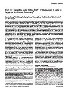
an increase in apoptotic cells [118]. However, contradictory results exist regarding the effect of corticosteroids on the survival of human DCs in vitro. Kim et al. [119] demonstrated that corticosteroids induce caspase-independent apoptosis in immature, monocyte-derived DCs, whereas several other studies describing the effect on monocyte-derived DCs did not find any apparent apoptosis [120 –122]. One consistent finding is the inhibitory effect of corticosteroids on the development of immature DCs from monocytes (Fig. 2). We have found that addition of corticosteroids to monocyte cultures for as little as the first 48 h completely prevented normal DC development upon a 7-day culture period in GM-CSF and IL-4 (Fig. 3) [122]. The corticosteroid-treated cells retain a monocyte/macrophage phenotype with high CD14 expression and no expression of CD1a, as a typical DC marker [122–124], but also lack expression of CD68, as one of the typical macrophage markers [123]. Corticosteroid treatment increased the endocytic and pinocytic activity of the cells, partially as a result of an up-regulation of mannose receptors [123, 125]. Furthermore, these cells were strongly hampered in their T cell stimulatory capacity [122, 123], which could be explained by an impaired up-regulation of costimulatory and MHC molecules upon stimulation with proinflammatory cytokines, LPS or CD40L, and a reduced production of proinflammatory cytokines, including IL-6, TNF-␣, IL-1, and IL-12 [122, 123]. Next to the inhibitory effect on the differentiation of monocytes into DCs leading to functional alterations, corticosteroids also inhibit maturation of DCs when applied to already differentiated, immature DCs (Fig. 2). Inhibitory effects were found on the up-regulation of costimulatory and HLA molecules and
Fig. 3. Incubation (48 h) with Dex causes complete inhibition of DC development. Human peripheral blood monocytes were cultured for 7 days with IL-4 and GM-CSF in the presence or absence of 1 M Dex, which was present during the entire period, the last 5 days, or only the first 2 days of the culture period. The cells treated with Dex for only the first 48 h of culture were harvested after this 48-h culture period, were extensively washed, and were further cultured in IL-4, GMCSF, and the glucocorticoid receptor antagonist RU486 to prevent a possible carryover effect of Dex. Fluorescein-activated cell sorter analysis was performed on day 7. Specific stainings for CD1a and CD14 are represented by the filled histograms and control stainings, by the open histograms. The gray bars in the culture scheme indicate the presence of Dex.
Woltman and van Kooten Functional modulation of dendritic cells
433
on the production of proinflammatory cytokines, including IL12, IL-6, and TNF-␣, whereas the production of the antiinflammatory cytokine IL-10 was supported by corticosteroid treatment [126, 127]. Comparable with the studies published on the inhibitory effects of IL-10 on DC maturation, corticosteroids strongly inhibit maturation induced by LPS [121, 127, 128], haptens [129], and proinflammatory cytokines [120, 123, 128, 130] but demonstrate differential effects ranging from complete to no inhibition on CD40L-induced maturation [121, 123, 126, 128]. In addition, lung DCs exposed to corticosteroids ex vivo demonstrated a hampered allostimulatory capacity and reduced expression levels of costimulatory molecules [131]. Next to the effect on monocyte-derived DCs, we have also described the effect of corticosteroids on the differentiation of interstitial DCs and LCs from CD34⫹ HPC [132]. Addition of glucocorticoids to the cultures of CD34⫹ HPC containing GM-CSF, TNF-␣, and stem cell factor prevented the development of CD14-derived interstitial DCs by specific induction of apoptosis in CD14⫹–DC precursors and blockade of spontaneous and IL-4-induced differentiation of CD14⫹–DC precursors into fully differentiated CD1a⫹ interstitial DCs, which can be compared with the inhibitory effect of corticosteroids on the differentiation of monocytes into DCs. The effect of corticosteroids on differentiation and function of LCs has been scarcely described. In vivo studies suggested a direct effect of corticosteroids on LCs, as topical steroid therapy decreased the number of LCs in skin [133–135] and nasal epithelium [136 –138] and almost completely inhibited the influx of LCs during allergen provocation [139, 140]. However, the mechanism by which corticosteroids alter the distribution of LCs is not known. It has been shown that LC numbers in the epidermis are modulated by cytokines such as GM-CSF and TNF-␣, which are produced by keratinocytes [141]. As corticosteroids block the production of cytokines and chemokines by keratinocytes [142], the regulation of LC numbers upon corticosteroid treatment might reflect an indirect effect via an inhibitory effect on neighboring cells. Furthermore, LCs still present in topical steroid-treated skin were shown to exhibit normal, alloantigen-presenting capacities [134]. In line with these observations, the development and function, including IL-12 production and T cell stimulation, of LCs generated from CD34⫹ HPC were not influenced by corticosteroids [132]. Vitamin D3 and its analogs
1␣,25-Dihydroxyvitamin D3 [1,25(OH)2D3], the biologically active metabolite of vitamin D3, is a steroid hormone that not only regulates bone and calcium/phosphate metabolism but also exerts a number of other biological activities, including modulation of the immune response via specific receptors expressed in APC and activated T cells. Immunosuppression by 1,25(OH)2D3 and its analogs has been demonstrated to prolong the survival of cardiac [143, 144] and small bowel [145] allografts in animal models and has been reported to inhibit not only acute but also chronic allograft rejection [146]. As already documented for IL-10 and corticosteroids, 1,25(OH)2D3 is another immunosuppressive compound that blocks the differentiation of monocytes into functional, immature DCs (Fig. 2) [83, 147–149]. The addition of 1,25(OH)2D3 434
Journal of Leukocyte Biology Volume 73, April 2003
or the nonhypercalcemic analog TX527 to monocyte cultures gives rise to the development of CD14highCD1alow cells with dendritic morphology but without the capacity to stimulate allogeneic T cells or antigen-specific T cell lines [147–150], apparently as a result of their inability to up-regulate costimulatory and MHC molecules upon contact with T cells, as suggested by the impaired response to CD40L, TNF-␣, or LPS in vitro [147, 149, 150]. When only present during maturation, 1,25(OH)2D3 was found to inhibit hapten- [129], LPS-, and CD40L-induced maturation of monocyte-derived DCs, as demonstrated by an impaired up-regulation of costimulatory and MHC molecules and a diminished capacity to stimulate allogeneic T cells (Fig. 2) [83, 147] and promoted DC apoptosis [83]. Coculture of alloreactive CD4 T cells with 1,25(OH)2D3treated DCs resulted in T cell hyporesponsiveness, as demonstrated by their reduced IFN-␥ secretion upon restimulation by untreated, mature DCs in a second MLR [83]. Whether 1,25(OH)2D3 also affects LCs is debatable. In vivo and in vitro treatment of human LCs with 1,25(OH)2D3 or its analog calcipotriol did not change the expression of MHC class II antigens [151, 152]. Treatment of cultured epidermal cells with 1,25(OH)2D3 or calcipotriol resulted in LCs with a reduced capacity to stimulate T cells [151, 152], but this might be secondary to its inhibitory effects on keratinocytes, which produce GM-CSF required for LC maturation [153]. In addition to its effect on LC function, conflicting results exist regarding the effect on LC numbers present in the skin upon treatment with calcipotriol [151, 153]. Calcineurin inhibitors
Cyclosporine A (CsA) and FK506 (tacrolimus) belong to the family of calcineurin inhibitors and have proved to be potent immunosuppressive drugs. They are widely used as primary maintenance immunosuppression after transplantation by virtue of their strong inhibitory effect on T cells. CsA and FK506 in complex with their intracellular receptors, cyclophilins and FK506-binding proteins (FKBPs), respectively, bind and inhibit the activation of calcineurin phosphatase [154]. Calcineurin appears to play a crucial role in the transduction of T cell calcium-dependent signaling events following TCR triggering, including transcription of IL-2 and several other cytokine genes [155–157]. Although CsA and FK506 possess similar mechanisms of action, FK506 is 50 –100 times more potent than CsA in inhibiting T cell activation in vitro. A direct effect of calcineurin inhibitors on the function of DCs has been a matter of controversy for several years. Experiments have been performed using mainly monocyte-derived DCs and LCs. As analysis of APC function requires the presence of T cells, which are very sensitive to CsA and even more so to FK506, the major problem lies in the possibility of CsA or FK506 carryover from DCs after treatment with the drugs. That DCs can function as a potential reservoir of drugs was confirmed by the finding that DCs contain FK506 in their intracellular compartments after exposure to the drug [122], which can be secreted into the supernatants [122, 158]. In vitro studies with monocyte-derived DCs revealed that calcineurin inhibitors, when present during differentiation, reduce the production of TNF-␣ induced by LPS, Staphylococcal enterotoxin B, or CD40L [122, 159]. No significant changes were http://www.jleukbio.org
observed in the expression of costimulatory and human leukocyte antigen (HLA) molecules upon culture with physiological concentrations of CsA or FK506 [122, 129, 159]. The lack of effect on the expression of costimulatory and HLA molecules supports the hypothesis that the reduced T cell proliferation found in allogeneic MLRs with CsA or FK506-treated DCs is a result of a carryover effect of the drugs. Only one study described the effect of calcineurin inhibitors on CD34-derived DCs. The presence of FK506 supported the generation of CD1a⫹ DCs without affecting the expression of costimulatory and HLA molecules [160]. Also, this study claimed a decreased T cell stimulatory capacity of FK506treated DCs, but the authors did not exclude a carryover effect. As for monocyte-derived DCs, conflicting results are published regarding the effect of CsA and FK506 on LCs. LCs isolated from normal skin derived from psoriasis patients treated with CsA displayed a reduced ability to stimulate T cells [161], which can be the result of a direct and indirect effect of the drug on the function of LCs. Also, ex vivo treatment of LCs seemed to reduce T cell stimulatory capacity [104, 162, 163], but as demonstrated by Peguet-Navarro et al. [158], this might be caused by carryover. Although several studies demonstrate a lack of effect of systemically or topically applied calcineurin inhibitors on the number of LCs in human skin [164 –167], there might be an effect on other epidermal DC populations [166, 167]. Other immunosuppressive drugs
Relatively new immunosuppressive drugs, such as sirolimus (rapamycin, Rapa), mycophenolate mofetil (MMF), leflunomide, FTY720, and CAMPATH antibodies, used to prevent acute and chronic allograft rejection, were also all developed to interfere with the function of lymphocytes. Although “promising” results are obtained with these immunosuppressive drugs with respect to allograft survival, and the major role for DCs in the induction and amplification of immune responses is recognized, very little is known about their effects on DCs. Rapa, an immunosuppressive drug introduced by virtue of its antiproliferative effect on T lymphocytes [168], seems to be effective in preventing allograft rejection [169, 170]. Rapa is structurally related to FK506, and it binds to and competes with FK506 for the same immunophilin FKBP-12. However, only the Rapa–FKBP12 complex binds and inhibits the function of the serine/threonine kinase mammalian target of rapamycin (mTOR) [171, 172]. In addition to its suppressive effect on lymphocytes, we have found that Rapa induces apoptosis in monocyte-derived DCs and CD34-derived DCs without any special preference for LCs or non-LCs [173]. It is interesting that Rapa does not interfere with the survival of other APC such as monocytes and macrophages. Rapa seems to specifically interfere with GM-CSF signaling at the level of mTOR, which we have recently shown to be a critical kinase in the survival mechanism of monocyte-derived DCs but does not play a role in the survival of monocytes and macrophages [174]. In contrast to these human in vitro studies, Rapa does not induce apoptosis in murine bone marrow-derived DCs but does inhibit macropinocytosis and mannose receptor-mediated endocytosis by these murine DCs [175].
MMF is most commonly used as adjunct therapy in combination with calcineurin inhibitors and corticosteroids. MMF is a prodrug for mycophenolic acid (MPA) [176], which is a reversible inhibitor of inosine 5⬘-monophosphate dehydrogenase [177]. This is the rate-limiting enzyme in the de novo synthesis of purines. As proliferation of activated lymphocytes, unlike most other cell types, relies on the de novo purine synthesis, with a minor contribution of the salvage pathway, MPA selectively inhibits proliferation of T and B lymphocytes [176]. In addition, MPA decreases the intracellular guanine nucleotide pools, which hamper the glycosylation of membrane glycoproteins leading to less effective adhesion molecules (selectins) [178]. Although human studies describing the effect of MMF on DCs are lacking, it has been shown that MMF decreases the ability of mouse bone marrow-derived DCs to stimulate T cells in MLR. Accordingly, MMF-treated DCs demonstrated a decreased expression of costimulatory and adhesion molecules with a concurrent reduction of IL-12 production [179]. Moreover, mice treated with MMF in combination with 1,25(OH)2D3 developed transferable tolerance toward fully mismatched islet allografts [180]. Leflunomide is another immunosuppressive drug that inhibits T and B cell proliferation by inhibiting an enzyme in the de novo pathway. In vivo, leflunomide is rapidly converted to the metabolite A77 1726 that inhibits dihydro-orotate dehydrogenase, which is a rate-limiting enzyme for pyrimidine synthesis. Like MMF, leflunomide shows antiproliferative and antiadhesive effects. Although the migration of DCs might be influenced by in vivo administration of leflunomide [181], direct effects of this immunosuppressive drug on the function of DCs remain to be elucidated. FTY720 is a recently introduced immunosuppressive drug used to prevent allograft rejection [182, 183]. FTY720 modulates the lymphocyte chemotactic response to chemokines, which results in increased homing of lymphocytes to secondary lymphatic organs and prevention of recruitment of effector lymphocytes to the allograft without impairment of immune responses to systemic infection [184 –186]. Whether FTY720 also affects the migration of DCs is not known yet. The CAMPATH series of monoclonal antibodies recognizes CD52, a phophatidylinositol-linked antigen, and is recently introduced in the field of transplantation. CD52 is highly expressed on lymphocytes, monocytes, and blood DCs and is a good target for cell lysis by antibody with human complement. Peripheral blood myeloid DCs are depleted after treatment with CAMPATH antibodies in vivo [187, 188]. Tissue DCs and mature monocyte-derived DCs do not express CD52 and are therefore unlikely to be depleted by these antibodies [188].
THERAPEUTIC IMPLICATIONS Although the immune regulatory activities of DCs are fully recognized, a detailed analysis of DC function in response to various treatments is required before DCs can be used effectively for the selective prevention of allograft rejection. So far, most promising results have been obtained with the administration of immature DCs. However, as DCs and T cells reciprocally affect one another, prevention of activation of immature
Woltman and van Kooten Functional modulation of dendritic cells
435
DCs with different approaches may potentate their tolerogenicity. One might think about the administration of donor-derived APC developed in the presence of IL-10, corticosteroids, or 1,25(OH)2D3, as these cells give rise to the development of regulatory T cells and are completely insensitive for maturation signals or administration of immature donor-derived DCs simultaneously with costimulation blockade, which was shown to be very successful in animal models [109, 189]. Proinflammatory signals, including CD40L, LPS, IL-1, TNF-␣, CpG, and haptens, which are responsible for DC maturation, are known to signal via NF-B. The NF-B family includes inducible transcription factors regulating the expression of many genes involved in inflammation, including cytokines, chemokines, and adhesion, and costimulatory molecules [190]. Several studies have demonstrated that NF-B is activated in mature DCs [191], but this in itself did not show the critical role of the transcription factor. Mice studies demonstrated the pivotal role for RelB in the development of myeloid CD8␣⫺ DCs [192, 193]. Studies specifically blocking the activity of NF-B in DCs by transfection of decoy doublestranded oligodeoxynucleotides (ODN) containing consensus NF-B-binding sites [194, 195] or by adenoviral delivery of I-B␣ [196] demonstrated the pivotal role for NF-B in the induction of T cell proliferation in MLR, the development of regulatory T cells, and the production of IL-12 by DCs [197, 198]. Furthermore, administration of NF-B decoy ODN-transduced, donor-derived DCs showed a significant prolongation of organ allograft survival [194]. In addition, mouse studies demonstrated that distinct NF-B subunits have specific functions in the regulation of DC development, survival, and cytokine production [199]. Therefore, the observation that IL-10, corticosteroids, and 1,25(OH)2D3, as described in the current review, and aspirin [200 –202], Nacetyl-L-cysteine [203], and vascular endothelial growth factor [204] inhibit DC development and maturation is not very surprising, as these compounds are thought to exert their anti-inflammatory effects through inhibition of NF-B. Genetic engineering of donor- or recipient-derived DCs, using viral vectors as was done for IL-10 [91–94] and TGF- [93, 108] or nonviral vectors as was done for NF-B inhibition [194, 195], has shown promising results. For optimal allogeneic T cell hyporesponsiveness, it might be worthwhile to coadminister costimulation blocking agents to potentiate the tolerogenic capacities of modified DCs, as was demonstrated in several animal models [109, 189, 195].
CONCLUSIONS The immune regulatory activities of DCs are now fully recognized. Whether active antigen-specific tolerance to transplanted organs induced by modulated DCs is a realistic goal remains to be elucidated. However, sustained allograft acceptance should not be attained by the induction of active suppression/tolerance per se but might also be achieved when we would succeed in infinite prevention of alloimmune reactivity. This might be obtained by reducing the immunogenicity of DCs, which may include changes in longevity, differentiation, maturation, and trafficking of different DC subsets. Therefore, 436
Journal of Leukocyte Biology Volume 73, April 2003
a detailed analysis of the mechanisms used by the different immunosuppressive agents and better understanding of the factors that determine development and function of DCs may provide tools for the selective prevention or therapy of undesired immune responses such as observed in allograft rejection and autoimmune diseases via in vivo or ex vivo manipulation of DCs.
REFERENCES 1. Hart, D. N. (1997) Dendritic cells: unique leukocyte populations which control the primary immune response. Blood 90, 3245–3287. 2. Banchereau, J., Briere, F., Caux, C., Davoust, J., Lebecque, S., Liu, Y. J., Pulendran, B., Palucka, K. (2000) Immunobiology of dendritic cells. Annu. Rev. Immunol. 18, 767– 811. 3. Banchereau, J., Steinman, R. M. (1998) Dendritic cells and the control of immunity. Nature 392, 245–252. 4. Dubois, B., Bridon, J. M., Fayette, J., Barthelemy, C., Banchereau, J., Caux, C., Briere, F. (1999) Dendritic cells directly modulate B cell growth and differentiation. J. Leukoc. Biol. 66, 224 –230. 5. Gerosa, F., Baldani-Guerra, B., Nisii, C., Marchesini, V., Carra, G., Trinchieri, G. (2002) Reciprocal activating interaction between natural killer cells and dendritic cells. J. Exp. Med. 195, 327–333. 6. Drakesmith, H., Chain, B., Beverley, P. (2000) How can dendritic cells cause autoimmune disease? Immunol. Today 21, 214 –217. 7. Fong, L., Engleman, E. G. (2000) Dendritic cells in cancer immunotherapy. Annu. Rev. Immunol. 18, 245–273. 8. Coates, P. T., Thomson, A. W. (2002) Dendritic cells, tolerance induction and transplant outcome. Am. J. Transplant. 2, 299 –307. 9. Morelli, A. E., Hackstein, H., Thomson, A. W. (2001) Potential of tolerogenic dendritic cells for transplantation. Semin. Immunol. 13, 323–335. 10. Holt, P. G., Haining, S., Nelson, D. J., Sedgwick, J. D. (1994) Origin and steady-state turnover of class II MHC-bearing dendritic cells in the epithelium of the conducting airways. J. Immunol. 153, 256 –261. 11. McWilliam, A. S., Nelson, D., Thomas, J. A., Holt, P. G. (1994) Rapid dendritic cell recruitment is a hallmark of the acute inflammatory response at mucosal surfaces. J. Exp. Med. 179, 1331–1336. 12. Flores-Romo, L. (2001) In vivo maturation and migration of dendritic cells. Immunology 102, 255–262. 13. Caux, C., Vanbervliet, B., Massacrier, C., Ait-Yahia, S., Vaure, C., Chemin, K., Dieu-Nosjean And, M. C., Vicari, A. (2002) Regulation of dendritic cell recruitment by chemokines. Transplantation 73, S7–11. 14. Sallusto, F., Schaerli, P., Loetscher, P., Schaniel, C., Lenig, D., Mackay, C. R., Qin, S., Lanzavecchia, A. (1998) Rapid and coordinated switch in chemokine receptor expression during dendritic cell maturation. Eur. J. Immunol. 28, 2760 –2769. 15. Janeway Jr., C. A., Bottomly, K. (1994) Signals and signs for lymphocyte responses. Cell 76, 275–285. 16. Bernard, A., Lamy, A. L., Alberti, I. (2002) The two-signal model of T-cell activation after 30 years. Transplantation 73, S31–S35. 17. Kalinski, P., Hilkens, C. M., Wierenga, E. A., Kapsenberg, M. L. (1999) T-cell priming by type-1 and type-2 polarized dendritic cells: the concept of a third signal. Immunol. Today 20, 561–567. 18. Banchereau, J., Schuler-Thurner, B., Palucka, A. K., Schuler, G. (2001) Dendritic cells as vectors for therapy. Cell 106, 271–274. 19. Ludewig, B., Junt, T., Hengartner, H., Zinkernagel, R. M. (2001) Dendritic cells in autoimmune diseases. Curr. Opin. Immunol. 13, 657– 662. 20. Feili-Hariri, M., Dong, X., Alber, S. M., Watkins, S. C., Salter, R. D., Morel, P. A. (1999) Immunotherapy of NOD mice with bone marrowderived dendritic cells. Diabetes 48, 2300 –2308. 21. Shinomiya, M., Fazle Akbar, S. M., Shinomiya, H., Onji, M. (1999) Transfer of dendritic cells (DC) ex vivo stimulated with interferon-gamma (IFN-gamma) down-modulates autoimmune diabetes in non-obese diabetic (NOD) mice. Clin. Exp. Immunol. 117, 38 – 43. 22. Link, H., Huang, Y. M., Masterman, T., Xiao, B. G. (2001) Vaccination with autologous dendritic cells: from experimental autoimmune encephalomyelitis to multiple sclerosis. J. Neuroimmunol. 114, 1–7. 23. Menges, M., Rossner, S., Voigtlander, C., Schindler, H., Kukutsch, N. A., Bogdan, C., Erb, K., Schuler, G., Lutz, M. B. (2002) Repetitive injections of dendritic cells matured with tumor necrosis factor alpha induce antigen-specific protection of mice from autoimmunity. J. Exp. Med. 195, 15–21.
http://www.jleukbio.org
24. Larsen, C. P., Morris, P. J., Austyn, J. M. (1990) Migration of dendritic leukocytes from cardiac allografts into host spleens. A novel pathway for initiation of rejection. J. Exp. Med. 171, 307–314. 25. Gould, D. S., Auchincloss, H. J. (1999) Direct and indirect recognition: the role of MHC antigens in graft rejection. Immunol. Today 20, 77– 82. 26. Saiki, T., Ezaki, T., Ogawa, M., Matsuno, K. (2001) Trafficking of hostand donor-derived dendritic cells in rat cardiac transplantation: allosensitization in the spleen and hepatic nodes. Transplantation 71, 1806 – 1815. 27. Lechler, R. I., Batchelor, J. R. (1982) Restoration of immunogenicity to passenger cell-depleted kidney allografts by the addition of donor strain dendritic cells. J. Exp. Med. 155, 31– 41. 28. McKenzie, J. L., Beard, M. E., Hart, D. N. (1984) Depletion of donor kidney dendritic cells prolongs graft survival. Transplant. Proc. 16, 948 – 951. 29. Morelli, A. E., Thomson, A. W. (2000) Role of dendritic cells in the immune response against allografts. Curr. Opin. Nephrol. Hypertens. 9, 607– 613. 30. Lu, L., Thomson, A. W. (2002) Manipulation of dendritic cells for tolerance induction in transplantation and autoimmune disease. Transplantation 73, S19 –S22. 31. Lechler, R., Ng, W. F., Steinman, R. M. (2001) Dendritic cells in transplantation—friend or foe? Immunity 14, 357–368. 32. Fairchild, P. J., Waldmann, H. (2000) Dendritic cells and prospects for transplantation tolerance. Curr. Opin. Immunol. 12, 528 –535. 33. Garrovillo, M., Ali, A., Depaz, H. A., Gopinathan, R., Oluwole, O. O., Hardy, M. A., Oluwole, S. F. (2001) Induction of transplant tolerance with immunodominant allopeptide-pulsed host lymphoid and myeloid dendritic cells. Am. J. Transplant. 1, 129 –137. 34. Oluwole, O. O., Depaz, H. A., Gopinathan, R., Ali, A., Garrovillo, M., Jin, M. X., Hardy, M. A., Oluwole, S. F. (2001) Indirect allorecognition in acquired thymic tolerance: induction of donor-specific permanent acceptance of rat islets by adoptive transfer of allopeptide-pulsed host myeloid and thymic dendritic cells. Diabetes 50, 1546 –1552. 35. Shortman, K., Liu, Y. J. (2002) Mouse and human dendritic cell subtypes. Nat. Rev. Immunol. 2, 151–161. 36. Caux, C., Massacrier, C., Vanbervliet, B., Dubois, B., Durand, I., Cella, M., Lanzavecchia, A., Banchereau, J. (1997) CD34⫹ hematopoietic progenitors from human cord blood differentiate along two independent dendritic cell pathways in response to granulocyte-macrophage colonystimulating factor plus tumor necrosis factor alpha: II. Functional analysis. Blood 90, 1458 –1470. 37. Caux, C., Dezutter-Dambuyant, C., Schmitt, D., Banchereau, J. (1992) GM-CSF and TNF-alpha cooperate in the generation of dendritic Langerhans cells. Nature 360, 258 –261. 38. Palucka, K. A., Taquet, N., Sanchez-Chapuis, F., Gluckman, J. C. (1998) Dendritic cells as the terminal stage of monocyte differentiation. J. Immunol. 160, 4587– 4595. 39. Sallusto, F., Lanzavecchia, A. (1994) Efficient presentation of soluble antigen by cultured human dendritic cells is maintained by granulocyte/ macrophage colony-stimulating factor plus interleukin 4 and downregulated by tumor necrosis factor alpha. J. Exp. Med. 179, 1109 –1118. 40. Randolph, G. J., Beaulieu, S., Lebecque, S., Steinman, R. M., Muller, W. A. (1998) Differentiation of monocytes into dendritic cells in a model of transendothelial trafficking. Science 282, 480 – 483. 41. Randolph, G. J., Inaba, K., Robbiani, D. F., Steinman, R. M., Muller, W. A. (1999) Differentiation of phagocytic monocytes into lymph node dendritic cells in vivo. Immunity 11, 753–761. 42. Ferrero, E., Bondanza, A., Leone, B. E., Manici, S., Poggi, A., Zocchi, M. R. (1998) CD14⫹ CD34⫹ peripheral blood mononuclear cells migrate across endothelium and give rise to immunostimulatory dendritic cells. J. Immunol. 160, 2675–2683. 43. Pastore, S., Fanales-Belasio, E., Albanesi, C., Chinni, L. M., Giannetti, A., Girolomoni, G. (1997) Granulocyte macrophage colony-stimulating factor is overproduced by keratinocytes in atopic dermatitis. Implications for sustained dendritic cell activation in the skin. J. Clin. Invest. 99, 3009 –3017. 44. Ito, T., Inaba, M., Inaba, K., Toki, J., Sogo, S., Iguchi, T., Adachi, Y., Yamaguchi, K., Amakawa, R., Valladeau, J., Saeland, S., Fukuhara, S., Ikehara, S. (1999) A CD1a⫹/CD11c⫹ subset of human blood dendritic cells is a direct precursor of Langerhans cells. J. Immunol. 163, 1409 –1419. 45. Geissmann, F., Prost, C., Monnet, J. P., Dy, M., Brousse, N., Hermine, O. (1998) Transforming growth factor beta1, in the presence of granulocyte/ macrophage colony-stimulating factor and interleukin 4, induces differentiation of human peripheral blood monocytes into dendritic Langerhans cells. J. Exp. Med. 187, 961–966.
46. Nestle, F. O., Zheng, X. G., Thompson, C. B., Turka, L. A., Nickoloff, B. J. (1993) Characterization of dermal dendritic cells obtained from normal human skin reveals phenotypic and functionally distinctive subsets. J. Immunol. 151, 6535– 6545. 47. Larregina, A. T., Morelli, A. E., Spencer, L. A., Logar, A. J., Watkins, S. C., Thomson, A. W., Falo Jr., L. D. (2001) Dermal-resident CD14⫹ cells differentiate into Langerhans cells. Nat. Immunol. 2, 1151–1158. 48. Pulendran, B., Banchereau, J., Maraskovsky, E., Maliszewski, C. (2001) Modulating the immune response with dendritic cells and their growth factors. Trends Immunol. 22, 41– 47. 49. Vremec, D., Pooley, J., Hochrein, H., Wu, L., Shortman, K. (2000) CD4 and CD8 expression by dendritic cell subtypes in mouse thymus and spleen. J. Immunol. 164, 2978 –2986. 50. Traver, D., Akashi, K., Manz, M., Merad, M., Miyamoto, T., Engleman, E. G., Weissman, I. L. (2000) Development of CD8alpha-positive dendritic cells from a common myeloid progenitor. Science 290, 2152– 2154. 51. Nakano, H., Yanagita, M., Gunn, M. D. (2001) CD11c(⫹)B220(⫹)Gr1(⫹) cells in mouse lymph nodes and spleen display characteristics of plasmacytoid dendritic cells. J. Exp. Med. 194, 1171–1178. 52. Asselin-Paturel, C., Boonstra, A., Dalod, M., Durand, I., Yessaad, N., Dezutter-Dambuyant, C., Vicari, A., O’Garra, A., Biron, C., Briere, F., Trinchieri, G. (2001) Mouse type I IFN-producing cells are immature APCs with plasmacytoid morphology. Nat. Immunol. 2, 1144 –1150. 53. Bjorck, P. (2001) Isolation and characterization of plasmacytoid dendritic cells from Flt3 ligand and granulocyte-macrophage colony-stimulating factor-treated mice. Blood 98, 3520 –3526. 54. Martin, P., Del Hoyo, G. M., Anjuere, F., Arias, C. F., Vargas, H. H., Fernandez, L., Parrillas, V., Ardavin, C. (2002) Characterization of a new subpopulation of mouse CD8alpha⫹ B220⫹ dendritic cells endowed with type 1 interferon production capacity and tolerogenic potential. Blood 100, 383–390. 55. Ferrero, I., Held, W., Wilson, A., Tacchini-Cottier, F., Radtke, F., MacDonald, H. R. (2002) Mouse CD11c(⫹) B220(⫹) Gr1(⫹) plasmacytoid dendritic cells develop independently of the T-cell lineage. Blood 100, 2852–2857. 56. Gilliet, M., Boonstra, A., Paturel, C., Antonenko, S., Xu, X. L., Trinchieri, G., O’Garra, A., Liu, Y. J. (2002) The development of murine plasmacytoid dendritic cell precursors is differentially regulated by FLT3-ligand and granulocyte/macrophage colony-stimulating factor. J. Exp. Med. 195, 953–958. 57. Jonuleit, H., Schmitt, E., Schuler, G., Knop, J., Enk, A. H. (2000) Induction of interleukin 10-producing, nonproliferating CD4(⫹) T cells with regulatory properties by repetitive stimulation with allogeneic immature human dendritic cells. J. Exp. Med. 192, 1213–1222. 58. Dhodapkar, M. V., Steinman, R. M., Krasovsky, J., Munz, C., Bhardwaj, N. (2001) Antigen-specific inhibition of effector T cell function in humans after injection of immature dendritic cells. J. Exp. Med. 193, 233–238. 59. Rastellini, C., Lu, L., Ricordi, C., Starzl, T. E., Rao, A. S., Thomson, A. W. (1995) Granulocyte/macrophage colony-stimulating factor-stimulated hepatic dendritic cell progenitors prolong pancreatic islet allograft survival. Transplantation 60, 1366 –1370. 60. Lutz, M. B., Suri, R. M., Niimi, M., Ogilvie, A. L., Kukutsch, N. A., Rossner, S., Schuler, G., Austyn, J. M. (2000) Immature dendritic cells generated with low doses of GM-CSF in the absence of IL-4 are maturation resistant and prolong allograft survival in vivo. Eur. J. Immunol. 30, 1813–1822. 61. Fu, F., Li, Y., Qian, S., Lu, L., Chambers, F., Starzl, T. E., Fung, J. J., Thomson, A. W. (1996) Costimulatory molecule-deficient dendritic cell progenitors (MHC class II⫹, CD80dim, CD86-) prolong cardiac allograft survival in nonimmunosuppressed recipients. Transplantation 62, 659 – 665. 62. Termeer, C. C., Hennies, J., Voith, U., Ahrens, T., Weiss, J. M., Prehm, P., Simon, J. C. (2000) Oligosaccharides of hyaluronan are potent activators of dendritic cells. J. Immunol. 165, 1863–1870. 63. Rescigno, M., Granucci, F., Citterio, S., Foti, M., Ricciardi-Castagnoli, P. (1999) Coordinated events during bacteria-induced DC maturation. Immunol. Today 20, 200 –203. 64. Hartmann, G., Weiner, G. J., Krieg, A. M. (1999) CpG DNA: a potent signal for growth, activation, and maturation of human dendritic cells. Proc. Natl. Acad. Sci. USA 96, 9305–9310. 65. Cella, M., Salio, M., Sakakibara, Y., Langen, H., Julkunen, I., Lanzavecchia, A. (1999) Maturation, activation, and protection of dendritic cells induced by double-stranded RNA. J. Exp. Med. 189, 821– 829. 66. Koski, G. K., Lyakh, L. A., Cohen, P. A., Rice, N. R. (2001) CD14⫹ monocytes as dendritic cell precursors: diverse maturation-inducing
Woltman and van Kooten Functional modulation of dendritic cells
437
67.
68.
69.
70.
71.
72.
73.
74.
75.
76.
77.
78.
79.
80.
81.
82.
83.
84.
85.
438
pathways lead to common activation of NF-kappab/RelB. Crit. Rev. Immunol. 21, 179 –189. Denk, A., Wirth, T., Baumann, B. (2000) NF-kappaB transcription factors: critical regulators of hematopoiesis and neuronal survival. Cytokine Growth Factor Rev. 11, 303–320. Kalinski, P., Schuitemaker, J. H., Hilkens, C. M., Kapsenberg, M. L. (1998) Prostaglandin E2 induces the final maturation of IL-12-deficient CD1a⫹CD83⫹ dendritic cells: the levels of IL-12 are determined during the final dendritic cell maturation and are resistant to further modulation. J. Immunol. 161, 2804 –2809. Gagliardi, M. C., Sallusto, F., Marinaro, M., Langenkamp, A., Lanzavecchia, A., De Magistris, M. T. (2000) Cholera toxin induces maturation of human dendritic cells and licences them for Th2 priming. Eur. J. Immunol. 30, 2394 –2403. Yamamoto, S., Kiyono, H., Yamamoto, M., Imaoka, K., Fujihashi, K., Van Ginkel, F. W., Noda, M., Takeda, Y., McGhee, J. R. (1997) A nontoxic mutant of cholera toxin elicits Th2-type responses for enhanced mucosal immunity. Proc. Natl. Acad. Sci. USA 94, 5267–5272. Mazzoni, A., Young, H. A., Spitzer, J. H., Visintin, A., Segal, D. M. (2001) Histamine regulates cytokine production in maturing dendritic cells, resulting in altered T cell polarization. J. Clin. Invest. 108, 1865–1873. Caron, G., Delneste, Y., Roelandts, E., Duez, C., Bonnefoy, J. Y., Pestel, J., Jeannin, P. (2001) Histamine polarizes human dendritic cells into Th2 cell-promoting effector dendritic cells. J. Immunol. 167, 3682–3686. Panina-Bordignon, P., Mazzeo, D., Lucia, P. D., D’Ambrosio, D., Lang, R., Fabbri, L., Self, C., Sinigaglia, F. (1997) Beta2-agonists prevent Th1 development by selective inhibition of interleukin 12. J. Clin. Invest. 100, 1513–1519. Whelan, M., Harnett, M. M., Houston, K. M., Patel, V., Harnett, W., Rigley, K. P. (2000) A filarial nematode-secreted product signals dendritic cells to acquire a phenotype that drives development of Th2 cells. J. Immunol. 164, 6453– 6460. Sayegh, M. H., Akalin, E., Hancock, W. W., Russell, M. E., Carpenter, C. B., Linsley, P. S., Turka, L. A. (1995) CD28 –B7 blockade after alloantigenic challenge in vivo inhibits Th1 cytokines but spares Th2. J. Exp. Med. 181, 1869 –1874. Onodera, K., Hancock, W. W., Graser, E., Lehmann, M., Sayegh, M. H., Strom, T. B., Volk, H. D., Kupiec-Weglinski, J. W. (1997) Type 2 helper T cell-type cytokines and the development of “infectious” tolerance in rat cardiac allograft recipients. J. Immunol. 158, 1572–1581. Mottram, P. L., Han, W. R., Purcell, L. J., McKenzie, I. F., Hancock, W. W. (1995) Increased expression of IL-4 and IL-10 and decreased expression of IL-2 and interferon-gamma in long-surviving mouse heart allografts after brief CD4-monoclonal antibody therapy. Transplantation 59, 559 –565. Bushell, A., Niimi, M., Morris, P. J., Wood, K. J. (1999) Evidence for immune regulation in the induction of transplantation tolerance: a conditional but limited role for IL-4. J. Immunol. 162, 1359 –1366. Qian, S., Li, W., Li, Y., Fu, F., Lu, L., Fung, J. J., Thomson, A. W. (1996) Systemic administration of cellular interleukin-10 can exacerbate cardiac allograft rejection in mice. Transplantation 62, 1709 –1714. Qin, L., Chavin, K. D., Ding, Y., Tahara, H., Favaro, J. P., Woodward, J. E., Suzuki, T., Robbins, P. D., Lotze, M. T., Bromberg, J. S. (1996) Retrovirus-mediated transfer of viral IL-10 gene prolongs murine cardiac allograft survival. J. Immunol. 156, 2316 –2323. Allavena, P., Piemonti, L., Longoni, D., Bernasconi, S., Stoppacciaro, A., Ruco, L., Mantovani, A. (1998) IL-10 prevents the differentiation of monocytes to dendritic cells but promotes their maturation to macrophages. Eur. J. Immunol. 28, 359 –369. Buelens, C., Verhasselt, V., De Groote, D., Thielemans, K., Goldman, M., Willems, F. (1997) Interleukin-10 prevents the generation of dendritic cells from human peripheral blood mononuclear cells cultured with interleukin-4 and granulocyte/macrophage-colony-stimulating factor. Eur. J. Immunol. 27, 756 –762. Penna, G., Adorini, L. (2000) 1alpha,25-Dihydroxyvitamin D3 inhibits differentiation, maturation, activation, and survival of dendritic cells leading to impaired alloreactive T cell activation. J. Immunol. 164, 2405–2411. Allavena, P., Piemonti, L., Longoni, D., Bernasconi, S., Stoppacciaro, A., Ruco, L., Mantovani, A. (1997) IL-10 prevents the generation of dendritic cells from CD14⫹ blood monocytes, promotes the differentiation to mature macrophages and stimulates endocytosis of FITC-dextran. Adv. Exp. Med. Biol. 417, 323–327. Brossart, P., Zobywalski, A., Grunebach, F., Behnke, L., Stuhler, G., Reichardt, V. L., Kanz, L., Brugger, W. (2000) Tumor necrosis factor alpha and CD40 ligand antagonize the inhibitory effects of interleukin 10
Journal of Leukocyte Biology Volume 73, April 2003
86.
87.
88.
89.
90.
91.
92.
93.
94.
95.
96.
97.
98.
99.
100.
101.
102.
103.
on T-cell stimulatory capacity of dendritic cells. Cancer Res. 60, 4485– 4492. Longoni, D., Piemonti, L., Bernasconi, S., Mantovani, A., Allavena, P. (1998) Interleukin-10 increases mannose receptor expression and endocytic activity in monocyte-derived dendritic cells. Int. J. Clin. Lab. Res. 28, 162–169. Buelens, C., Willems, F., Delvaux, A., Pierard, G., Delville, J. P., Velu, T., Goldman, M. (1995) Interleukin-10 differentially regulates B7–1 (CD80) and B7–2 (CD86) expression on human peripheral blood dendritic cells. Eur. J. Immunol. 25, 2668 –2672. Bellinghausen, I., Brand, U., Steinbrink, K., Enk, A. H., Knop, J., Saloga, J. (2001) Inhibition of human allergic T-cell responses by IL-10-treated dendritic cells: differences from hydrocortisone-treated dendritic cells. J. Allergy Clin. Immunol. 108, 242–249. Steinbrink, K., Wolfl, M., Jonuleit, H., Knop, J., Enk, A. H. (1997) Induction of tolerance by IL-10-treated dendritic cells. J. Immunol. 159, 4772– 4780. Corinti, S., Albanesi, C., la Sala, A., Pastore, S., Girolomoni, G. (2001) Regulatory activity of autocrine IL-10 on dendritic cell functions. J. Immunol. 166, 4312– 4318. Takayama, T., Tahara, H., Thomson, A. W. (2001) Differential effects of myeloid dendritic cells retrovirally transduced to express mammalian or viral interleukin-10 on cytotoxic T lymphocyte and natural killer cell functions and resistance to tumor growth. Transplantation 71, 1334 – 1340. Takayama, T., Nishioka, Y., Lu, L., Lotze, M. T., Tahara, H., Thomson, A. W. (1998) Retroviral delivery of viral interleukin-10 into myeloid dendritic cells markedly inhibits their allostimulatory activity and promotes the induction of T-cell hyporesponsiveness. Transplantation 66, 1567–1574. Lu, L., Lee, W. C., Takayama, T., Qian, S., Gambotto, A., Robbins, P. D., Thomson, A. W. (1999) Genetic engineering of dendritic cells to express immunosuppressive molecules (viral IL-10, TGF-beta, and CTLA4Ig). J. Leukoc. Biol. 66, 293–296. Takayama, T., Morelli, A. E., Onai, N., Hirao, M., Matsushima, K., Tahara, H., Thomson, A. W. (2001) Mammalian and viral IL-10 enhance C–C chemokine receptor 5 but down-regulate C–C chemokine receptor 7 expression by myeloid dendritic cells: impact on chemotactic responses and in vivo homing ability. J. Immunol. 166, 7136 –7143. Steinbrink, K., Jonuleit, H., Muller, G., Schuler, G., Knop, J., Enk, A. H. (1999) Interleukin-10-treated human dendritic cells induce a melanomaantigen-specific anergy in CD8(⫹) T cells resulting in a failure to lyse tumor cells. Blood 93, 1634 –1642. Steinbrink, K., Graulich, E., Kubsch, S., Knop, J., Enk, A. H. (2002) CD4(⫹) and CD8(⫹) anergic T cells induced by interleukin-10-treated human dendritic cells display antigen-specific suppressor activity. Blood 99, 2468 –2476. Enk, A. H., Angeloni, V. L., Udey, M. C., Katz, S. I. (1993) Inhibition of Langerhans cell antigen-presenting function by IL-10. A role for IL-10 in induction of tolerance. J. Immunol. 151, 2390 –2398. Gorczynski, R. M., Bransom, J., Cattral, M., Huang, X., Lei, J., Xiaorong, L., Min, W. P., Wan, Y., Gauldie, J. (2000) Synergy in induction of increased renal allograft survival after portal vein infusion of dendritic cells transduced to express TGFbeta and IL-10, along with administration of CHO cells expressing the regulatory molecule OX-2. Clin. Immunol. 95, 182–189. Riedl, E., Strobl, H., Majdic, O., Knapp, W. (1997) TGF-beta 1 promotes in vitro generation of dendritic cells by protecting progenitor cells from apoptosis. J. Immunol. 158, 1591–1597. Caux, C., Massacrier, C., Dubois, B., Valladeau, J., Dezutter-Dambuyant, C., Durand, I., Schmitt, D., Saeland, S. (1999) Respective involvement of TGF-beta and IL-4 in the development of Langerhans cells and nonLangerhans dendritic cells from CD34⫹ progenitors. J. Leukoc. Biol. 66, 781–791. Yang, D., Howard, O. M., Chen, Q., Oppenheim, J. J. (1999) Cutting edge: immature dendritic cells generated from monocytes in the presence of TGF-beta 1 express functional C–C chemokine receptor 6. J. Immunol. 163, 1737–1741. Geissmann, F., Revy, P., Regnault, A., Lepelletier, Y., Dy, M., Brousse, N., Amigorena, S., Hermine, O., Durandy, A. (1999) TGF-beta 1 prevents the noncognate maturation of human dendritic Langerhans cells. J. Immunol. 162, 4567– 4575. Bonham, C. A., Lu, L., Banas, R. A., Fontes, P., Rao, A. S., Starzl, T. E., Zeevi, A., Thomson, A. W. (1996) TGF-beta 1 pretreatment impairs the allostimulatory function of human bone marrow-derived antigen-presenting cells for both naive and primed T cells. Transpl. Immunol. 4, 186 –191.
http://www.jleukbio.org
104. Demidem, A., Taylor, J. R., Grammer, S. F., Streilein, J. W. (1991) Comparison of effects of transforming growth factor-beta and cyclosporin A on antigen-presenting cells of blood and epidermis. J. Invest. Dermatol. 96, 401– 407. 105. Sato, K., Kawasaki, H., Nagayama, H., Enomoto, M., Morimoto, C., Tadokoro, K., Juji, T., Takahashi, T. A. (2000) TGF-beta 1 reciprocally controls chemotaxis of human peripheral blood monocyte-derived dendritic cells via chemokine receptors. J. Immunol. 164, 2285–2295. 106. Raju, G. P., Belland, S. E., Eisen, H. J. (1994) Prolongation of cardiac allograft survival with transforming growth factor-beta 1 in rats. Transplantation 58, 392–396. 107. Carel, J. C., Sheehan, K. C., Schreiber, R. D., Lacy, P. E. (1993) Prevention of rejection of transforming growth factor beta-treated rat-tomouse islet xenografts by monoclonal antibody to tumor necrosis factor. Transplantation 55, 456 – 458. 108. Asiedu, C., Dong, S. S., Pereboev, A., Wang, W., Navarro, J., Curiel, D. T., Thomas, J. M. (2002) Rhesus monocyte-derived dendritic cells modified to over-express TGF-beta1 exhibit potent veto activity. Transplantation 74, 629 – 637. 109. Lu, L., Li, W., Fu, F., Chambers, F. G., Qian, S., Fung, J. J., Thomson, A. W. (1997) Blockade of the CD40 –CD40 ligand pathway potentiates the capacity of donor-derived dendritic cell progenitors to induce longterm cardiac allograft survival. Transplantation 64, 1808 –1815. 110. Tomura, M., Nakatani, I., Murachi, M., Tai, X. G., Toyo-oka, K., Fujiwara, H. (1997) Suppression of allograft responses induced by interleukin-6, which selectively modulates interferon-gamma but not interleukin-2 production. Transplantation 64, 757–763. 111. Mitani, H., Katayama, N., Araki, H., Ohishi, K., Kobayashi, K., Suzuki, H., Nishii, K., Masuya, M., Yasukawa, K., Minami, N., Shiku, H. (2000) Activity of interleukin 6 in the differentiation of monocytes to macrophages and dendritic cells. Br. J. Haematol. 109, 288 –295. 112. Menetrier-Caux, C., Montmain, G., Dieu, M. C., Bain, C., Favrot, M. C., Caux, C., Blay, J. Y. (1998) Inhibition of the differentiation of dendritic cells from CD34(⫹) progenitors by tumor cells: role of interleukin-6 and macrophage colony-stimulating factor. Blood 92, 4778 – 4791. 113. Chomarat, P., Banchereau, J., Davoust, J., Palucka, A. K. (2000) IL-6 switches the differentiation of monocytes from dendritic cells to macrophages. Nat. Immunol. 1, 510 –514. 114. Wilckens, T., De Rijk, R. (1997) Glucocorticoids and immune function: unknown dimensions and new frontiers. Immunol. Today 18, 418 – 424. 115. Moser, M., De Smedt, T., Sornasse, T., Tielemans, F., Chentoufi, A. A., Muraille, E., Van Mechelen, M., Urbain, J., Leo, O. (1995) Glucocorticoids down-regulate dendritic cell function in vitro and in vivo. Eur. J. Immunol. 25, 2818 –2824. 116. Koopman, G., Dalgleish, A. G., Bhogal, B. S., Haaksma, A. G., Heeney, J. L. (2001) Changes in dendritic cell subsets in the lymph nodes of rhesus macaques after application of glucocorticoids. Hum. Immunol. 62, 208 –214. 117. Shodell, M., Siegal, F. P. (2001) Corticosteroids depress IFN-alphaproducing plasmacytoid dendritic cells in human blood. J. Allergy Clin. Immunol. 108, 446 – 448. 118. Brokaw, J. J., White, G. W., Baluk, P., Anderson, G. P., Umemoto, E. Y., McDonald, D. M. (1998) Glucocorticoid-induced apoptosis of dendritic cells in the rat tracheal mucosa. Am. J. Respir. Cell Mol. Biol. 19, 598 – 605. 119. Kim, K. D., Choe, Y. K., Choe, I. S., Lim, J. S. (2001) Inhibition of glucocorticoid-mediated, caspase-independent dendritic cell death by CD40 activation. J. Leukoc. Biol. 69, 426 – 434. 120. Matasic, R., Dietz, A. B., Vuk-Pavlovic, S. (1999) Dexamethasone inhibits dendritic cell maturation by redirecting differentiation of a subset of cells. J. Leukoc. Biol. 66, 909 –914. 121. Vanderheyde, N., Verhasselt, V., Goldman, M., Willems, F. (1999) Inhibition of human dendritic cell functions by methylprednisolone. Transplantation 67, 1342–1347. 122. Woltman, A. M., Fijter, J. W., Kamerling, S. W. A., Paul, L. C., Daha, M. R., Van Kooten, C. (2000) The effect of calcineurin inhibitors and corticosteroids on the differentiation of human dendritic cells. Eur. J. Immunol. 30, 1807–1812. 123. Piemonti, L., Monti, P., Allavena, P., Sironi, M., Soldini, L., Eugenio Leone, B., Socci, C., Di Carlo, V. (1999) Glucocorticoids affect human dendritic cell differentiation and maturation. J. Immunol. 162, 6473– 6481. 124. Canning, M. O., Grotenhuis, K., De Wit, H. J., Drexhage, H. A. (2000) Opposing effects of dehydroepiandrosterone and dexamethasone on the generation of monocyte-derived dendritic cells. Eur. J. Endocrinol. 143, 687– 695.
125. Piemonti, L., Monti, P., Allavena, P., Leone, B. E., Caputo, A., Di Carlo, V. (1999) Glucocorticoids increase the endocytic activity of human dendritic cells. Int. Immunol. 11, 1519 –1526. 126. Rea, D., Van Kooten, C., van Meijgaarden, K. E., Ottenhoff, T. H., Melief, C. J., Offringa, R. (2000) Glucocorticoids transform CD40-triggering of dendritic cells into an alternative activation pathway resulting in antigen-presenting cells that secrete IL-10. Blood 95, 3162–3167. 127. Vieira, P. L., Kalinski, P., Wierenga, E. A., Kapsenberg, M. L., de Jong, E. C. (1998) Glucocorticoids inhibit bioactive IL-12p70 production by in vitro-generated human dendritic cells without affecting their T cell stimulatory potential. J. Immunol. 161, 5245–5251. 128. de Jong, E. C., Vieira, P. L., Kalinski, P., Kapsenberg, M. L. (1999) Corticosteroids inhibit the production of inflammatory mediators in immature monocyte-derived DC and induce the development of tolerogenic DC3. J. Leukoc. Biol. 66, 201–204. 129. Singh, S., Aiba, S., Manome, H., Tagami, H. (1999) The effects of dexamethasone, cyclosporine, and vitamin D(3) on the activation of dendritic cells stimulated by haptens. Arch. Dermatol. Res. 291, 548 – 554. 130. Manome, H., Aiba, S., Singh, S., Yoshino, Y., Tagami, H. (2000) Dexamethasone and cyclosporin A affect the maturation of monocyte-derived dendritic cells differently. Int. Arch. Allergy Immunol. 122, 76 – 84. 131. Verhoeven, G. T., Van Haarst, J. M., De Wit, H. J., Simons, P. J., Hoogsteden, H. C., Drexhage, H. A. (2000) Glucocorticoids hamper the ex vivo maturation of lung dendritic cells from their low autofluorescent precursors in the human bronchoalveolar lavage: decreases in allostimulatory capacity and expression of CD80 and CD86. Clin. Exp. Immunol. 122, 232–240. 132. Woltman, A. M., Massacrier, C., De Fijter, J. W., Caux, C., Van Kooten, C. (2002) Corticosteroids prevent generation of CD34-derived dermal DC, but do not inhibit Langerhans cell development. J. Immunol. 168, 6181– 6188. 133. Belsito, D. V., Flotte, T. J., Lim, H. W., Baer, R. L., Thorbecke, G. J., Gigli, I. (1982) Effect of glucocorticosteroids on epidermal Langerhans cells. J. Exp. Med. 155, 291–302. 134. Ashworth, J., Booker, J., Breathnach, S. M. (1988) Effects of topical corticosteroid therapy on Langerhans cell antigen presenting function in human skin. Br. J. Dermatol. 118, 457– 469. 135. Berman, B., France, D. S., Martinelli, G. P., Hass, A. (1983) Modulation of expression of epidermal Langerhans cell properties following in situ exposure to glucocorticosteroids. J. Invest. Dermatol. 80, 168 –171. 136. Fokkens, W. J., Godthelp, T., Holm, A. F., Blom, H., Klein-Jan, A. (1997) Allergic rhinitis and inflammation: the effect of nasal corticosteroid therapy. Allergy 52, 29 –32. 137. Holm, A. F., Godthelp, T., Fokkens, W. J., Severijnen, E. A., Mulder, P. G., Vroom, T. M., Rijntjes, E. (1999) Long-term effects of corticosteroid nasal spray on nasal inflammatory cells in patients with perennial allergic rhinitis. Clin. Exp. Allergy 29, 1356 –1366. 138. Holm, A. F., Fokkens, W. J., Godthelp, T., Mulder, P. G., Vroom, T. M., Rijntjes, E. (1995) Effect of 3 months’ nasal steroid therapy on nasal T cells and Langerhans cells in patients suffering from allergic rhinitis. Allergy 50, 204 –209. 139. Meltzer, E. O. (1997) The pharmacological basis for the treatment of perennial allergic rhinitis and non-allergic rhinitis with topical corticosteroids. Allergy 52, 33– 40. 140. Till, S. J., Jacobson, M. R., O’Brien, F., Durham, S. R., KleinJan, A., Fokkens, W. J., Juliusson, S., Lowhagen, O. (2001) Recruitment of CD1a⫹ Langerhans cells to the nasal mucosa in seasonal allergic rhinitis and effects of topical corticosteroid therapy. Allergy 56, 126 –131. 141. O’Sullivan, G. M., Halliday, G. M. (1997) Modulation of MHC class II⫹ Langerhans cell numbers in corticosteroid treated epidermis by GM-CSF in combination with TNF-alpha. Exp. Dermatol. 6, 236 –242. 142. Albanesi, C., Pastore, S., Fanales-Belasio, E., Girolomoni, G. (1998) Cetirizine and hydrocortisone differentially regulate ICAM-1 expression and chemokine release in cultured human keratinocytes. Clin. Exp. Allergy 28, 101–109. 143. Lemire, J. M., Archer, D. C., Khulkarni, A., Ince, A., Uskokovic, M. R., Stepkowski, S. (1992) Prolongation of the survival of murine cardiac allografts by the vitamin D3 analogue 1,25-dihydroxy-delta 16-cholecalciferol. Transplantation 54, 762–763. 144. Hullett, D. A., Cantorna, M. T., Redaelli, C., Humpal-Winter, J., Hayes, C. E., Sollinger, H. W., Deluca, H. F. (1998) Prolongation of allograft survival by 1,25-dihydroxyvitamin D3. Transplantation 66, 824 – 828. 145. Johnsson, C., Tufveson, G. (1994) MC 1288 —a vitamin D analogue with immunosuppressive effects on heart and small bowel grafts. Transpl. Int. 7, 392–397. 146. Raisanen-Sokolowski, A. K., Pakkala, I. S., Samila, S. P., Binderup, L., Hayry, P. J., Pakkala, S. T. (1997) A vitamin D analog, MC1288, inhibits
Woltman and van Kooten Functional modulation of dendritic cells
439
147.
148.
149.
150.
151.
152.
153.
154.
155.
156.
157.
158.
159.
160.
161.
162.
163.
164.
165.
166.
440
adventitial inflammation and suppresses intimal lesions in rat aortic allografts. Transplantation 63, 936 –941. Piemonti, L., Monti, P., Sironi, M., Fraticelli, P., Leone, B. E., Dal Cin, E., Allavena, P., Di, C. V. (2000) Vitamin D3 affects differentiation, maturation, and function of human monocyte-derived dendritic cells. J. Immunol. 164, 4443– 4451. Canning, M. O., Grotenhuis, K., de Wit, H., Ruwhof, C., Drexhage, H. A. (2001) 1-alpha,25-Dihydroxyvitamin D3 (1,25(OH)(2)D(3)) hampers the maturation of fully active immature dendritic cells from monocytes. Eur. J. Endocrinol. 145, 351–357. Berer, A., Stockl, J., Majdic, O., Wagner, T., Kollars, M., Lechner, K., Geissler, K., Oehler, L. (2000) 1,25-Dihydroxyvitamin D(3) inhibits dendritic cell differentiation and maturation in vitro. Exp. Hematol. 28, 575–583. van Halteren, A. G. S., van Etten, E., de Jong, E. C., Bouillon, R., Roep, B. O., Mathieu, C. (2002) Redirection of human autoreactive T-cells upon interaction with dendritic cells modulated by TX527, an analog of 1,25 dihydroxyvitamin D3. Diabetes 51, 2119 –2125. Dam, T. N., Moller, B., Hindkjaer, J., Kragballe, K. (1996) The vitamin D3 analog calcipotriol suppresses the number and antigen-presenting function of Langerhans cells in normal human skin. J. Investig. Dermatol. Symp. Proc. 1, 72–77. Bagot, M., Charue, D., Lescs, M. C., Pamphile, R. P., Revuz, J. (1994) Immunosuppressive effects of 1,25-dihydroxyvitamin D3 and its analogue calcipotriol on epidermal cells. Br. J. Dermatol. 130, 424 – 431. Jensen, A. M., Llado, M. B., Skov, L., Hansen, E. R., Larsen, J. K., Baadsgaard, O. (1998) Calcipotriol inhibits the proliferation of hyperproliferative CD29 positive keratinocytes in psoriatic epidermis in the absence of an effect on the function and number of antigen-presenting cells. Br. J. Dermatol. 139, 984 –991. Liu, J., Albers, M. W., Wandless, T. J., Luan, S., Alberg, D. G., Belshaw, P. J., Cohen, P., MacKintosh, C., Klee, C. B., Schreiber, S. L. (1992) Inhibition of T cell signaling by immunophilin-ligand complexes correlates with loss of calcineurin phosphatase activity. Biochemistry 31, 3896 –3901. Clipstone, N. A., Crabtree, G. R. (1992) Identification of calcineurin as a key signalling enzyme in T-lymphocyte activation. Nature 357, 695– 697. O’Keefe, S. J., Tamura, J., Kincaid, R. L., Tocci, M. J., O’Neill, E. A. (1992) FK-506- and CsA-sensitive activation of the interleukin-2 promoter by calcineurin. Nature 357, 692– 694. Bierer, B. E., Hollander, G., Fruman, D., Burakoff, S. J. (1993) Cyclosporin A and FK506: molecular mechanisms of immunosuppression and probes for transplantation biology. Curr. Opin. Immunol. 5, 763–773. Peguet-Navarro, J., Slaats, M., Thivolet, J. (1991) Lack of demonstrable effect of cyclosporin A on human epidermal Langerhans cell function. Arch. Dermatol. Res. 283, 198 –202. Szabo, G., Gavala, C., Mandrekar, P. (2001) Tacrolimus and cyclosporine A inhibit allostimulatory capacity and cytokine production of human myeloid dendritic cells. J. Investig. Med. 49, 442– 449. Shimizu, K., Fujii, S., Fujimoto, K., Kawa, K., Yamada, A., Kawano, F. (2000) Tacrolimus (FK506) treatment of CD34⫹ hematopoietic progenitor cells promote the development of dendritic cells that drive CD4⫹ T cells toward Th2 responses. J. Leukoc. Biol. 68, 633– 640. Cooper, K. D., Baadsgaard, O., Ellis, C. N., Duell, E., Voorhees, J. J. (1990) Mechanisms of cyclosporine A inhibition of antigen-presenting activity in uninvolved and lesional psoriatic epidermis. J. Invest. Dermatol. 94, 649 – 656. Dupuy, P., Bagot, M., Michel, L., Descourt, B., Dubertret, L. (1991) Cyclosporin A inhibits the antigen-presenting functions of freshly isolated human Langerhans cells in vitro. J. Invest. Dermatol. 96, 408 – 413. Teunissen, M. B., De Jager, M. H., Kapsenberg, M. L., Bos, J. D. (1991) Inhibitory effect of cyclosporin A on antigen and alloantigen presenting capacity of human epidermal Langerhans cells. Br. J. Dermatol. 125, 309 –316. Fransson, J., Heffler, L. C., Tengvall, L. M., Scheynius, A. (1998) Culture of human epidermal Langerhans cells in a skin equivalent. Br. J. Dermatol. 139, 598 – 604. Urabe, A., Haftek, M., Kanitakis, J., Schmitt, D., Thivolet, J. (1989) Cyclosporin A does not modify Langerhans’ cell number and distribution in normal human skin. Acta Derm. Venereol. 69, 249 –252. Baker, B. S., Griffiths, C. E., Lambert, S., Powles, A. V., Leonard, J. N., Valdimarsson, H., Fry, L. (1988) The effects of cyclosporine A on T lymphocyte and dendritic cell subpopulations in psoriasis. Transplant. Proc. 20, 72–77.
Journal of Leukocyte Biology Volume 73, April 2003
167. Wollenberg, A., Sharma, S., von Bubnoff, D., Geiger, E., Haberstok, J., Bieber, T. (2001) Topical tacrolimus (FK506) leads to profound phenotypic and functional alterations of epidermal antigen-presenting dendritic cells in atopic dermatitis. J. Allergy Clin. Immunol. 107, 519 – 525. 168. McKeon, F. (1991) When worlds collide: immunosuppressants meet protein phosphatases. Cell 66, 823– 826. 169. Kahan, B. D. (2000) Efficacy of sirolimus compared with azathioprine for reduction of acute renal allograft rejection: a randomised multicentre study. N. Engl. J. Med. 356, 194 –202. 170. Kahan, B. D. (2000) Efficacy of sirolimus compared with azathioprine for reduction of acute renal allograft rejection: a randomised multicentre study. The Rapamune US Study Group. Lancet 356, 194 –202. 171. Sabers, C. J., Martin, M. M., Brunn, G. J., Williams, J. M., Dumont, F. J., Wiederrecht, G., Abraham, R. T. (1995) Isolation of a protein target of the FKBP12-rapamycin complex in mammalian cells. J. Biol. Chem. 270, 815– 822. 172. Chung, J., Kuo, C. J., Crabtree, G. R., Blenis, J. (1992) RapamycinFKBP specifically blocks growth-dependent activation of and signaling by the 70 kd S6 protein kinases. Cell 69, 1227–1236. 173. Woltman, A. M., De Fijter, J. W., Kamerling, S. W., van der Kooij, S. W., Paul, L. C., Daha, M. R., Van Kooten, C. (2001) Rapamycin induces apoptosis in monocyte- and CD34-derived dendritic cells, but not in monocytes and macrophages. Blood 98, 174 –180. 174. Woltman, A. M., van der Kooij, S. W., Coffer, P. J., Offringa, R., Daha, M. R., Van Kooten, C. (2003) Rapamycin specifically interferes with GM-CSF signaling in human dendritic cells leading to apoptosis via increased p27KIP1 expression. Blood, in press. 175. Hackstein, H., Taner, T., Logar, A. J., Thomson, A. W. (2002) Rapamycin inhibits macropinocytosis and mannose receptor-mediated endocytosis by bone marrow-derived dendritic cells. Blood 100, 1084 –1087. 176. Fulton, B., Markham, A. (1996) Mycophenolate mofetil. A review of its pharmacodynamic and pharmacokinetic properties and clinical efficacy in renal transplantation. Drugs 51, 278 –298. 177. Sintchak, M. D., Fleming, M. A., Futer, O., Raybuck, S. A., Chambers, S. P., Caron, P. R., Murcko, M. A., Wilson, K. P. (1996) Structure and mechanism of inosine monophosphate dehydrogenase in complex with the immunosuppressant mycophenolic acid. Cell 85, 921–930. 178. Laurent, A. F., Dumont, S., Poindron, P., Muller, C. D. (1996) Mycophenolic acid suppresses protein N-linked glycosylation in human monocytes and their adhesion to endothelial cells and to some substrates. Exp. Hematol. 24, 59 – 67. 179. Mehling, A., Grabbe, S., Voskort, M., Schwarz, T., Luger, T. A., Beissert, S. (2000) Mycophenolate mofetil impairs the maturation and function of murine dendritic cells. J. Immunol. 165, 2374 –2381. 180. Gregori, S., Casorati, M., Amuchastegui, S., Smiroldo, S., Davalli, A. M., Adorini, L. (2001) Regulatory T cells induced by 1 alpha,25-dihydroxyvitamin D3 and mycophenolate mofetil treatment mediate transplantation tolerance. J. Immunol. 167, 1945–1953. 181. Dimitrijevic, M., Milenkovic, M., Milosavljevic, P., Colic, M. (1998) Effects of leflunomide on experimental autoimmune myocarditis with special reference to the dendritic cells. Transplant. Proc. 30, 4149 – 4150. 182. Pinschewer, D. D., Ochsenbein, A. F., Odermatt, B., Brinkmann, V., Hengartner, H., Zinkernagel, R. M. (2000) FTY720 immunosuppression impairs effector T cell peripheral homing without affecting induction, expansion, and memory. J. Immunol. 164, 5761–5770. 183. Brinkmann, V., Pinschewer, D. D., Feng, L., Chen, S. (2001) FTY720: altered lymphocyte traffic results in allograft protection. Transplantation 72, 764 –769. 184. Brinkmann, V., Chen, S., Feng, L., Pinschewer, D., Nikolova, Z., Hof, R. (2001) FTY720 alters lymphocyte homing and protects allografts without inducing general immunosuppression. Transplant. Proc. 33, 530 –531. 185. Chiba, K., Yanagawa, Y., Kataoka, H., Kawaguchi, T., Ohtsuki, M., Hoshino, Y. (1999) FTY720, a novel immunosuppressant, induces sequestration of circulating lymphocytes by acceleration of lymphocyte homing. Transplant. Proc. 31, 1230 –1233. 186. Yanagawa, Y., Sugahara, K., Kataoka, H., Kawaguchi, T., Masubuchi, Y., Chiba, K. (1998) FTY720, a novel immunosuppressant, induces sequestration of circulating mature lymphocytes by acceleration of lymphocyte homing in rats. II. FTY720 prolongs skin allograft survival by decreasing T cell infiltration into grafts but not cytokine production in vivo. J. Immunol. 160, 5493–5499. 187. Klangsinsirikul, P., Carter, G. I., Byrne, J. L., Hale, G., Russell, N. H. (2002) Campath-1G causes rapid depletion of circulating host dendritic cells (DCs) before allogeneic transplantation but does not delay donor DC reconstitution. Blood 99, 2586 –2591.
http://www.jleukbio.org
188. Buggins, A. G. S., Mufti, G. J., Salisbury, J., Codd, J., Westwood, N., Arno, M., Fishlock, K., Pagliuca, A., Devereux, S. (2002) Peripheral blood but not tissue dendritic cells express CD52 and are depleted by treatment with alemtuzumab. Blood 100, 1715–1720. 189. Niimi, M., Shirasugi, N., Ikeda, Y., Kan, S., Takami, H., Hamano, K. (2001) Operational tolerance induced by pretreatment with donor dendritic cells under blockade of CD40 pathway. Transplantation 72, 1556 –1562. 190. Li, Q., Verma, I. M. (2002) NF-kappaB regulation in the immune system. Nat. Rev. Immunol. 2, 725–734. 191. Neumann, M., Fries, H., Scheicher, C., Keikavoussi, P., Kolb-Maurer, A., Brocker, E., Serfling, E., Kampgen, E. (2000) Differential expression of Rel/NF-kappaB and octamer factors is a hallmark of the generation and maturation of dendritic cells. Blood 95, 277–285. 192. Burkly, L., Hession, C., Ogata, L., Reilly, C., Marconi, L. A., Olson, D., Tizard, R., Cate, R., Lo, D. (1995) Expression of relB is required for the development of thymic medulla and dendritic cells. Nature 373, 531– 536. 193. Wu, L., D’Amico, A., Winkel, K. D., Suter, M., Lo, D., Shortman, K. (1998) RelB is essential for the development of myeloid-related CD8alpha-dendritic cells but not of lymphoid-related CD8alpha⫹ dendritic cells. Immunity 9, 839 – 847. 194. Giannoukakis, N., Bonham, C. A., Qian, S., Chen, Z., Peng, L., Harnaha, J., Li, W., Thomson, A. W., Fung, J. J., Robbins, P. D., Lu, L. (2000) Prolongation of cardiac allograft survival using dendritic cells treated with NF-B decoy oligodeoxyribonucleotides. Mol. Ther. 1, 430 – 437. 195. Bonham, C. A., Peng, L., Liang, X., Chen, Z., Wang, L., Ma, L., Hackstein, H., Robbins, P. D., Thomson, A. W., Fung, J. J., Qian, S., Lu, L. (2002) Marked prolongation of cardiac allograft survival by dendritic cells genetically engineered with NF-kappaB oligodeoxyribonucleotide decoys and adenoviral vectors encoding CTLA4-Ig. J. Immunol. 169, 3382–3391. 196. Foxwell, B. M. J., Yoshimura, S., Bondeson, J., Brennan, F. M., Feldmann, M. (2001) High efficiency gene transfer is an efficient way of
197.
198.
199. 200.
201.
202. 203.
204.
defining therapeutic targets: a functional genomics approach. Ann. Rheum. Dis. 60 (Suppl. 3), iii13–iii17. Yoshimura, S., Bondeson, J., Foxwell, B. M., Brennan, F. M., Feldmann, M. (2001) Effective antigen presentation by dendritic cells is NF-kappaB dependent: coordinate regulation of MHC, co-stimulatory molecules and cytokines. Int. Immunol. 13, 675– 683. Yoshimura, S., Bondeson, J., Brennan, F. M., Foxwell, B. M., Feldmann, M. (2001) Role of NFkappaB in antigen presentation and development of regulatory T cells elucidated by treatment of dendritic cells with the proteasome inhibitor PSI. Eur. J. Immunol. 31, 1883–1893. Ouaaz, F., Arron, J., Zheng, Y., Choi, Y., Beg, A. A. (2002) Dendritic cell development and survival require distinct NF-kappaB subunits. Immunity 16, 257–270. Ho, L. J., Chang, D. M., Shiau, H. Y., Chen, C. H., Hsieh, T. Y., Hsu, Y. L., Wong, C. S., Lai, J. H. (2001) Aspirin differentially regulates endotoxin-induced IL-12 and TNF-alpha production in human dendritic cells. Scand. J. Rheumatol. 30, 346 –352. Hackstein, H., Morelli, A. E., Larregina, A. T., Ganster, R. W., Papworth, G. D., Logar, A. J., Watkins, S. C., Falo, L. D., Thomson, A. W. (2001) Aspirin inhibits in vitro maturation and in vivo immunostimulatory function of murine myeloid dendritic cells. J. Immunol. 166, 7053–7062. Matasic, R., Dietz, A. B., Vuk-Pavlovic, S. (2000) Cyclooxygenaseindependent inhibition of dendritic cell maturation by aspirin. Immunology 101, 53– 60. Verhasselt, V., Vanden Berghe, W., Vanderheyde, N., Willems, F., Haegeman, G., Goldman, M. (1999) N-acetyl-L-cysteine inhibits primary human T cell responses at the dendritic cell level: association with NF-kappaB inhibition. J. Immunol. 162, 2569 –2574. Oyama, T., Ran, S., Ishida, T., Nadaf, S., Kerr, L., Carbone, D. P., Gabrilovich, D. I. (1998) Vascular endothelial growth factor affects dendritic cell maturation through the inhibition of nuclear factor-kappa B activation in hemopoietic progenitor cells. J. Immunol. 160, 1224 – 1232.
Woltman and van Kooten Functional modulation of dendritic cells
441