www.impactaging.com
AGING, January 2015, Vol. 7 No 1 Research Paper
Functional properties of bone marrow derived multipotent mesenchymal stromal cells are altered in heart failure patients, and could be corrected by adjustment of expansion strategies 1 Renata I. Dmitrieva , Alla V. Revittser1,2, Maria A. Klukina1, Yuri V. Sviryaev1, Ludmila S. Korostovtseva1, Anna A. Kostareva1, Andrey Yu. Zaritskey1, and Evgeny V. Shlyakhto1 1
Federal Almazov Medical Research Centre, St. Petersburg, Russia St. Petersburg State Polytechnical University, Branch of Medical Physics and Bioengineering, Russia
2
Key words: heart failure, bone marrow multipotent mesenchymal stromal cells, proliferation, differentiation, myofibroblas, natriuretic peptides system, senescence, hypoxia, cell therapy Received: 06/10/14; Accepted: 01/07/15; Published: 01/09/15 Correspondence to: Renata I. Dmitrieva, PhD; E‐mail:
[email protected] Copyright: Dmitrieva et al. This is an open‐access article distributed under the terms of the Creative Commons Attribution License, which permits unrestricted use, distribution, and reproduction in any medium, provided the original author and source are credited
Abstract: Background: Bone marrow multipotent mesenchymal stromal cells (BM‐MMSC) considered as a prospective substrate for cell therapy applications, however adult stem cells could be affected by donor‐specific factors: age, gender, medical history. Our aim was to investigate how HF affects the functional properties of BM‐MMSC. Materials and methods: BM‐MMSC from 10 healthy donors (HD), and 16 donors with chronic HF were evaluated for proliferative activity, ability to differentiate, replicative senescence, expression of genes that affect regeneration and fibrosis. The effect of culturing conditions on efficiency of BM‐MMSC expansion was determined. Results: HF‐derived BM‐MMSC demonstrated early decrease of proliferative activity and upregulation of genes that control both, regeneration and fibrosis: Tgf‐β pathway, synthesis of ECM, remodeling enzymes, adhesion molecules. We assume that these effects were related to increase of frequency of myofibroblast‐like CD146+/SMAα+ CFU‐F in HF samples; (ii) low seeding density and hypoxia resulted in predominant purification and expansion of CD146+/SMAα‐ CFU‐Fs. (iii) the activity of NPs system was downregulated in HF BM‐MMSC; Conclusions: downregulation of NP signaling in combination with upregulation of Tgf‐β pathway in BM‐MMSC would result in pro‐fibrotic phenotype and make these cells non‐effective for therapeutic applications; the corrections in culturing strategy resulted in 23‐27 increase of expansion efficiency.
INTRODUCTION Chronic heart failure (HF) is one of the most common causes of death worldwide, and the only radical treatment for severe chronic HF remains to be heart transplantation. However, in many cases the transplantation is not a solution due to the limited availability of donors, high cost and problems associated with immunosuppression [1]. Therefore, it is necessary to search for new therapeutic approaches to restore the structure and function of cardiac muscle. In the past two decades cell therapy has been considered as the prospective therapeutic approach to the treatment of cardiovascular diseases including HF [1-9].
www.impactaging.com
The cells intended for cell therapy must have certain characteristics: they should be relatively easy available, safe, and demonstrate efficiency in stimulation of reparation of cardiac muscle [8]. Different cell types were tested in regeneration protocols: multipotent mesenchymal stromal cells from bone marrow and adipose tissue, bone marrow mononuclear cells, skeletal muscle myoblasts, fetal cardiomyocytes, fibroblasts, and others [2, 8, 10-12]. By now BM-MMSC remain to be the most attractive, and one of the best characterized substrates for clinical applications: these cells could be rapidly and efficiently expanded in vitro and this type of cells is known to be immunologically privileged. The latter makes possible to consider BM-MMSC for
14 AGING, January 2015, Vol. 7 No.1
allogeneic transplantation [13-17]. Though there are clinical evidence that the low immunogenicity of MMSC enables the use of allogeneic MMSC transplantation [18], many researchers still believe that the ideal substrate for cell therapy are autologous cells [7, 8]. However, it was demonstrated in several animal-based studies that donorspecific factors could attenuate stem cell functions and reduce regenerative potential [19][20]. Influence of donor’s age and gender on the properties of BM-MMSC has been studied actively in recent years in many laboratories [19-24], but the studies of impact of chronic cardiovascular disorders, including HF, on multipotent progenitor cells are limited [24-27]. In the present work the direct impact of HF on functional properties of BMMMSC was estimated, and the possibilities to compensate for these effects by correcting ex vivo expansion strategies of BM-MMSC were studied.
RESULTS BM-MMSC characterization All cells used in this project were analyzed routinely for expression of stromal cell-associated markers CD105, CD90, CD166 and CD73 and for hematopoietic lineage
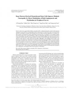
cells markers CD34, CD19, CD45 to make sure that cells meet minimal criteria for defining multipotent mesenchymal stromal cells [12]. All BM-MMSC samples were CD105/CD90/CD166/CD73 positive and negative for CD34/CD19/CD45 at all tested passages. Additionally, the ability of BM-MMSC to differentiate to osteo- and adipo- lineages was tested. Representative images are demonstrated on Figure 1. BM-MMSC proliferative activity Proliferative activity is an important feature of cell samples that intended for therapeutic protocols. The decline in proliferative activity could indicate that cells either enter the state of replicative senescence [28, 29] or they just stop growing and begin to differentiate [30]. To estimate the proliferative activity in BM-MMSC we calculated population doubling time (PD) for each sample in successive passages, and found out that at the first steps of in vitro expansion PD was less than 24 h in practically all tested samples, but increased gradually during expansion, and could reach more than 7 days by P3. The significant difference in proliferative activity between HD and HF samples was detected after about 14 in vitro doublings (Figure 2A).
Figure 1. Representative results of routine analysis of BM‐MMSC samples quality. (A) Immunophenotype; Performed on Guava EasyCyte 8 using ExpressPro software. (B,C) Ability to differentiate: (B) Adipogenic differentiation (x200, OilRedO staining) and (C) osteogenic differentiation (x200, Alizarin staining).
www.impactaging.com
15 AGING, January 2015, Vol. 7 No.1
Figure 2. Analysis of in vitro proliferation activity of HD and HF BM‐MMSC cells seeded at 3000 cells/cm2 and expanded in normoxia (pO2 =20%). (A) The population doubling time (PD) at passage 3; Data are presented as mean +/‐SEM; n=16 for HF, and n=10 for HD; ** p