and 4 years of real-world experience with a desktop workstation ... -4 clinical image display application, we perform usability studies on all .... build up of images.
Functional Requirements of a Desktop Clinical Image Display Application Bradley J. Erickson, William J. Ryan, Dale G. Gehring
HILE MUCH ATIENTION has focused on the user interface of picture archiving and communication system (PACS) interpretation workstations, there has been relatively little emphasis on the functional requirements of the display application for clinicians. In some arenas, such as the intensive care unit, it may be appropriate to deploy a full PACS workstation for clinician use. But even there, it is not clear that the interpretation toolset and interface are optimal for the clinician. The need for ubiquitous image display and the very rapid pace of outpatient clinical practice result in different functional requirements for this setting. This report wiII describe the Mayo multi-year experience with a desktop display application, focusing on the performance and feature set necessary for clinician (nonradiologist) image display.
W
out loud'? describing what he/she expects to happen, and to express feelings about how the interface performs. Typically, five physicians are used for each study.
Real-World Deployment Data Collection The usability laboratory methods are excellent for optimizing the user interface for candidate features. However, the majority of candidate features come from the user base. Further, there are aspects of real-world deployment that do not conform well to laboratory testing. For example, a clean laboratory setting with a "cooperative" subject may not disclose real-world performance problems, nor actual clinician tolerance for slow execution speed. Therefore, we provide forums for feedback on a regular basis, and occasionally perform user surveys. In addition to the verbal and written communications described above, we have also designed the application to record every operation (eg, button clicks and key strokes) performed by every user'. This allows us to collect precise, objective usage information by user for each feature, over any arbitrary period of time.
RESULTS AND DISCUSSION METHODS The requirements identified here have been derived from two sources: formal usability laboratory testing of possible features and 4 years of real-world experience with a desktop workstation application developed to display radiologic images for clinicians. The requirements coming from these two sources are often very different. While they are usually complementary, occasional areas of conflict have been seen.
Usability Laboratory Studies In order to continue to improve the QREADS I -4 clinical image display application, we perform usability studies on all major new features considered for implementation. After each release, a list of candidate features for the next release is developed, based on concerns/suggestions by users, as well as the development group. Features that were deferred from prior lists are also considered. The development group then studies the size and feasibility of each candidate feature, and tries to assess the value. This is then used to prioritize potential features for a test version for usability testing. The highest priority features that can be implemented in the ensuing 3 to 4 months are then implemented for usability testing. For each candidate feature, one or more test scenarios are created. which are used in usability laboratory testing. For usability testing, the user is given little or no instruction on how to operate the application, but rather is given a description of the tasks they are to accomplish. The users range in experience from little computer use to fairly extensive computer use. A group of scenarios are created that exercise the new features, which the typical clinician user can complete in about 60 minutes. The entire session is videotaped, with an inset of the video from the computer screen. The user is encouraged to "talk
The requirements can be separated into hardware and software requirements. In both categories, there is significant variation in requirements, which depends on the users and the environment. In the case of hardware requirements, we found that in most clinical situations, a single consumergrade display was adequate. However, for some clinical areas (such as the Intensive Care Unit) where images are provided prior to interpretation, and in surgical areas where many images are routinely viewed, dual-monitor systems were deemed necessary. This experience seems fairly typical of other institutions, and wiII not be explored further here. Image display speed is an important requirement. Most clinicians felt that the first images of an examination should be displayed within 2 seconds of the command to open the examination. Any image requested after that should be displayed
From the Departments of Radiology and Information Services, Mayo Clinic. Rochester, MN. Address reprint requests to Bradley J. Erickson. MD. P/lD. Department ofRadiology. Mayo Clinic, 200 First St SIV, Rochester, MN 55905. E-mail: bjetemayo.edu. . Copyright © 2001 by IV.B. Saunders Company 0897-188910111402·1039$35.0010 doi: 1O.1053/jdim.2001.23849
Journal of Digital/maging, Vol 14. No 2, Suppl1 (June), 2001: pp 149-152
149
ERICKSON, RYAN, AND GEHRING
150
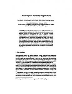
Fig 1. Initial survey screen of clinical display application.
within 1 second. All image manipulations should be completed in less than 1 second. The standard display manipulation tools like brightness/contrast and zoom/pan have been described by others; the need for these is apparent. We do note that most clinicians expect buttons for computed tomography (CT) presets (abdomen, head, bone, and lung), although these buttons confused some clinicians. Enabling and disabling buttons provides useful feedback about what can and cannot be done. Clinicians uniformly emphasized the need for fast (sub-second) response with image manipulations. In most outpatient settings, clinical efficiency is essential, and therefore, integration with the other clinical tools is a key feature to maintaining or improving productivity. The Clinical Context Object Workgroup (CCOW) of the Health Level 7 (HL-7) standard provides this capability, and it, or an equivalent capability, should be included in the desktop display application. We have also found that clinicians typically want to view the radiologist's interpretation along with the images. This is particularly true when the report refers to specific images. The ability to directly connect the two (eg, double click on the image reference in the report to bring up the associated image) is difficult now, but may be easier after adoption of structured reporting. Flexibility of the display format is also essential. We have found it beneficial· to begin with a "survey" view that allows clinicians to get the "gestalt" of the examination, and then move on to focus on specific images. This initial view (Fig I) also shows the radiologic report, and the list of all
examinations for the patient. After this initial view, more detailed viewing is accomplished in the fullscreen mode (Fig 2). In-this case, the layout should be flexible, and the navigation should be clear. This can be difficult when dealing with large examinations and limited screen space. Consistency of behavior appears to be more important than "cleverness," as most clinicians do not use the display application frequently enough to learn the complex patterns of "clever" buttons. By "clever" buttons, we mean buttons that change their behavior according to the situation. For example, we experimented with the behavior of the "Next" button under varying display formats. When displaying a magnetic resonance image (MRI) with different numbers of images per series, and displaying the images in a "series per stack" format, we thought it might be most sensible that the Next button do nothing to view areas displaying the final image in that stack, while advancing the slice number in areas with more images. However, this was inconsistent with its behavior in the survey and tile formats, where one always advances on to the next series. Therefore, we did decide to have the "Series per Area" button simply start the arrangement, but the user interface actually showed the matrix size that resulted. The result is a consistent behavior that users found less confusing, while also providing a mechanism that allowed clinicians to quickly achieve the desired layouts. The ability to correctly localize a cross-sectional image was an important requirement, particularly for spine surgeons, where axial MRI or CT images of lumbar and cervical levels look similar. In order
Fig 2.
Full-screen mode of clinical display application.
CLINICAL IMAGE VIEWER FUNCTIONAL REQUIREMENTS
to be certain of the level of some pathology, such as a disk protrusion, a line representing the location of the axial slice on a perpendicular view is needed. A mechanism for easy comparison with older examinations, particularly CT and MRI, is also very important. The first part of this requirement is the ability to easily open and view two or more examinations at once. In addition, when screen space is limited, the display application should have a (user-selectable) function to semiautomatically align corresponding slices from different series and examinations. Once they are aligned, the user can navigate the one series, and the application wiII step the required number of slices to display the matching slice. This is trivial when slice spacing is the same for the series, but when spacing changes, this is more challenging. One must also have a mechanism to allow the user to override or adjust the match, for cases where the patient moved. In some arenas (such as orthopedics and oncology) measurement tools are helpful. Many of the tools provided on diagnostic workstations are not necessary for examination room image display. It is critical that the images be displayed with optimal window settings, and that buttons for recovering from mis-steps be quick and simple. Clinicians do not constitute a uniform group of users, and the requirements vary substantially from individual to individual, even within a specialty area (though there are clear tendencies for some groups like surgeons to require more tools). This appears' to drive the need for a user-configurable interface, allowing "simple," "medium," and "advanced" tool sets. Another mechanism for increasing clinician efficiency is "Key Images." When a case with many images is interpreted (eg, CT, MRI, ultrasound, fluoroscopy), there are typically 5 to 10 images that convey most of the critical features, and the remaining images are substantially less important for most purposes. Providing a way for the radiologist to identify these key images, and have the clinical display application initially display only those images could improve clinical efficiency (and reduce network load). This requires that the PACS communicate which are the key images, and that the clinical display system support this. This feature must also allow for the clinician to view the entire images set, and also be able to add and remove key
151
images. We have identified this as an important feature, but have not been able to implement it due to limitations of the source systems. In many cases, the radiologist wiII measure certain structures like tumors. While the display application must display these annotations, it is also necessary to allow the clinician to make and store measurements, with an audit trail. The measurement tools should include distance (line) area (rectangle and free-hand), and pixel measurements. We are also experimenting with a tool that would provide the maximum minor axis measurement after the user has defined the major axis of a mass/structure. The Digital Imaging and Communications in Medicine (DICOM) header has a significant amount of textual information that is not typically displayed. We have found that most clinicians only want to see the date, anatomic markers (left/right, etc), and series/image numbers. While radiologists would expect to see much more extensive information (such as repetition and echo times for MRI, kVp and rnA for radiography), most clinicians do not find this information useful, and prefer a less cluttered display. For those cases where this information is needed, we provide a pop-up window that displays this textual information. As other electronic tools become more ubiquitous, the ability to communicate electronic images becomes more important. We therefore provide the ability to copy an image to the system clipboard. We made an intentional decision to not provide either the ability to store these as files, or to print them. Our medical electronic environment consists of about 10,000 personal computers that are maintained centrally. The large size of images could quickly fill the local disk, and the lack of local "ownership" raised the concern that these machines could be compromised by unintentional build up of images. Similarly, the ability to print images would significantly increase the burden on the network and printers, and could also be a security risk if an image was inadvertently printed to the wrong device. There are exception areas where we are considering printing-the operating room areas, and providing films for patients to take back to referring physicians. This is one example where we noted the conflicting requirementssome areas seemed to need printing, while in other areas it was important to disable this feature.
152
ERICKSON, RYAN, AND GEHRING
CONCLUSION
The requirements for a clinical viewing station have some similarities to requirements of an interpretation workstation, but have some important differences. The user base is a heterogeneous group, and the tasks the clinicians perform vary substantially, increasing the variation in the requirements. However, it is possible to deter-
mine a list of requirements that satisfy most clinicians under most circumstances. In addition to the standard image manipulation tools, we have found that integration with the other clinical tools is essential to maintaining or increasing their efficiency. Providing for varying levels of functionality/complexity is also useful, because of the variation in the user base.
REFERENCES 1. Erickson B, Ryan W, Gehring D, et al: Clinician usage patterns of a desktop radiology information display application. I Digit Imaging 11:137-141, 1998 (suppl I) 2. Erickson BI, Ryan WI, Gehring DG, et al: Image display for clinicians on medical record workstations. I Digit Imaging 10:38-40, 1997 (suppl 1) 3. Erickson BI, Ryan WI, Gehring DG, et al: Clinician image
review patterns in an out-patient setting, in Horii, Blaine (eds): SPIE Medical Imaging. San Diego, CA, SPIE, 1998 4. Eversman W, Pavlicek W, Zavalkovskiy BV, et al: Performance and function of a desktop viewer at Mayo Clinic Scottsdale. I Digit Imaging 13:147-152,2000 (suppl 1) 5. Nielsen I: Usability Engineering (ed 1). San Diego, CA, Academic Press, 1993, p 362