Biochimica et Biophysica Acta 1837 (2014) 178–185
Contents lists available at ScienceDirect
Biochimica et Biophysica Acta journal homepage: www.elsevier.com/locate/bbabio
Functional role of the MrpA- and MrpD-homologous protein subunits in enzyme complexes evolutionary related to respiratory chain complex I☆ Vamsi K. Moparthi a,1, Brijesh Kumar a,2, Yusra Al-Eryani a, Eva Sperling a, Kamil Górecki a, Torbjörn Drakenberg b, Cecilia Hägerhäll a,⁎ a b
Department of Biochemistry and Structural Biology, Center for Molecular Protein Science, Lund University, PO Box 124, S-221 00 Lund, Sweden Dept. of Biophysical Chemistry, Center for Molecular Protein Science, Lund University, PO Box 124, S-221 00 Lund, Sweden
a r t i c l e
i n f o
Article history: Received 4 July 2013 Received in revised form 18 September 2013 Accepted 24 September 2013 Available online 1 October 2013 Keywords: NADH:quinone oxidoreductase Na+/H+ antiporter 11-subunit complex I Membrane-bound hydrogenase Mrp-complex
a b s t r a c t NADH:quinone oxidoreductase or complex I is a large membrane bound enzyme complex that has evolved from the combination of smaller functional building blocks. Intermediate size enzyme complexes exist in nature that comprise some, but not all of the protein subunits in full size 14-subunit complex I. The membrane spanning complex I subunits NuoL, NuoM and NuoN are homologous to each other and to two proteins from one particular class of Na+/H+ antiporters, denoted MrpA and MrpD. In complex I, these ion transporter protein subunits are prime candidates for harboring important parts of the proton pumping machinery. Using a model system, consisting of Bacillus subtilis MrpA and MrpD deletion strains and a low copy expression plasmid, it was recently demonstrated that NuoN can rescue the strain deleted for MrpD but not that deleted for MrpA, whereas the opposite tendency was seen for NuoL. This demonstrated that the MrpA-type and MrpD-type proteins have unique functional specializations. In this work, the corresponding antiporter-like protein subunits from the smaller enzymes evolutionarily related to complex I were tested in the same model system. The subunits from 11-subunit complex I from Bacillus cereus behaved essentially as those from full size complex I, corroborating that this enzyme should be regarded as a bona fide complex I. The hydrogenase-3 and hydrogenase-4 antiporter-like proteins on the other hand, could substitute equally well for MrpA or MrpD at pH 7.4, suggesting that these enzymes have intermediate forms of the antiporter-like proteins, which seemingly lack the functional specificity. © 2013 The Authors. Published by Elsevier B.V. All rights reserved.
1. Introduction NADH:quinone oxidoreductase or complex I (EC 1.6.99.3) is the largest enzyme in the respiratory chain of mitochondria and bacteria. The mammalian enzyme comprises 45 protein subunits, but of these, fourteen protein subunits make up a conserved core structure also found in bacteria. Seven subunits protrude from the membrane and contain a flavin prosthetic group and eight iron-sulfur clusters that guide electrons from the oxidation of NADH towards the quinone binding site [1]. The remaining seven subunits make up the membrane domain of the enzyme complex. These proteins are lacking prosthetic ☆ This is an open-access article distributed under the terms of the Creative Commons Attribution-NonCommercial-No Derivative Works License, which permits non-commercial use, distribution, and reproduction in any medium, provided the original author and source are credited. ⁎ Corresponding author. Fax: +46 46 2224116. E-mail address:
[email protected] (C. Hägerhäll). 1 Present address: DOE Joint Genome Institute, University of California, Berkeley, United States. 2 Present address: School of Biological Sciences, University of Auckland, Auckland, New Zealand.
groups, but nevertheless, this domain must harbor important parts of the energy coupling machinery. The structure of the soluble domain of complex I has been solved to 3.1 Å resolution [2,3], whereas recently, the membrane spanning domain has also been revealed to about 3.0 Å [4,5]. Already in structures solved at lower resolution, the three homologous proteins NuoL, NuoM and NuoN stood out as three virtually identical structural entities, except for a NuoL C-terminal extension in the form of a membrane-parallel helix extending from the distally located NuoL subunit towards the proximal NuoN [6,7]. The complex I protein subunits can be divided into functional modules [1]. This reflects that the evolution of the large enzyme complex has occurred from smaller functional building blocks [8]. The NADH dehydrogenase or N-module is composed of the NuoE, NuoF, and NuoG protein subunits, and can be regarded as an electron input device. The Q-module enzyme comprises the NuoB, NuoC, NuoD, and NuoI protein subunits, where NuoB and D resemble the small and large subunit of NiFe hydrogenase [9]. The metal site in the hydrogenase corresponds to a quinone-binding site in complex I [10], explaining the module name. Finally the P-module, for proton pumping, is made up of all the hydrophobic proteins NuoA, NuoH, NuoJ, NuoK, NuoL, NuoM, and NuoN [1]. The three large proteins NuoL, NuoM and NuoN show primary
0005-2728/$ – see front matter © 2013 The Authors. Published by Elsevier B.V. All rights reserved. http://dx.doi.org/10.1016/j.bbabio.2013.09.012
V.K. Moparthi et al. / Biochimica et Biophysica Acta 1837 (2014) 178–185
sequence similarity to two protein subunits in the Mrp Na+/H+ antiporter complex, MrpA and MrpD [8,11]. Therefore, these proteins come out as main suspects in providing ion channels for the proton pumping machinery. Such putative proton pathways have also been visualized in the structure [4]. The Mrp antiporter complex is composed of seven proteins in total, including MrpA and MrpD. It has been demonstrated that all the seven Mrp-complex protein subunits must be present to have a functional Na+/H+ antiporter [12,13]. Complex I homologous protein assemblies are also found in several different types of smaller enzyme complexes containing fewer units than the 14 proteins in the “standard” bacterial enzyme that are also conserved in the mitochondrial complex I. One such smaller enzyme is predicted to be a bona fide complex I enzyme, lacking the N-module ([14], Table 1). This enzyme is the putative ancestor of all present day complex I and could probably interact with different electron donors/ acceptors [15], whereas the 14-subunit enzymes operate solely with NADH/NAD+. Other homologous smaller enzyme complexes are membrane-spanning hydrogenases that are typically defined by the presence of the NiFe active site cysteine ligands in the NuoD homologous subunit. These membrane-bound hydrogenases come in several varieties. Hydrogenase-4 contain three MrpA- and MrpD-homologous proteins, i.e. three putative ion channels, whereas hydrogenase-3 have only one such subunit (Table 1). Both these hydrogenase varieties exist in Escherichia coli. The smaller hydrogenase-3 enzyme, with protein subunits denoted Hyc [16], is functioning together with FdhH, a formate dehydrogenase protein for which the structure was solved already in 1997 [17]. The hydrogenase-3 enzyme was shown to be the main hydrogen producing enzyme in E. coli whereas the soluble hydrogenase-1 and -2 enzymes were responsible for respiratory hydrogen oxidation [18]. The hydrogenase-4 in E. coli is encoded by the hyf operon and was postulated to also operate with FdhH and carry out energy conservation by performing formate dependent proton translocation [19]. The Hyf enzyme complex has never been biochemically characterized. The hyf operon was found to be silent in wild type E. coli but its expression could be activated [20]. It was also demonstrated that hyf, in spite of activation, cannot functionally substitute for the loss of hyc. A CO-induced hydrogenase has been described in
179
Rhodospirillum rubrum, which generates energy from the oxidation of CO to CO2 coupled to the production of H2 [21]. The Ech hydrogenase was first described in Methanosarcina barkeri [22] and has since been found in several methanogenic archaea [9]. Other archeal hydrogenases, denoted Mbh and Mbx, were first described in the hyperthermophile Pyrococcus furiosus [23–25]. Their evolution and relation to complex I were recently reviewed in [26]. By understanding more about the building blocks and their primordial function, important clues to the molecular function of present day complex can be deduced (see Fig. 1). A survey of the distribution of different types of complex I in the tree of life recently showed that a compact 11-subunit complex I was found both in the archaeal and the eubacterial kingdoms, whereas the full size, 14- subunit complex I was only found in some eubacterial phyla [14]. This demonstrated that such an 11-subunit complex I was the last common ancestor of all present day complex I enzymes. Membrane-bound hydrogenases have also been suggested to be ancestors of complex I [8,27,28]. Indeed, a membrane bound hydrogenase is a likely evolutionary progenitor of complex I but this enzyme must have differed from the present day membrane-bound hydrogenases in several respects [15,29]. The antiporter-like ion transporter proteins in membrane-bound hydrogenases were lacking primary sequence features that were present in both the bona fide antiporter proteins MrpA and MrpD and in the complex I proteins NuoL, NuoM and NuoN, and therefore must have evolved separately in hydrogenases, and only after the split from the last common ancestor [29]. A model system, where the function of the Mrp-type ion transporter proteins can be assessed, was recently developed. Deletion of mrpA or mrpD from the Bacillus subtilis chromosome resulted in Na+ and pH sensitive growth phenotype, which could be explored in complementation tests. The properties of the complex I subunits NuoL, NuoM and NuoN from E. coli complex I were investigated using the model system. NuoL from E. coli expressed in the B. subtilis ΔmrpA brought back the wild type growth properties, whereas expression of E. coli NuoN in the
Table 1 List of homologous subunits. E. coli FHL-1 (Hyc)
E. coli FHL-2 (Hyf)
B. subtilis M. barkerii R. rubrum Mrp CO-induced Ech hydrogenase hydrogenase antiporter
HyfI HyfG(Nter) HyfG(Cter) HyfH
CooL CooU
EchC EchD
CooH
EchE
NuoI
HycG HycE (N-ter) HycE (C-ter) HycF
CooX
EchF
NuoA NuoH NuoJ
– HycD –
– HyfC –
– CooK –
EchB –
E. coli complex I
B. cereus complex I
N module NuoE NuoF NuoG
– – -
Q module NuoB NuoC1
NuoB NuoC
NuoD1
NuoD
NuoI P module NuoA NuoH NuoJ
NuoK NuoK – NuoL/M/N NuoL/M/N HycC
HyfE – HyfB/D/F CooM2
– EchA
MrpA C-term MrpC MrpA/D
1 In E. coli, NuoC and NuoD are in fact also fused into one subunit [41], just as in HycE and HyfG, but this is not a general feature of complex I. 2 The CooM protein comprises two antiporter-like polypeptides fused into one polypeptide.
Fig. 1. Schematic representation of the protein subunits in the respective enzyme complexes. Subunit nomenclature is also summarized in Table 1. The bona fide Mrpantiporter contains two homologous subunits, MrpA and MrpD. The MrpEFG subunits are not related to any present-day complex I proteins. Hydrogenase-3 and hydrogenase4 contain one or three homologous proteins of the MrpA/D type respectively. In addition, both types of hydrogenases interact with a formate dehydrogenase unit in E. coli (not shown). The membrane-spanning part of 11-subunit complex I and classical 14-subunit complex I is fully conserved and contains three MrpA/D type proteins. In some complex I enzymes, such as that from E. coli, the NuoC and NuoD homologues are fused into one subunit, just as in the corresponding hydrogenase subunits HycE and HyfG ([41] see also Table 1).
180
V.K. Moparthi et al. / Biochimica et Biophysica Acta 1837 (2014) 178–185
same deletion strain did not improve the growth at all. The opposite was true for the B. subtilis ΔmrpD strain, that was efficiently complemented by NuoN, and to a much lesser extent by NuoL [30]. It was also noticed that the complementation observed at pH 7.4 did not require Mrpcomplex formation but merely the presence of all subunit types. At pH 8.4, where no complementation with complex I subunits was seen, Mrp-complex formation was shown to be necessary for function [30]. The simplest explanation for the observed growth properties was that each protein comprised a single ion channel rather than a complete antiporter. Since Na+ translocation had previously been observed in NuoL [31] MrpA and NuoL were tentatively assigned as Na+ channels whereas MrpD and NuoN were proposed to be H+ channels. In this work we have addressed the issue of ion-specificity further, by measuring the internal sodium concentration in cells by 23Na+-NMR. The compact, 11-subunit complex I is not present in B. subtilis, but is found in the close relative Bacillus cereus. Membrane-bound hydrogenases can be of several types. Examples of antiporter-like proteins are HycC from hydrogenase-3, HyfB, HyfD and HyF from hydrogenase-4 [19,32], CooM from CO-induced hydrogenase, actually comprising two subunits fused into one [21] and EchA from Ech-hydrogenase [33], see Fig. 1 and Table 1. In this work, the previous phylogenetic analyses [29] were repeated, since the number of available sequences had increased significantly since the completion of that work in 2002. Subsequently, the B. subtilis model system was used to investigate the ion translocation abilities of the individual NuoL, NuoM and NuoN complex I subunits from 11-subunit complex I from B. cereus, to elucidate if the functional properties are indeed conserved. In addition, the HycC protein subunit from E. coli hydrogenase-3 and the HyfB, HyfD and HyfF subunits from E. coli hydrogenase-4 were cloned and expressed in the model system, to investigate if the theoretical predictions regarding their functionality could be verified experimentally. 2. Materials and methods 2.1. Construction of the phylogenetic tree of ion-transporter subunits from complex I and their homologues in membrane-bound hydrogenases Protein primary sequences of HycC subunit of hydrogenase-3, HyfB, HyfD, HyfF subunits from hydrogenase-4, EchA subunit from Ech hydrogenase, and NuoL, NuoM and NuoN subunit of 11 subunit, 14 subunit, and 12 subunit complex I enzymes from both Archaea and Eubacteria were collected from TIGR and NCBI and aligned using Clustal W with default settings. The aligned dataset was analyzed, edited and converted into MEGA format by using Data Analysis in Molecular Biology and Evolution, DAMBE ver 4.13. Unrooted phylogenetic trees were created using MEGA version 4.0 with boot-strap support of 100 replicates in the Neighbor-Joining method. The tree is drawn to scale, with branch lengths in the same units as those of the evolutionary distances used to infer the phylogenetic tree. The evolutionary distances were computed using the Poisson correction method and are in the units of the number of amino acid substitutions per site. All positions containing gaps and missing data were eliminated from the dataset (complete deletion option). For substitutions in the dataset the included option was used for the whole sample. Pattern among lineage was set to homogeneous and rates among site were set to uniform rates. Phylogenetic analyses were conducted in MEGA4. Protein accession numbers are listed in the supplementary material. 2.2. 23Na+-NMR experiments The 23Na+-NMR experiments were done essentially as in [34]. Briefly, the cells were grown without extra Na+ added to the media to OD 0.5, and then challenged with 80 mM NaCl for 1–2 h. After that, the cells were harvested, washed with Na+-free buffer and resuspended at 0.32 g cells/ml buffer. To 400 μl of the cell suspension D2O and the shift reagent Tm(DOTP)5- were added to concentrations 10% and
0.8 mM, respectively. At least 512 scans were accumulated for each spectrum, and the internal and external Na+ concentrations were calculated from the signal intensities. The internal signal was multiplied by 2.5, to compensate for the extreme broadening of the cell interior (see discussion in [34]). 2.3. Cloning and construction of expression plasmids Bacterial strains, plasmids and primers used are listed in Table 2. E. coli cells and B. subtilis cells were routinely grown aerobically at 37 °C and 200 rpm in LB medium as before. For solid media, 1.5% agar was added. B. subtilis strains were grown aerobically at 37°C and 200 rpm in nutrient sporulation medium with phosphate (NSMP [35]) and were kept on Tryptose Blood Agar Base (TBAB) plates. Antibiotics were added in the following concentrations when appropriate: 100 μg/ml ampicillin, 12.5 μg/ml chloramphenicol and 10 μg/ml kanamycin for E. coli, 5 μg/ml chloramphenicol and 2 μg/ml phleomycin for B. subtilis. All standard molecular biological techniques were done as before [30]. Primers (Table 2) were synthesized by MWG Biotech AB, Germany and Fermentas Life Sciences. PCR products were purified from agarose gel with the Jetsorb gel extraction kit (Genomed) and restriction enzyme digested vectors and DNA fragments were purified by DNA Clean Up (Genomed) or Jetsorb. Chromosomal DNA from B. cereus was prepared as described by Marmur [36], and for E. coli chromosomal DNA the method of Wilson [37] or the Gene Jet™ Genomic DNA Purification Kit (Fermentas) was used. DNA sequencing reactions were carried out using Big Dye™ (Applied Biosystems) and the subsequent sequence analysis was performed at the Biomolecular Resource Facility, Lund University. B. cereus chromosomal DNA was used as a template for amplification of nuoL, nuoM and nuoN, whereas E. coli chromosomal DNA was used for amplification of hycC, hyfB, hyfD and hyfF using the primers shown in Table 2. In the upstream primer, a unique restriction-enzyme recognition site was created, for nuoL a HindIII site and for nuoM, and nuoN a XbaI site. In the downstream primer sequence no restriction-enzyme recognition site was introduced, since a DNA polymerase leaving blunt ends was used. The stop codon was removed from the respective gene sequences, to produce fusion proteins to cytochrome c550 when the gene fragments were cloned into pTRC19 [38] and/or pTCH [39]. The constructs were transformed into E. coli JM109 [40]. The resulting, correct constructs were named pVM12, pVM13, pVM14, pVM18, pVM19, pVM21 and pVM22 (Table 2). These plasmids were subsequently used as template for the amplification of nuoLcytc, nuoMcytc, nuoNcytc, hycCcytc hyfBcytc, hyfDcytc and hyfFcytc respectively, for subcloning of the cytochrome-tagged polypeptides into the shuttle vector pCW6. In the downstream primers, a unique PstI site was introduced, except in nuoMcytc that contains an internal PstI site, and therefore SalI was used instead. In the upstream primers a XbaI site was created in all the constructs (Table 2). The pCW6 was purified from E. coli GM3819 to obtain unmethylated plasmid DNA that can be digested with XbaI. After confirming the correct constructs by DNA sequencing, the resulting constructs were named pVM15, pVM16, pVM17, pVM20, pVM23, pVM24 and pVM25. 2.4. B. subtilis growth studies The B. subtilis strains were grown essentially as described previously [30]. The same 250 ml baffled E-flasks were used. The flasks have an attached side-arm that allowed convenient measurement of the optical density (OD) of the culture without opening the flasks or changing the growth medium volume throughout the experiment. According to the manufacturer, the NB contains about 7 mM Na+ when used as recommended (8 g/L). The NaCl concentrations indicated in each experiment always refer to the amount of NaCl added to the growth medium. As before, the B. subtilis strains to be studied were taken from the −80°C freezer and incubated on plates with no added NaCl, at 37°C for 8 h,
V.K. Moparthi et al. / Biochimica et Biophysica Acta 1837 (2014) 178–185
181
Table 2 Bacterial strains, plasmids and primers used in this work. Bacterial strain
Genotype
Reference/Source
B. subtilis 168A B. subtilis ΔmrpA B. subtilis ΔmrpD B. cereus ATCC 14579 E. coli JM109
Wild type, trpC2 ΔmrpA bler ΔmrpD bler Wild type endA1 glnV44 thi-1 relA1 gyrA96 recA1 mcrB+ Δ(lac-proAB) e14-[F′ traD36 pro AB+ lacIq lacZΔM15] hsdR17(rk− mk+) recA1, endA1, gyrA96, thi, hsdR17, supE44, relA1 (lac) dam-16::Kan thr-1 leuB6 thi-1 argE3 hisG4 proA2 lacY1 galK2 mtl-1 xyl-5 ara-14 rpsL31 tsx-33 supE44 rfbD1 kdgK51
Bacillus Genetic Stock Center (type train) [30] [30] DSMZ (Type strain, DSM31, ATCC 14579) [40]
E. coli XL1-Blue E. coli GM3819 (Dam−) Plasmids
Relevant properties
pCW6 pVM6 pVM11 pVM12 pVM13 pVM14 pVM15 pVM16 pVM17 pVM18 pVM20 pVM19 pVM21 pVM22 pVM23 pVM24 pVM25
Cmr mrpDcytc Cmr mrpAcytc Cmr nuoLBccytc, Ampr nuoMBccytc, Ampr nuoNBccytc, Ampr nuoLBccytc, Cmr nuoMBccytc, Cmr nuoNBccytc, Cmr hycCcytc, Ampr hycCcytc, Cmr hyfBcytc Ampr hyfDcytc, Ampr hyfFcytc, Ampr hyfBcytc, Cmr hyfDcytc, Cmr hyfFcytc, Cmr
Promega
Claes von Wachenfeldt [38] [38] This work This work This work This work This work This work This work This work This work This work This work This work This work This work
Primers
Restriction enzyme site
Primer sequence
nuoLBc_for nuoLBc_rev nuoMBc_for nuoMBc_rev nuoNBc_for nuoNBc_rev hycC_for hycC_rev hyfB_for hyfB_rev hyfD_for hyfD_rev hyfF_for hyfF_rev nuoLBc_for_pCW6 nuoMBc_for_pCW6 nuoNBc_for_pCW6 nuoLBc_rev_pCW6 nuoMN_Bc_rev_pCW6 hycC_for_pCW6 hycC_rev_pCW6 hyfB_for_pCW6 hyfD_for_pCW6 hyfF_for_pCW6 hyfBDF_rev_pCW6
HindIII – XbaI – XbaI – XbaI – XbaI – HindIII – HindIII – XbaI XbaI XbaI SalI PstI XbaI PstI XbaI XbaI XbaI PstI
5′ CGTACGAAGCTTCAGGTGGA 3′ 5′ TCGTAAATCCCCTCCCGTAT 3′ 5′ GTGGTTTCTCTAGATGTACT 3′ 5′ CTTCACCCCCAATGTTTTCA 3′ 5′ AAACATTGGGGGTCTAGAAGG 3′ 5′ TTGTACCACGTTCCCTAAGAAGA 3′ 5′ CAGCAAAGCTTAGCGTGAGC 3′ 5′ GGCTCCTCGTGAAACAATAATC 3′ 5′ GAAAGATCTAGAGCGCCTTG 3′ 5′ GACGGCAATAGCGATTAGCA 3′ 5′ CTCATTAAGCTTGTGACCTGGCTTG 3′ 5′ CTGCAACCAGGTCGCGGCGATTAC 3′ 5′ GTCATTAAGCTTGTGTTACTGGC 3′ 5′ ACGTTCACTGTGTTTCTCCGA 3′ 5′ CGCTCAAAGTCTAGAATTGTGACCG 3′ 5′ GTCATTATTTTGCTCTAGACGGG 3′ 5′ GATGGATATCTAGACGTTATTTAGC 3′ 5′ CCGCTACTGTCGACAATCGTTTA 3′ 5′ CCGCTACTGTCTGCAGTCGTTTA 3′ 5′ CACGGACTCTAGAGATCTCACCTTG 3′ 5′ CCGCTACTGTCTGCAGTCGTTTA 3′ 5′ CTGGTTTTCTAGAGGCGGGGGAGGATC 3′ 5′ CCTTGCTGCTCTAGAATGGGTCT 3′ 5′ CCCACGGCACTCTAGACGTG 3′ 5′ CGGGTCTGCAGGCAGCAGTC 3′
and were then used immediately to inoculate the liquid cultures. Most of the growth studies were repeated several times, but all experiments were repeated in at least five independent experiments. The generation time g is the time it takes for bacteria to divide. The g was calculated using Eqs. (1) and (2), where OD1 and OD2 are optical densities from the logarithmic growth phase, t1 and t2, and k is a growth constant. k¼
ð ln ðOD2Þ− ln ðOD1ÞÞ ðt2 −t1 Þ
ð1Þ
g¼
ln 2 : k
ð2Þ
3. Results 3.1. The role of cation transport in the deletion strain phenotype The MrpA, MrpD, NuoL, NuoM and NuoN are all homologous proteins, but the primary sequence of MrpA is more similar to NuoL, whereas MrpD is grouping with NuoN [29]. This coincided with the recently determined functional similarity of MrpA and NuoL, and MrpD and NuoN [30]. The unambiguous role of Na+ in the observed deletion strain growth defect was demonstrated by measuring the internal Na+ concentration after a two hour 80 mM sodium challenge using 23 Na+-NMR and a shift agent [34]. Assuming that the NMR sample volume consists of 20% cells, the B. subtilis ΔmrpA strain contained 56 ± 17 mM Na+ and the B. subtilis ΔmrpD strain contained 57 ±
yd .h
M oo
2
S. fle
C yc tH n cC e SE..col Hy C c Hy pe D.s yfB nH fB T.te Hy e l S.f E.col HyfB
C.k or L H.bu tL
C.jej L N.pha L A.aeo L R.xyl L D.eth L poL F z a M.m rL N.eu L pL B.ja .spe L N nt ra L i . L f L B . er c B.
Y.ent HyfB M.the HyfB A R.p .spe HyfB al H yf B
S.aur MrpD
.b ov P L .v ib E. L c T. t ol L he L
C.eff MrpD
M
I.ioi
B. s ub M rpD R.le gM C.g r p D lu Mr pD T. G. ro for C s .b M ur MrpD rp Mr D pD N.e ur B. A. M ja p ae o M E. M co lM
A.spe MrpD B.sui MrpD MrpD
C.
M
j je
t in L.
M
ib P.v M he T.t lM xy R. M pe N.s er M M B.c B.fra M z Fpo M.ma N.pha M D.eth M P.vib M
M.bov M
N
N r N spe . ce N B. oN N ae .eth A. D
N
C.jej N
C.kor N H.but N
M.maz F poN T.the N N.ph aN
.fr a
L . in t N M.b ov N R.x yl N B.j N. ap e E. ur N N co lN
B
A.ful FpoN
0.1
Hy cC
C
R.rub CooM2 R.pa l Co o M2 D.vu l Co oM2 D T.c.car C ooM ar 2 Co oM 2 E. S.fle H yfF Y co M .en l Hy fF .th tH e yf H F yf F
E.co l Hy fD S.fle HyfD Y.ent HyfD HyfD R.pal D Hyf pe A.s fD Hy yfD en eH T .t h t pA M. i Mr rpA B .su M pe pA A.s i Mr I.io A rp M A r p ur A f M S.a rp ef M lu .g
C.
C
C
fF Hy n te . T F yf eH sp HyfF . A al R.p
B. su T fo b r M .ros rp M Mrp rp A R.l D .e A A eg C.b M.m th Ec ur M Mrp hA az E A r pA c hA M.b A.fu ar EchA l Fp oL
G.
R.rub CooM1 R.pal CooM 1 D.vu l Co oM1
V.K. Moparthi et al. / Biochimica et Biophysica Acta 1837 (2014) 178–185
1 ooM ar C T.cD.car CooM1 C.hyd CooM1
182
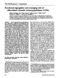
Fig. 2. Phylogenetic tree of the MrpA and MrpD-type proteins comprising NuoL, M and N of complex I and the homologous subunits from membrane-bound hydrogenases. The sample is chosen to represent the most distant branches of the tree of life. NuoL from organisms containing 14-subunit, 12-subunit or 11-subunit complex I is shown in green, violet and empty red circles respectively. NuoM from organisms containing 14-subunit, 12-subunit and 11-subunit complex I is shown in green, violet and red filled circles. NuoN from organisms containing from 14- subunit, 12-subunit and 11-subunit complex I is shown in green, violet and red filled triangles (upward pointing). HycC from hydrogenase-3 is shown with filled blue triangles (downward pointing), and EchA from Ech-hydrogenases with empty downward pointing triangles. The N-terminal part of CooM from carbon monoxide-induced hydrogenase is denoted CooM1 and represented by black filled diamonds whereas the C-terminal part is labeled CooM2 and is shown as empty diamonds. HyfB, HyfD and HyfF subunits from hydrogenase-4 are shown as filled red, blue and black squares respectively. MrpA and MrpD from the Mrp-antiporter are shown as filled (upward pointing) black triangles and filled, light blue (downward pointing) triangles respectively. Full names of organisms and protein sequence accession numbers are given in the supplementary material.
5 mM Na+. When MrpA was expressed from a plasmid in the ΔmrpA strain, substantially less Na+ was retained (19 ± 13 mM), demonstrating that the antiporter is now functional. Expression of MrpA in the ΔmrpD strain, on the other hand, did not alter the intracellular Na+ content of that strain at all, illustrating that both subunits must be present to have a functional Mrp-antiporter. In this work the analyses were repeated for B. subtilis ΔmrpA cells expressing the complex I subunits NuoL and NuoN. As expected, expression of NuoL significantly decreased the internal Na+ concentration (36 ± 5 mM) whereas
expression of NuoN caused a very small effect, if any (48 ± 8 mM). This corroborates the functional difference of MrpA/NuoL and MrpD/ NuoN, as originally predicted from sequence. 3.2. Repeated phylogenetic analyses As mentioned in the introduction, it was also noted that hydrogenases contained transporter protein subunits with lower sequence conservation, which could not be accurately assigned as MrpA-type or
V.K. Moparthi et al. / Biochimica et Biophysica Acta 1837 (2014) 178–185
MrpD-type [29]. These previous results were however derived from a much smaller dataset, and therefore the phylogenetic analyses were repeated with a larger dataset of currently available primary sequences of the proteins in question. A phylogenetic tree, showing representative members of the dataset, is shown in Fig. 2. NuoL, NuoM and NuoN subunits from all types of complex I, including the compact 11-subunit enzyme and hydrogenase subunits from enzymes having just one, two or three different putative ion transporter subunits (Table 1), were compared. In the unrooted phylogenetic tree (Fig. 2), transporter-protein subunits NuoL, NuoM, and NuoN from all the different types of complex I formed clusters comprising each subunit, corroborating the previous notion that the compact 11-subunit complex I enzymes should indeed be regarded as a bona fide complex I [14], in spite of the missing N-module. Just as previously observed, two distinct domains formed in the tree, comprising MrpA-type and MrpD type polypeptides. The 11-subunit complex I subunits did not deviate from this general pattern, and just like the classical complex I subunits, the NuoM and NuoN ended up in the MrpD domain and NuoL in the MrpA domain, predicting that the proteins have the same function. Among hydrogenase-4 (i.e. Hyf proteins) that have three transporter subunits just like complex I, HyfB is consistently the longer polypeptide, suggesting that it originated from NuoL, but in the phylogenetic tree HyfB sequences are ending up in an intermediate position, close to the HycC cluster. The HyfD sequence on the other hand, is grouping closer to NuoL on the MrpA-type side. Hydrogenase-3 enzymes (i.e. Ech and Hyc) that have only one transporter subunit still form separate groups, not related to the type of hydrogenase. The EchA from the methanogens forms a group closer to NuoL in the MrpA-type cluster, whereas the HycC polypeptides are found together with HyfB and HyfF. Taken together, the new results seem to corroborate the previous findings that the transporter subunits of hydrogenases are separately evolved forms, presumably with less functional specificity than the bona fide Mrp antiporter subunits and the bona fide complex I subunits. 3.3. Complementation of B. subtilis ΔmrpA and ΔmrpD strains with 11-subunit complex I ion transporter proteins Subsequently, the predictions were tested experimentally. The B. subtilis Mrp deletion strains, previously developed as a model system to study the function of the homologous ion transporter subunits from E. coli complex I [30], were implemented to investigate the function of the ion-transporter proteins from the 11-subunit complex I from B. cereus. To be able to monitor and compare the amounts of protein produced, the genes encoding for NuoL, NuoM and NuoN subunits were tagged with cytochrome c as before [38]. The cytochrome tagged fusion constructs were subsequently sub-cloned to a low copy number plasmid, containing the IPTG inducible Pspac promoter, thereby producing low, physiologically relevant amounts of proteins in bacteria, as was previously done for the E. coli NuoL, NuoM and NuoN subunits. The B. cereus complex I subunits were restoring the growth of B. subtilis deletion strains in a similar way as the corresponding E. coli complex I subunits, albeit with slightly lower efficiency in some cases (see Table 3). Expression of NuoL in B. subtilis ΔmrpA at pH 7.5 improved the growth substantially, but expression of NuoM or NuoN was not helpful at all to this strain (see Fig. 3A). At the same pH, wild type growth properties were regained by expression of NuoN in B. subtilis ΔmrpD, whereas NuoL expression was less efficient (see Fig. 3B). NuoM was not complementing the growth very well in either B. subtilis ΔmrpA or ΔmrpD (Fig. 3A and B). Just as was previously seen in the E. coli proteins, the functional specificity was lost at a more acidic pH. At pH 6.5, all the B. cereus proteins could rescue both deletion strains to some extent. Likewise, at pH 8.4, none of the antiporter like Nuo proteins could complement the growth of the deletion strains
183
Table 3 Growth properties of B. subtilis deletion strains, expressing antiporter like proteins under different growth conditions.
At pH 7.4 E. coli complex I NuoL NuoM NuoN B. cereus complex I NuoL NuoM NuoN E. coli hydrogenase-3 HycC E. coli hydrogenase-4 HyfB HyfD HyfF B. subtilis Mrp MrpA MrpD At pH 6.5 E. coli complex I NuoL NuoM NuoN B. cereus complex I NuoL NuoM NuoN E. coli hydrogenase-3 HycC E. coli hydrogenase-4 HyfB HyfD HyfF B. subtilis Mrp MrpA MrpD At pH 8.4 E. coli complex I NuoL NuoM NuoN B. cereus complex I I NuoL NuoM NuoN E. coli hydrogenase-3 HycC E. coli hydrogenase-4 HyfB HyfD HyfF B. subtilis Mrp MprA MrpD
B. subtilis ΔmrpA at 80 mM Na+
B. subtilis ΔmrpD at 60 mM Na+
Max OD
g (min)
lag (h)
Max OD
g (min)
lag (h)
1.74 1.24 0.43
73 155 na
0 0 na
1.58 0.40 1.77
115 na 76
2 na 2
1.46 0.57 0.51
102 na na
0 na na
1.50 0.56 1.7
112 na 95
2 na 2
1.77
75
2
1.65
111
2
1.38 1.80 1.73
108 91 83
1 1 1
1.53 1.19 1.32
178 277 195
1 1 1
1.83 0.27
46 na
0 na
0.26 1.88
na 41
na 2
1.61 1.52 1.65
79 91 85
0 0 0
1.67 1.62 1.80
98 87 89
2 1 0
1.78 1.60 1.76
115 118 100
0 0 0
1.75 1.77 1.81
102 123 96
2 1 0
1.71
75
0
1.80
88
0
1.65 1.73 1.69
90 96 96
0 0 0
1.74 1.80 1.78
89 98 76
0 0 0
1.74 1.14
71 103
0 1
1.02 1.81
110 64
2 0
0.12 0.13 0.10
na na na
na na na
0.14 0.12 0.14
na na na
na na na
0.15 0.12 0.10
na na na
na na na
0.13 0.09 0.07
na na na
na na na
0.11
na
na
0.15
na
na
0.11 0.10 0.10
na na na
na na na
0.11 0.11 0.11
na na na
na na na
1.71 0.10
64 na
3 na
0.12 1.67
na 56
na 6
(Table 3). The functional comparison of NuoL, NuoM and NuoN proteins from classical, 14-subunit complex I from E. coli and 11-subunit complex I from B. cereus (see Section 3.2, Fig. 3) corroborated the theoretical predictions (Section 3.2, Fig. 2) and supports the notion that 11-subunit complex I is a real, bona fide complex I enzyme. In analogy with the previous study [30], it can be concluded that the compact 11-subunit complex I from B. cereus most likely also contain a Na+ transporter subunit, NuoL, and two H+ transporter subunits, NuoM and NuoN. The recently discovered structural homologue NuoH [5] is not expected to conduct any ion transport, since any primary sequence similarity has since long been obliterated.
184
V.K. Moparthi et al. / Biochimica et Biophysica Acta 1837 (2014) 178–185
Fig. 3. Growth properties of B. subtilis deletion strains expressing B. cereus complex I proteins. Complementation of B. subtilis ΔmrpA by NuoL is shown in panel A and complementation of B. subtilis ΔmrpD by NuoN in panel B. The cells were grown aerobically in rich media (see Section 2.) at pH 7.4 and 80 mM NaCl (A) or 60 mM NaCl (B). In panel A, the growth of B. subtilis mrpA/pVM15 expressing NuoL, is shown with filled circles, B. subtilis ΔmrpA/pVM16 expressing NuoM is shown with filled upward facing triangles and B. subtilis ΔmrpA/pVM17, expressing NuoN is shown with filled downward facing triangles. B. subtilis mrpA/pVM11, and B. subtilis ΔmrpA/pVM6, expressing MrpA or MrpD respectively are shown with filled squares and filled diamonds for comparison. In Panel B, the growth of B. subtilis ΔmrpD/pVM15 expressing NuoL is shown with filled circles, B. subtilis ΔmrpD/pVM16 expressing NuoM is shown with filled upward facing triangles, B. subtilis ΔmrpD/pVM17, expressing NuoN is shown with filled downward facing triangles. B. subtilis ΔmrpD/pVM11, and B. subtilis ΔmrpD/pVM6, expressing MrpA and MrpD are shown in filled squares and diamonds respectively.
3.4. Complementation of B. subtilis ΔmrpA and ΔmrpD strains with ion transporter proteins from hydrogenase-3 and -4 To test the predictions also for a present day hydrogenase, the HycC subunit from E. coli formate:hydrogen lyase (a hydrogenase-3 enzyme, see Fig. 1 and Table 1), was cloned, cytochrome tagged and the construct was moved to the low copy number expression plasmid. The construct was transformed into B. subtilis ΔmrpA and B. subtilis ΔmrpD and the growth properties were investigated as before. Interestingly, HycC was able to complement both strains (Fig. 4), and to about the same extent at neutral and more acidic pH (Table 3). This suggests that the HycC
Fig. 4. Growth properties of B. subtilis deletion strains expressing the E. coli hydrogenase-3 subunit, HycC. Complementation of B. subtilis ΔmrpA by HycC is shown in panel A and complementation of B. subtilis ΔmrpD by HycC is shown in panel B. The cells were grown as in Fig. 3. In panel A, the growth of B. subtilis ΔmrpA/pVM20 expressing HycC, is shown with filled stars. The growth of B. subtilis ΔmrpA/pVM11, expressing MrpA (filled squares) and B. subtilis ΔmrpA/pVM6 expressing MrpD (filled diamonds) are shown for comparison. In panel B, B. subtilis ΔmrpD/pVM20 expressing HycC, is shown with filled stars. As in A, the growth of B. subtilis ΔmrpD/pVM11 and B. subtilis ΔmrpD/pVM6 is shown for comparison using the same symbols as before for the cells expressing harboring MrpA and MrpD respectively.
subunit exhibits as little ion specificity at pH 7.4 as the other ion transporters did at pH 6.5. As expected, the hydrogenase protein was not able to influence the growth of the deletion strains at pH 8.4, since these conditions seemingly require Mrp complex formation [30]. Very similar results were obtained for the three Hyf-proteins, showing no particular preference substituting for MrpA or MrpD (Table 3). Taken together, these results are in agreement with those predicted from sequence, i.e. that the hydrogenase ion transporter proteins seem to have much less functional specialization. 4. Discussion The 11-subunit complex I, lacking the NuoE, F and G subunits, was previously shown to be the evolutionary progenitor of all complex I [14] since it was found scattered all over the phylogenetic “tree of life”, both
V.K. Moparthi et al. / Biochimica et Biophysica Acta 1837 (2014) 178–185
in archaea and in eubacteria. We have now demonstrated that the antiporter-like membrane spanning subunits NuoL, M and N of the compact complex I also behaves in the same way as the corresponding subunits from full size complex I, using a previously tested model system. This warrants the conclusion that 11-subunit complex I is a bona fide complex I enzyme, in spite that a different electron donor module is used. We have previously concluded that the simplest explanation for the observed growth properties of the bacteria in the model system was that each protein comprised a single ion channel rather than a complete antiporter [30]. In this work we have demonstrated that NuoL could act as a Na+ channel whereas NuoN could not, further supporting the different functional properties of these homologous polypeptides. The assignment of MrpA/NuoL as a Na+ channel and MrpD/NuoN as a H+ channel was corroborated by the Na+-NMR experiments, suggesting that this functional specificity of the subunits is indeed related to their ion specificity. Sequence comparisons [29] suggested that modern-day hydrogenases were lacking the signature features of MrpA or MrpD sequences, although all the proteins in the family are homologous. In the current work it was shown that the corresponding subunits from both hydrogenase-3 and hydrogenase-4 enzymes could substitute equally well either for MrpA or MrpD, corroborating the theoretical predictions (Fig. 2). In complex I we observe such loss of ion specificity at more acidic pH ([30], Table 3), but in hydrogenase subunits this feature seems to be always present. Either the functional specificity is not needed in a hydrogenase enzyme, or it is regulated differently than in complex I. In any case, the reasons for this difference remain to be elucidated. In addition, hydrogenases from other sources should be tested, to unravel if the observation is valid for all hydrogenases and if so, if the loss of ion specificity has happened multiple times over the course of evolution.
[14]
[15]
[16] [17]
[18]
[19]
[20]
[21] [22]
[23]
[24]
[25]
[26]
[27]
Acknowledgements We thank Claes von Wachenfeldt for providing the pCW6 plasmid. The financial support of the Carl Tryggers Foundation, The Crafoord Foundation and the Swedish National Research Council (Vetenskapsrådet, grant number 2010-5058) is gratefully acknowledged.
[28]
[29]
Appendix A. Supplementary data
[30]
Supplementary data to this article can be found online at http://dx. doi.org/10.1016/j.bbabio.2013.09.012.
[31]
References
[32]
[1] U. Brandt, Energy converting NADH: quinone oxidoreductase (complex I), Annu. Rev. Biochem. 75 (2006) 69–92. [2] L.A. Sazanov, P. Hinchliffe, Structure of the hydrophilic domain of respiratory complex I from Thermus thermophilus, Science 311 (2006) 1430–1436. [3] J.M. Berrisford, L.A. Sazanov, Structural basis for the mechanism of respiratory complex I, J. Biol. Chem. 284 (2009) 29773–29783. [4] R.G. Efremov, L.A. Sazanov, Structure of the membrane domain of respiratory complex I, Nature 476 (2011) 414–420. [5] R. Baradaran, J.M. Berrisford, G.S. Minhas, L.A. Sazanov, Crystal structure of the entire respiratory complex I, Nature 494 (2013) 443–448. [6] R.G. Efremov, R. Baradaran, L.A. Sazanov, The architecture of respiratory complex I, Nature 465 (2010) U441–U461. [7] C. Hunte, V. Zickermann, U. Brandt, Functional modules and structural basis of conformational coupling in mitochondrial complex I, Science 329 (2010) 448–451. [8] T. Friedrich, H. Weiss, Modular evolution of the respiratory NADH ubiquinone oxidoreductase and the origin of its modules, J. Theor. Biol. 187 (1997) 529–540. [9] R. Hedderich, Energy-converting [NiFe] hydrogenases from archaea and extremophiles: ancestors of complex I, J. Bioenerg. Biomembr. 36 (2004) 65–75. [10] M.A. Tocilescu, V. Zickermann, K. Zwicker, U. Brandt, Quinone binding and reduction by respiratory complex I, BBA Bioenergetics 1797 (2010) 1883–1890. [11] T.H. Swartz, S. Ikewada, O. Ishikawa, M. Ito, T.A. Krulwich, The Mrp system: a giant among monovalent cation/proton antiporters? Extremophiles 9 (2005) 345–354. [12] Y. Kajiyama, M. Otagiri, J. Sekiguchi, S. Kosono, T. Kudo, Complex formation by the mrpABCDEFG gene products, which constitute a principal Na+/H+ antiporter in Bacillus subtilis, J. Bacteriol. 189 (2007) 7511–7514. [13] M. Morino, S. Natsui, T.H. Swartz, T.A. Krulwich, M. Ito, Effect of single gene deletions of mrpA-G and mrpE point mutations on activity of the Mrp Na+/H+ antiporter of
[33]
[34]
[35] [36] [37]
[38]
[39]
[40]
[41]
185
alkaliphilic Bacillus and formation of hetero-oligomeric Mrp complex, BBA Bioenergetics 1777 (2008) S26–S27. V.K. Moparthi, C. Hägerhäll, The evolution of respiratory chain complex I from a smaller last common ancestor consisting of 11 protein subunits, J. Mol. Evol. 72 (2011) 484–497. V.K. Moparthi, C. Hägerhäll, Recruitment of the antiporter module — a key event in complex I evolution, in: L.A. Sazanov (Ed.), A Structural Perspective on Complex I, Springer, 2011, pp. 123–143. M. Sauter, R. Bohm, A. Bock, Mutational analysis of the operon (hyc) determining hydrogenase 3 formation in Escherichia coli, Mol. Microbiol. 6 (1992) 1523–1532. J.C. Boyington, V.N. Gladyshev, S.V. Khangulov, T.C. Stadtman, P.D. Sun, Crystal structure of formate dehydrogenase H: catalysis involving Mo, molybdopterin, selenocysteine, and an Fe4S4 cluster, Science 275 (1997) 1305–1308. M.D. Redwood, I.P. Mikheenko, F. Sargent, L.E. Macaskie, Dissecting the roles of Escherichia coli hydrogenases in biohydrogen production, FEMS Microbiol. Lett. 278 (2008) 48–55. S.C. Andrews, B.C. Berks, J. McClay, A. Ambler, M.A. Quail, P. Golby, J.R. Guest, A 12-cistron Escherichia coli operon (hyf) encoding a putative proton-translocating formate hydrogenlyase system, Microbiology UK 143 (1997) 3633–3647. W.T. Self, A. Hasona, K.T. Shanmugam, Expression and regulation of a silent operon, hyf, coding for hydrogenase 4 isoenzyme in Escherichia coli, J. Bacteriol. 186 (2004) 580–587. S.W. Singer, M.B. Hirst, P.W. Ludden, CO-dependent H2 evolution by Rhodospirillum rubrum: role of CODH: CooF complex, BBA Bioenergetics 1757 (2006) 1582–1591. J. Meuer, S. Bartoschek, J. Koch, A. Kunkel, R. Hedderich, Purification and catalytic properties of Ech hydrogenase from Methanosarcina barkeri, Eur. J. Biochem. 265 (1999) 325–335. R. Sapra, M.F. Verhagen, M.W. Adams, Purification and characterization of a membrane-bound hydrogenase from the hyperthermophilic archaeon Pyrococcus furiosus, J. Bacteriol. 182 (2000) 3423–3428. R. Sapra, K. Bagramyan, M.W. Adams, A simple energy-conserving system: proton reduction coupled to proton translocation, Proc. Natl. Acad. Sci. U. S. A. 100 (2003) 7545–7550. P.J. Silva, E.C. van den Ban, H. Wassink, H. Haaker, B. de Castro, F.T. Robb, W.R. Hagen, Enzymes of hydrogen metabolism in Pyrococcus furiosus, Eur. J. Biochem. 267 (2000) 6541–6551. G.J. Schut, E.S. Boyd, J.W. Peters, M.W. Adams, The modular respiratory complexes involved in hydrogen and sulfur metabolism by heterotrophic hyperthermophilic archaea and their evolutionary implications, FEMS Microbiol. Rev. 37 (2013) 182–203. T. Friedrich, D. Scheide, The respiratory complex I of bacteria, archaea and eukarya and its module common with membrane-bound multisubunit hydrogenases, FEBS Lett. 479 (2000) 1–5. B.C. Marreiros, A.P. Batista, A.M. Duarte, M.M. Pereira, A missing link between complex I and group 4 membrane-bound [NiFe] hydrogenases, Biochim. Biophys. Acta 1827 (2013) 198–209. C. Mathiesen, C. Hägerhäll, Transmembrane topology of the NuoL, M and N subunits of NADH: quinone oxidoreductase and their homologues among membrane-bound hydrogenases and bona fide antiporters, Biochim. Biophys. Acta 1556 (2002) 121–132. V.K. Moparthi, B. Kumar, C. Mathiesen, C. Hägerhäll, Homologous protein subunits from Escherichia coli NADH:quinone oxidoreductase can functionally replace MrpA and MrpD in Bacillus subtilis, Biochim. Biophys. Acta Bioenerg. 1807 (2011) 427–436. J. Steuber, The C-terminally truncated NuoL subunit (ND5 homologue) of the Na+-dependent complex I from Escherichia coli transports Na+, J. Biol. Chem. 278 (2003) 26817–26822. M. Finel, Organization and evolution of structural elements within complex I, Biochim. Biophys. Acta Bioenerg. 1364 (1998) 112–121. A. Kunkel, J.A. Vorholt, R.K. Thauer, R. Hedderich, An Escherichia coli hydrogenase-3-type hydrogenase in methanogenic archaea, Eur. J. Biochem. 252 (1998) 467–476. K. Gorecki, C. Hägerhäll, T. Drakenberg, The Na+ transport in Gram-positive bacteria defect in the Mrp antiporter complex measured with 23Na-NMR, Anal. Biochem. (2013), http://dx.doi.org/10.1016/j.ab.2013.10.003. P. Fortnagel, E. Freese, Analysis of sporulation mutants.2. Mutants blocked in citric acid cycle, J. Bacteriol. 95 (1968) 1431–1438. J. Marmur, A procedure for the isolation of deoxyribonucleic acid from bacteria, J. Mol. Biol. 3 (1961) 208–218. K. Wilson, Preparations of genomic DNA from bacteria, in: D.D. Moore (Ed.), Current Protocols in Molecular Biology, Preparation and Analysis of DNA, Wiley Interscience, 1994, pp. 2.4.1–2.4.5. T. Gustavsson, M. Trane, V.K. Moparthi, E. Miklovyte, L. Moparthi, K. Gorecki, T. Leiding, S.P. Arskold, C. Hägerhäll, A cytochrome c fusion protein domain for convenient detection, quantification, and enhanced production of membrane proteins in Escherichia coli—expression and characterization of cytochrome-tagged complex I subunits, Protein Sci. 19 (2010) 1445–1460. E. Virzintiene, M. Trane, C. Hägerhäll, Revised transmembrane orientation of the NADH:quinone oxidoreductase subunit NuoA, FEBS Lett. 585 (2011) 3277–3283. C. Yanisch-Perron, J. Vieira, J. Messing, Improved M13 phage cloning vectors and host strains: nucleotide sequences of the M13mp18 and pUC19 vectors, Gene 33 (1985) 103–119. H. Leif, V.D. Sled, T. Ohnishi, H. Weiss, T. Friedrich, Isolation and characterization of the proton-translocating NADH: ubiquinone oxidoreductase from Escherichia coli, Eur. J. Biochem. 230 (1995) 538–548.