www.nature.com/scientificreports
OPEN
received: 02 October 2015 accepted: 16 February 2016 Published: 03 March 2016
Functioning free gracilis transfer to reconstruct elbow flexion and quality of life in global brachial plexus injured patients Yi Yang1, Jian-Tao Yang1, Guo Fu1, Xiang-Ming Li1,2, Ben-Gang Qin1, Yi Hou1, Jian Qi1, Ping Li1, Xiao-Lin Liu1 & Li-Qiang Gu1 In the study, the functional recovery and relative comprehensive quality of life of cases of global brachial plexus treated with free functioning muscle transfers were investigated. Patients who received functioning gracilis muscle transfer between August 1999 and October 2014 to reconstruct elbow flexion, wrist and fingers extension were recruited. The mean age of the patients was 26.36 (range, 16–42) years. The mean period of time from gracilis transfer to the last follow-up was 54.5 months (range, 12–185 months). Muscle power, active range of motion of the elbow flexion, wrist extension, and total active fingers extension were recorded. SDS, SAS and DASH questionnaires were given to estimate patients’ quality of life. 35.71% reported good elbow flexion and 50.00% reported excellent elbow flexion. The average ROM of the elbow flexion was 106.5° (range, 0–142°) and was 17.00° (range, 0–72°) for wrist extension. The average DASH score was 51.14 (range, 17.5–90.8). The prevalence of anxiety and depression were 42.86% and 45.24%. Thrombosis and bowstringing were the most common short and long-term complications. Based on these findings, free gracilis transfer using accessory nerve as donor nerve is a satisfactory treatment to reconstruct the elbow flexion and wrist extension in global-brachial-plexus-injured patients. Traumatic brachial plexus injury (BPI) is a devastating lesion that causes severe upper extremity disability, especially in those patients who suffer from global BPI. Unfortunately, there is a lack of effective therapeutic approaches for this injury. Global BPI can be treated within one year with nerve transfer procedures such as the contralateral C7 (CC7), phrenic, spinal accessory, intercostal nerve, and motor nerve of the cervical plexus1. In cases with unsatisfactory outcome of primary nerve reconstruction, or denervation period more than one year, functioning free muscle transfer (FFMT) is a reliable option to reconstruct the disabled limb function. FFMT was first introduced for facial reanimation2 or Volkmann’s contractures3. Since FFMT was introduced for elbow flexion in BPI reconstruction4, different muscles have been proposed to reconstruct upper extremity function after BPI; the gracilis remains the most widely used by different doctors5–7. Quality of life (QOL) in BPI patients after reconstructive surgery, with or without free muscle transfer, has been reported8–11 by using disabilities of the arm shoulder and hand (DASH) and 36-item short-form (SF-36), while the psychological state of BPI patients have not been described yet. Because mental health may play an important role in the long-term effect of QOL, it is important to pay attention to the patients’ psychological problems. The purpose of the present study was to investigate the functional recovery and relatively comprehensive QOL in global BPI patients after FFMT.
Materials and Methods
Patients. The preliminary diagnosis of BPI was based on detailed histories, physical examinations, electromyography (EMG) and MRI. The inclusion criteria were patients who received functioning gracilis muscle transfer 1
Department of Microsurgery and Orthopedic Trauma, the First Affiliated Hospital of Sun Yat-sen University, Guangzhou 510080, China. 2Department of Orthopedic Surgery, the First Affiliated Hospital of Henan University of Science and Technology, Luoyang 471003, China. Correspondence and requests for materials should be addressed to L.-Q.G. (email:
[email protected]) Scientific Reports | 6:22479 | DOI: 10.1038/srep22479
1
www.nature.com/scientificreports/
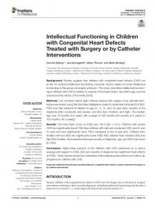
Figure 1. (A) Surgical design of the recipient site before operation; (B) Surgical design of the donor site before operation; (C) Showed the gracilis was dissected from the thigh; (D) The gracilis was placed subcutaneously in the anterio-medialis aspect of the arm; (E) The nerve of the gracilis was anastomosised to the accessory nerve (arrow); (F) Rubber sheets and tube were used for drainage after surgery.
(FGMT) to reconstruct elbow flexion, wrist extension, and fingers extension between August 1999 and October 2014 in our hospital. The exclusion criteria were patients whose follow-up had been less than 12 months and those who received an additional procedure to reinforce elbow flexion, wrist extension, and fingers extension. Age, gender, etiology, complications, follow-up times, and the outcomes were respectively recorded. All surgeries and perioperative management were done by the same surgeon (Li-Qiang Gu) and his medical team.
Surgical procedures. With the patient in the supine position, the operation was carried out under general
anesthesia. The gracilis with the surrounding fascia preserved was harvested with the anterior branch of the obturator nerve, together with its vascular supply, which is a branch of the profunda femoris artery; a skin paddle was typically harvested to facilitate postoperative flap monitoring as well. The entire gracilis, with tendon from pubic symphysis to the pes anserine insertion distal, was taken and positioned in the recipient site so that the nerve was as close as possible to the donor nerve. The gracilis was placed subcutaneously in the anterio-medialis aspect of the arm, passed under the pulley formed by the brachioradialis, extensor carpi radialis longus and extensor carpi radialis brevis muscle. The proximal part of the gracilis muscle was sutured to the acromion or the distal portion of the clavicle; the distal part was sutured to the extensor digitorum communis tendon and extensor pollicis longus tendon with double interlacing sutures. The muscle was perfused by the brachial artery, axillary artery, subclavian artery, etc. with T-shaped or end-to-end anastomosis12 and refluxed by comitant vein, such as cephalic vein, brachial vein, and axillary vein. The nerve to the gracilis was coapted to the spinal accessory nerve (SAN) (forty-five patients) and phrenic nerve (two patients) using 9-0 nylon sutures with the assistance of a 10 power surgical microscope (Fig. 1). The transferred muscle was returned to its resting length after suturing.
Post-operative care. The patient was required to wear a cast with elbow flexion of 90°, wrist in neutral posi-
tion and fingers full extension for 6 weeks after the surgery. Patients were required to do rehabilitation and electrical stimulation therapy regularly and to take neurotrophic drugs, as well as being followed up postoperatively on a regular basis.
Scientific Reports | 6:22479 | DOI: 10.1038/srep22479
2
www.nature.com/scientificreports/ Variables
frequency/mean
Percentage/standard deviation
Gender Female
2
4.76
Male
40
95.24
Left
29
69.05
Right
13
30.95
Motorcycle
32
76.16
Automobile
2
4.76
Fall
5
11.90
Traction
1
2.38
Trauma
2
4.76
Age*
26.36
6.77
BMI*
22.21
3.59
Follow-up times**
54.5
25.5 ~ 83.5
Investigated arm
Cause of injury
Table 1. Demographic characteristics of patients (N = 42). *Data were presented as mean ± standard deviation. **Data were presented as median and quartile range. The muscle power was recorded using modified MRC standard and the active range of motion (ROM) of the elbow flexion, wrist extension, and total active fingers extension (TAFE).
Questionnaires. The patients were given the DASH, Self-Rating Depression Scale (SDS), and Self-Rating Anxiety Scale (SAS) questionnaires (all in Chinese version) to estimate their QOL and the Numeric Rating Scale (NRS) was used to evaluate the pain. The ethics committee of the First Affiliated Hospital of Sun Yat-sen University approved the study, and all patients gave written informed consent. All methods were carried out in “accordance” with the approved guidelines. The DASH questionnaire has been validated for measuring upper limb function. This questionnaire is composed of 30 questions regarding patients’ symptoms and their ability to perform certain activities. Each question is scored on a scale from 1 to 5, with a minimum of 0 (no disability) and a maximum total score of 100(the severest disability)-a lower score represents better results while a higher score reflects a greater degree of disability. NRS represents the pain level on a scale from 0 to 10. (0 represents there is no pain; 1 to 3, mild pain; 4 to 6, moderate pain; and 7 to 10, severe pain). The SDS and SAS were used to estimate the patients’ depression and anxiety states. The Chinese versions of SAS and SDS scales have been confirmed reliable and validated by previous investigation13. Each questionnaire is composed of 20 items. Each item is scored on a scale of 1–4 (never, some of the time, relatively often, most of the time). The cutoff point of SAS scores ≥ 50 and SDS scores ≥ 53 reflected the existence of anxiety and depression, respectively. The SDS or SAS scores were classified into four categories of depression or anxiety severity: For the SDS scores, normal (below 53 points), presence of minimal to mild depression (53–62 points), presence of moderate to marked depression (62–72 points) and presence of severe to extreme depression (72 points and above); For the SAS scores, normal (below 50 points), presence of mild to moderate anxiety levels (50–59 points), severe anxiety levels (59–70 points) and presence of extreme depression (70 points and above). Statistical analysis. Continuous variables were presented as mean ± standard deviation (SD) and were
compared by means of independent sample t-test or ANOVA, when normal distribution is satisfied, otherwise it was expressed as median and quartile range and the difference between groups were compared by nonparametric tests. Ordinal categorical variables were expressed as frequency and were compared by Wilcoxon rank sum test or Kruskal-Wallis H test. A two-tailed p