NeuroImage 58 (2011) 709–715
Contents lists available at ScienceDirect
NeuroImage j o u r n a l h o m e p a g e : w w w. e l s ev i e r. c o m / l o c a t e / y n i m g
Gender differences in partial-volume corrected brain perfusion using brain MRI in healthy children Yasuyuki Taki a,⁎, Hiroshi Hashizume a, Yuko Sassa a, Hikaru Takeuchi a, Kai Wu b, Michiko Asano a, Kohei Asano a, Hiroshi Fukuda b, Ryuta Kawashima a, c, d a
Division of Developmental Cognitive Neuroscience, Institute of Development, Aging and Cancer, Tohoku University, Sendai 980-8575, Japan Department of Nuclear Medicine & Radiology, Institute of Development, Aging and Cancer, Tohoku University, Sendai 980-8575, Japan Department of Functional Brain Imaging, Institute of Development, Aging and Cancer, Tohoku University, Sendai 980-8575, Japan d Smart Ageing International Research Centre, Institute of Development, Aging and Cancer, Tohoku University, Sendai 980-8575, Japan b c
a r t i c l e
i n f o
Article history: Received 17 August 2010 Revised 15 November 2010 Accepted 8 July 2011 Available online 22 July 2011 Keywords: Arterial spin labeling Brain perfusion Children Gray matter Gender differences Magnetic resonance imaging
a b s t r a c t To investigate gender differences in brain perfusion, this study utilized pulsed arterial spin-labeling magnetic resonance imaging (MRI) in a large number of healthy children. Data on structural and perfusion MRI in the brain were collected from 202 healthy children aged 5–18 years. Gender differences in brain perfusion using partial volume correction (PVC), which was calculated by dividing the normalized perfusion MRI by the normalized gray-matter segments, were analyzed by applying voxel-based analysis and region-of-interest (ROI) analysis. Girls showed significantly higher brain perfusion with PVC in the bilateral medial aspect of the parietal lobes, including the posterior cingulate cortex and precuneus, as compared to boys using voxel-based analysis. In addition, brain perfusion with PVC in the bilateral posterior cingulate cortex, bilateral precuneus, and left thalamus was significantly higher in girls than in boys in the ROI analysis. In contrast, no regions were seen in which boys exhibited higher brain perfusion with PVC than girls in both analyses. The findings showed significant differences between boys and girls in brain perfusion with PVC, and these differences may contribute to gender differences in the cognitive ability of healthy children. © 2011 Elsevier Inc. All rights reserved.
Introduction Recent neuroimaging studies have identified gender differences in the brain, primarily in the neural structure of children (Sowell et al., 2007; Wilke et al., 2007) and adults (Filipek et al., 1994; Good et al., 2001a; Harasty et al., 1997; Li et al., 2004; Murphy et al., 1996; Schlaepfer et al., 1995; Sowell et al., 2007; Witelson et al., 1995; Xu et al., 2000). For example, girls exhibit a significantly larger volume of regional gray matter in the caudate nucleus and the anterior cingulate cortex, whereas boys show larger regional gray-matter volume in the posterior cingulate cortex and the insula (Wilke et al., 2007). Despite a growing wealth of knowledge concerning gender differences in neural structure, differences in brain perfusion between healthy boys and girls have not yet been well documented. Because brain perfusion is thought to reflect cerebral metabolic demand (Chugani, 1998), the evaluation of gender differences in brain perfusion in healthy children is important when attempting to understand differences in brain maturation and cognitive function.
⁎ Corresponding author at: Division of Developmental Cognitive Neuroscience, Institute of Development, Aging & Cancer, Tohoku University, 4-1 Seiryocho, Aobaku, 980-8575 Sendai, Japan. Fax: +81 22 717 8457. E-mail address:
[email protected] (Y. Taki). 1053-8119/$ – see front matter © 2011 Elsevier Inc. All rights reserved. doi:10.1016/j.neuroimage.2011.07.020
Until recently, the measurement of brain perfusion was performed by intravenous bolus injection of contrast agents or radioisotopes. Although these methods are important in acquiring quantitative measurements of brain perfusion, subjecting healthy children to these invasive methods is ethically problematic. More recently, arterial spin-labeling (ASL) perfusion magnetic resonance imaging (MRI) was developed for evaluating brain perfusion (Williams et al., 1992). In ASL, arterial blood water is magnetically labeled proximal to the tissue of interest, and the effects of this pre-labeling are determined by pairwise comparison with images acquired using control labeling. This technique provides reproducible and reliable quantitative measurements of brain perfusion in adults with various diseases and psychiatric disorders (Alsop et al., 2000; Chalela et al., 2000; Chao et al., 2010; Detre et al., 1998; Du et al., 2006). Because ASL is noninvasive and does not require intravenous injection of contrast agents or radioactive tracers, this method is more appropriate for the measurement of brain perfusion in healthy children. Moreover, because higher water content (Dobbing and Sands, 1973) and greater carotid arterial blood-flow velocity exist in children than in adults (Schoning et al., 1993), the limitations of low signal-to-noise ratio and transit effects in ASL are actually reduced in children (Wang and Licht, 2006). Conversely, because of the limited spatial resolution of ASL, the observed brain perfusion derived from ASL images may produce
710
Y. Taki et al. / NeuroImage 58 (2011) 709–715
partial-volume biases, as is the case with brain perfusion images derived from nuclear medicine (Inoue et al., 2005; Matsuda et al., 2003; Meltzer et al., 1990; Muller-Gartner et al., 1992; Rousset et al., 1998; Strul and Bendriem, 1999; Videen et al., 1988). In addition, because brain perfusion in the gray matter is significantly higher than in the white matter (Ito et al., 2005), observed brain perfusion images potentially reflect amounts of gray matter as well as genuine brain perfusion. For these reasons, the observed brain perfusion should be adjusted by the amount of gray matter to obtain a measure of genuine brain perfusion; this is more commonly known as partial-volume correction (PVC; Matsuda et al., 2003; Inoue et al., 2005). To date, a lack of information exists concerning gender differences in the brain perfusion of healthy children with or without the use of PVC. Therefore, this study examined gender differences in brain perfusion using PVC in a large number of healthy children with a wide age range using both voxel-based and region-of-interest (ROI) analyses. In ROI analysis, the gray-matter region of the cerebrum was divided into 22 ROIs, which corresponded to anatomical structures in each hemisphere, to reveal gender differences in brain perfusion with PVC in each ROI. In addition, to analyze whether the effects of gender on brain perfusion with PVC in each ROI were significant and whether there were significant interactions among gender, age, and hemisphere in brain perfusion with PVC in each ROI, we applied a general linear model to investigate the main effect of gender as well as the age × gender and age × hemisphere interactions to brain perfusion with PVC in each ROI. Regarding the ASL, the Quantitative STAR labeling of Arterial Regions (QUASAR) implementation of pulsed ASL was employed (Petersen et al., 2006a,b, 2010) because absolute brain perfusion can be quantified in well characterized units of mL/100 g/min. Furthermore, the QUASAR protocol exhibited robust test–retest reliability in a multicenter study (Petersen et al., 2010). In performing PVC brain perfusion, gray-matter density was used for the amount of gray matter and was calculated using a voxel-based morphometric analysis (Good et al., 2001b). We hypothesized that several regions that have significant gender differences in gray-matter volume, such as the cingulate cortex (Wilke et al., 2007), would also show gender differences in brain perfusion with PVC. Materials and methods Subjects All subjects were healthy Japanese children recruited by advertisements directed at kindergartens, elementary schools, junior high schools, and high schools within a local area. When the subjects and their parent(s) who were willing to participate replied by mail, a preliminary telephone interview, mail-in health questionnaire, and oral interview were given to identify any subjects with a history of malignant tumors, head trauma with a loss of consciousness lasting over 5 min, developmental disorders, epilepsy, psychiatric diseases, or claustrophobia. These subjects were excluded from the study. Of this group, brain magnetic resonance (MR) images were not collected from a total of eight subjects due to claustrophobia (3 subjects) and tiredness (5 subjects). Finally, brain MR images were collected from 274 subjects in the order in which the notifications of their intention to participate in the project arrived by mail. Trained examiners collected intelligence quotients (IQs) from subjects over the age of 16 years by administering the Japanese version of the Wechsler Adult Intelligence Scale (WAIS), 3rd edition (Fujita, et al., 2006). For subjects less than 16 years of age, the Japanese version of the Wechsler Intelligence Scale for Children (WISC), 3rd edition (Azuma, et al., 1998) was used. Full-scale IQs from the score of WAIS/WISC for each subject were calculated. Data from the MR images of 72 subjects were excluded because of motion artifacts in ASL, although most of those ASL images showed relatively slight motion artifacts. We excluded ASL image data with motion artifacts
on visual inspection and according to the voxel value (mL/100 g/min), which is indicative of actual brain perfusion. Specifically, if one or more voxels in the ASL image had a value higher than 150, we regarded the images as containing motion artifacts. Thus, the final sample consisted of 202 participants (95 boys, 107 girls). No significant differences in age and full-scale IQs existed between boys and girls (Table 1). According to the 1991 Declaration of Helsinki, written informed consent was obtained from each subject and his/her parents after receipt of a full explanation concerning the purpose and procedures of the study prior to MR image scanning. Approval for these experiments was obtained from the institutional review board of Tohoku University. Image acquisition All images were collected using a 3-T Philips Intera Achieva scanner. Three-dimensional, high-resolution, T1-weighted structural images were collected using a Magnetization Prepared Rapid Gradient Echo (MPRAGE) sequence. The parameters were as follows: 240 × 240 matrix, TR = 6.5 ms, TE = 3 ms, TI = 711 ms, FOV = 24 cm, 162 slices, 1.0 mm slice thickness, scan duration = 8 min, 3 s. Additionally, pulsed ASL brain perfusion images were collected, with the subject's eyes closed, by QUASAR implementation (Petersen et al., 2006a,b, 2010). The position of the slice was determined by putting the fourth of seven slices on the body of the corpus callosum in the coronal scout view. The parameters were the same as in a recent study, except slice thickness (Petersen et al., 2010), and were as follows: 64 × 64 matrix, TR = 4000 ms, TE = 22 ms, ΔTI = 300 ms, TI1 = 40 ms, 13 inversion times (40–3640 ms), flip-angle = 35/11.7 deg, SENSE = 2.5, 84 averages (48@Venc = 4 cm/s, 24@Venc = ∞, 12 low flip-angle), all implemented in a single sequence, FOV = 24 cm, 7 slices, 7.0-mm slice thickness (2.0-mm gap), SENSE = 2.5, scan duration = 5 min, 52 s. Image analysis of structural MRI A schematic of the image analysis is shown in Fig. 1. After image acquisition by MRI, all T1-weighted MR images were analyzed using Statistical Parametric Mapping 2 (SPM2; Wellcome Department of Cognitive Neurology, London, UK; (Friston et al., 1995)) in MATLAB (Math Works, Natick, MA, USA) and part of the MATLAB program “cg_vbm_optimized” (http://dbm.neuro.uni-jena.de/vbm.html). First, the T1-weighted MR images were transformed into the stereotactic space (Talairach and Tournoux, 1988) by registering each of the images to the custom template image that was derived from all subjects in this study using VBM tools for SPM2 (http://dbm.neuro. uni-jena.de/vbm/). Tissue segmentation from the transformed images to the gray matter, white matter, cerebrospinal fluid (CSF) space, and non-brain was then performed using the SPM2 default segmentation procedure. Next, the segmented gray-matter images were nonlinearly normalized to the custom template using 7 × 8 × 7 nonlinear basis functions in three orthogonal directions. These normalization parameters were reapplied to the T1-weighted whole-brain structural images of each subject to perform optimal spatial normalization. The Table 1 Subject characteristics according to gender. Factor
Boys (N = 95) Mean (SD)
Girls (N = 107) Range
Age 11.48 (2.93) 5.70, (years) 17.01 Full-scale 103.44 (12.96) 77, 136 IQ
Mean (SD) 11.91 (3.44)
t
p
0.955
0.341
Range
5.82, 18.36 100.36 (11.33) 71, 125
− 1.805 0.073
Y. Taki et al. / NeuroImage 58 (2011) 709–715
711
Fig. 1. Schematic of the magnetic resonance image analysis. In the figure, Co-registration indicates that perfusion images were co-registered to corresponding gray-matter segments in native space. The normalization parameter indicates that the T1-weighted image and perfusion image were spatially normalized using the corresponding normalization parameter derived from the procedure in which the segmented gray-matter images were non-linearly normalized to the custom template. Masking indicates that a gray-matter mask image consisting of voxels with a value higher than an empirically determined value of 0.2 in the gray-matter image was applied to the perfusion images to restrict analysis within the gray matter. Partial-volume correction (PVC) indicates that the masked brain perfusion images were divided by the corresponding masked, smoothed gray-matter segment to adjust for the amount of gray matter so as to obtain a measure of genuine brain perfusion.
optimally normalized T1-weighted images were segmented into gray matter, white matter, and CSF space. The optimally normalized graymatter segment was smoothed by an 8 mm FWHM Gaussian kernel. In addition, a gray-matter mask image, consisting of voxels with a value higher than an empirically determined value of 0.2 in the gray-matter image, was created. Smoothed gray-matter segments were masked using the corresponding gray-matter masks, and the masked, smoothed gray-matter segment was used for PVC.
Image analysis of perfusion MRI After image acquisition in ASL, all perfusion images were analyzed using SPM2 (Friston et al., 1995) in MATLAB. First, the perfusion image voxel size was resized to match that of the MPRAGE images. Next, perfusion images were co-registered to corresponding gray-matter segments in native space and nonlinearly normalized by applying the corresponding normalization parameter, which was derived from the normalization process of structural MR images. We checked all of the co-registered ASL images and corresponding graymatter segments, and no apparent errors were found on visual inspection. Then, a gray-matter mask image consisting of voxels with a value higher than an empirically determined value of 0.2 in the gray-matter image was applied to the perfusion images to restrict analysis within the gray matter. Brain perfusion with PVC was accomplished by dividing the masked brain perfusion images with the corresponding masked, smoothed gray-matter segment to adjust for the amount of gray matter so as to obtain a measure of genuine brain perfusion (Inoue et al., 2005; Matsuda et al., 2003). These brain perfusion images with PVC were used for voxel-based analysis. Finally, 22 ROIs, which corresponded to gray-matter structural regions of the cerebrum and deep gray-matter structures in each hemisphere, were selected using “WFU_PickAtlas” (Lancaster et al., 2000; Maldjian et al., 2003), and the brain perfusion image with PVC was obtained for each ROI bilaterally.
Statistical analyses In voxel-based analysis, statistical analyses were performed using SPM5 and VBM5 software (http://dbm.neuro.uni-jena.de/vbm/), an extension of SPM5. To illustrate the gender differences in brain perfusion with PVC, the full factorial model in SPM5 was used and included age and full-scale IQ as covariants. The significance level was set at p b 0.05 and corrected for a family-wise error rate. To reveal gender differences in brain perfusion with PVC for each ROI, an analysis of covariance, adjusted for age and full-scale IQ, was performed. The main effect of gender and the interaction effect of age × gender on brain perfusion with PVC were also analyzed using a general linear model. Regarding those interactions, if the effect of gender on brain perfusion with PVC changes depended on the age or hemisphere, then significant interactions would be observed in each ROI. These effects and interactions were estimated using effect size by calculating the partial η 2. Partial η 2 is one of the measures of effect size, and it is usually used to explain the effect size of an analysis such as ANOVA (Tabachnick and Fidell, 2006). Effect size represents the strength of the association in a statistical analysis, and it is not affected by sample size (Field and Hole, 2003). The significance level was determined using the Bonferroni correction to correct for multiple comparisons among ROIs. The total number of ROIs was 22 in each hemisphere; thus the significance level was set at p = 0.0023 (0.05/22). Results After adjusting for age and full-scale IQ, the voxel-based analyses indicate that girls show significantly higher brain perfusion with PVC in the gray-matter region of the bilateral medial parietal cortices, including the left precuneus (x, y, z = − 11, − 44, 47; t = 5.17) and the right posterior cingulate cortex (x, y, z = 18, − 68, 20; t = 4.84) when compared to boys (Fig. 2). No regions existed in which boys showed significantly higher brain perfusion with PVC as compared to girls.
712
Y. Taki et al. / NeuroImage 58 (2011) 709–715
Fig. 2. Brain regions in which brain perfusion with partial-volume correction (PVC) is significantly higher in girls than boys. The left side of the image represents the left side of the brain. Color scales indicate the t-score.
ROI analysis of brain perfusion with PVC for each of the 22 regions in the left hemisphere and the right hemisphere is shown in Tables 2 and 3, respectively. After correction for multiple comparisons, measurements of brain perfusion with PVC in the bilateral posterior cingulate cortex, the bilateral precuneus, and the left thalamus were
significantly higher in girls than boys. Additionally, a substantial difference in brain perfusion with PVC was observed in the right thalamus when comparing girls to boys (t = 2.892, p = 0.004). No regions existed in which brain perfusion with PVC was significantly or substantially higher in boys than girls.
Table 2 Mean and standard deviation of brain perfusion with partial-volume correction (PVC) in the left hemisphere for each gender.
Table 3 Mean and standard deviation of brain perfusion with partial-volume correction (PVC) in the right hemisphere for each gender.
Structure
Angular gyrus Anterior cingulate cortex Caudate nucleus Cingulate gyrus Cuneus Hippocampus Inferior frontal gyrus Inferior parietal lobule Insula Medial superior frontal gyrus Middle frontal gyrus Middle temporal gyrus Paracentral lobule Posterior cingulate cortex Postcentral gyrus Precentral gyrus Precuneus Superior frontal gyrus Superior occipital gyrus Superior temporal gyrus Supramarginal gyrus Thalamus
Brain perfusiona Boys (N = 95)
Girls (N = 107)
82.17 71.51 47.29 79.02 69.54 52.70 83.16 90.75 62.37 93.21 95.60 61.20 80.97 69.12 75.05 76.39 85.24 103.61 55.75 77.28 84.47 48.74
84.13 73.40 48.41 85.19 72.31 52.50 83.46 93.55 64.13 94.00 97.86 63.36 88.39 75.42 79.55 80.00 93.08 104.80 58.64 78.40 85.85 53.47
(20.91) (14.89) (9.77) (16.42) (18.36) (12.83) (19.48) (20.56) (10.62) (19.75) (23.18) (15.24) (19.37) (16.79) (17.78) (18.01) (20.95) (25.85) (24.98) (14.79) (20.41) (9.43)
(21.11) (17.02) (11.86) (19.30) (17.54) (13.99) (20.93) (21.40) (13.85) (21.74) (24.85) (14.52) (24.33) (16.29) (17.71) (16.91) (21.43) (24.45) (21.23) (15.64) (21.09) (12.10)
t
pb
Structure
1.098 0.902 0.815 2.580 1.458 − 0.108 0.295 1.306 0.961 0.406 0.846 1.500 2.603 3.308 2.107 1.767 3.270 0.541 1.164 0.700 0.763 3.215
0.274 0.368 0.416 0.011 0.146 0.914 0.768 0.193 0.338 0.685 0.399 0.135 0.010 0.001 0.036 0.079 0.001 0.589 0.246 0.485 0.447 0.002
Angular gyrus Anterior cingulate cortex Caudate nucleus Cingulate gyrus Cuneus Hippocampus Inferior frontal gyrus Inferior parietal lobule Insula Medial superior frontal gyrus Middle frontal gyrus Middle temporal gyrus Paracentral lobule Posterior cingulate cortex Postcentral gyrus Precentral gyrus Precuneus Superior frontal gyrus Superior occipital gyrus Superior temporal gyrus Supramarginal gyrus Thalamus
Brain perfusiona
t
pb
1.257 0.895 0.786 1.839 1.948 − 0.598 − 0.247 0.894 0.692 0.609 0.679 0.652 1.673 4.020 1.170 1.251 3.318 0.697 0.398 − 0.082 1.414 2.892
0.210 0.372 0.433 0.067 0.053 0.551 0.805 0.372 0.490 0.543 0.498 0.515 0.096 b 0.001 0.243 0.213 0.001 0.486 0.691 0.935 0.159 0.004
Boys (N = 95) Girls (N = 107)
a Regional brain perfusion with partial volume correction, mean (± SD). Unit is mL/100 g/min. b The p-value of the analysis of covariance adjusting for age and full-scale IQ.
75.61 66.83 43.76 82.65 75.48 57.18 72.84 84.54 57.19 84.82 80.63 62.16 92.76 73.50 73.43 68.23 92.29 89.13 64.78 72.71 80.67 49.65
(19.55) (13.53) (9.78) (16.34) (18.02) (13.39) (16.70) (18.94) (10.46) (18.20) (19.10) (14.73) (20.90) (17.50) (17.08) (15.73) (21.94) (20.70) (27.30) (13.15) (18.74) (8.99)
77.89 (19.24) 68.64 (17.17) 44.84 (10.74) 86.99 (20.47) 79.70 (19.25) 56.18 (14.86) 71.83 (18.21) 86.08 (19.45) 58.40 (12.76) 86.21 (20.77) 82.15 (20.87) 62.73 (14.41) 97.56 (25.12) 81.87 (18.25) 75.56 (17.00) 70.41 (15.75) 100.59 (23.29) 90.72 (22.10) 65.54 (24.98) 72.14 (15.57) 83.47 (19.34) 53.70 (11.91)
a Regional brain perfusion with partial volume correction, mean (± SD). Unit is mL/100 g/min. b The p-value of analysis of covariance adjusting for age and full-scale IQ.
Y. Taki et al. / NeuroImage 58 (2011) 709–715
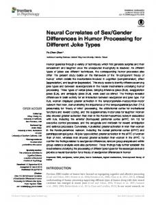
The main effects of gender, age, and hemisphere and the interaction effects of age × gender, age × hemisphere, and gender × hemisphere on brain perfusion with PVC are also shown in Table 4. After correction for multiple comparisons, measurements of brain perfusion with PVC in the cingulate gyrus (partial η 2, 0.030, p = 0.001), paracentral lobule (partial η 2, 0.027, p = 0.001), posterior cingulate cortex (partial η 2, 0.070, p b 0.001), precuneus (partial η 2, 0.059, p b 0.001), and thalamus (partial η 2, 0.046, p b 0.001) showed a significant main effect of gender. The posterior cingulate cortex also showed a significant age × gender interaction (partial η 2, 0.057, p b 0.001) on brain perfusion with PVC. To characterize the significant age × gender interaction of the posterior cingulate cortex, the boy and girl subjects were divided into younger (aged ≤ 12 years; boys: n = 52, girls: n = 53) and older groups (aged N 12 years; boys: n = 43, girls: n = 54); 12 years of age was the mean age for both genders. To correct for multiple comparisons, the significance level was set at p = 0.013 (0.05/4). Although no significant differences in brain perfusion with PVC were observed between the younger boy group and younger girl group in the bilateral hemispheres (left, t = 1.603, p = 0.112; right, t = 2.370, p = 0.020), measurements of brain perfusion with PVC of the older girl group were significantly higher in both hemispheres than those of the older boy group (left, t = 3.250, p = 0.002; right, t = 3.185, p = 0.002). The correlation between age and brain perfusion with PVC in each gender and in each hemisphere is shown in Fig. 3. No other regions were found that
Table 4 Effects on brain perfusion with partial-volume correction (PVC) in each region of interest. Included are the main effects of age, gender, and hemisphere and the interactions effects of age × gender, age × hemisphere, and gender × hemisphere. Data are shown by effect size using partial η2. Structure
Angular gyrus Anterior cingulate cortex Caudate nucleus Cingulate gyrus Cuneus Hippocampus Inferior frontal gyrus Inferior parietal lobule Insula Medial superior frontal gyrus Middle frontal gyrus Middle temporal gyrus Paracentral lobule Posterior cingulate cortex Postcentral gyrus Precentral gyrus Precuneus Superior frontal gyrus Superior occipital gyrus Superior temporal gyrus Supramarginal gyrus Thalamus
713
Fig. 3. Correlation between brain perfusion with partial-volume correction (PVC) and age in the posterior cingulate cortex in the (A) left hemisphere for boys, (B) left hemisphere for girls, (C) right hemisphere for boys, and (D) right hemisphere for girls.
showed a significant main effect of gender or an interaction effect of age × gender on brain perfusion with PVC.
Effect sizea Age (A)
Gender (G)
Hemisphere (H)
A×G
A×H
G×H
0.161⁎⁎ 0.044⁎
0.008 0.006
0.030⁎⁎ 0.023⁎
0.021 0.008
0.003 0.002
0.000 0.000
0.036⁎ 0.059⁎⁎ 0.103⁎⁎ 0.022 0.069⁎⁎
0.002 0.030⁎⁎ 0.015⁎ 0.001 0.001
0.029⁎⁎ 0.006 0.037⁎⁎ 0.023⁎ 0.084⁎⁎
0.034⁎ 0.024 0.032⁎ 0.035⁎ 0.012
0.003 0.001 0.001 0.004 0.006
0.000 0.001 0.000 0.000 0.000
0.127⁎⁎
0.009
0.033⁎⁎
0.038⁎
0.002
0.000
0.023 0.068⁎⁎
0.004 0.003
0.051⁎⁎ 0.042⁎⁎
0.026 0.017
0.008 0.003
0.000 0.000
0.078⁎⁎
0.006
0.117⁎⁎
0.011
0.008
0.000
0.146⁎⁎
0.006
0.000
0.025
0.002
0.001
0.072⁎⁎ 0.175⁎⁎
0.027⁎⁎ 0.070⁎⁎
0.056⁎⁎ 0.030⁎⁎
0.027 0.057⁎⁎
0.001 0.001
0.001 0.001
0.085⁎⁎ 0.094⁎⁎ 0.202⁎⁎ 0.099⁎⁎
0.016⁎ 0.015⁎ 0.059⁎⁎ 0.004
0.007 0.075⁎⁎ 0.034⁎⁎ 0.094⁎⁎
0.026 0.035⁎ 0.044⁎ 0.014
0.003 0.004 0.002 0.006
0.001 0.001 0.000 0.000
0.062⁎⁎
0.003
0.027⁎⁎
0.005
0.010
0.000
0.061⁎⁎
0.001
0.036⁎⁎
0.029⁎
0.004
0.001
0.126⁎⁎
0.009
0.007
0.041⁎
0.004
0.000
0.046⁎
0.046⁎⁎
0.001
0.026
0.001
0.000
Bonferroni critical α = 0.0023. a Partial η2. ⁎⁎ p b 0.0023. ⁎ p b 0.05.
Discussion To our knowledge, this is the first study to investigate brain perfusion with PVC by applying ASL perfusion MRI to demonstrate a gender difference in healthy children over a wide range of ages. Girls exhibited significantly higher brain perfusion with PVC than boys in the medial aspect of the parietal lobe in areas such as the precuneus and the posterior cingulate cortex. These results are at least partially consistent with the primary hypothesis, as previous research indicates that the medial aspect of the parietal lobe is a region that shows gender differences in brain structure (Wilke et al., 2007). Also, a significant age × gender interaction was detected in the posterior cingulate cortex but no regions showed significantly higher brain perfusion with PVC in boys than girls. The current findings demonstrate that girls have significantly higher brain perfusion with PVC in the medial aspect of the parietal cortex than boys. Recent studies showed that the medial aspect of the parietal cortex, including areas such as the precuneus and the posterior cingulate cortex, is involved in self-related information processing and distinguishing the self from others (Kircher et al., 2000; Ochsner et al., 2004; Vogeley et al., 2004). The ability to distinguish one's own perspective from that of others is related to understanding that the contents of others' minds are different from one's own. To take such a third-person perspective, a person has to be aware of what another person is thinking and how they intend to act, an awareness that has been referred to as theory of mind (Siegal and Varley, 2002). The medial aspects of the parietal cortex have been implicated in this process (Carrington and Bailey, 2009; Spreng et al., 2009). Because theory of mind is closely related with empathy (Vollm et al., 2006), the Empathizing Quotient (EQ), based on a questionnaire that includes a list of statements about real-life situations, experiences, and interests in which empathizing skills are required (Baron-Cohen and Wheelwright, 2004), may function as
714
Y. Taki et al. / NeuroImage 58 (2011) 709–715
a measure of theory of mind (Baron-Cohen, 2009). A recent study reported that EQ score is significantly higher in girls than boys (Auyeung et al., 2009), suggesting that the ability to utilize theory of mind is greater in girls than boys. Thus, the higher brain perfusion with PVC in the medial aspect of the parietal cortex in girls compared to boys may be related to the ability to engage theory of mind. In addition, the following two issues may be related to the gender differences in the medial aspect of the parietal cortex seen in brain perfusion with PVC. First, recent studies have shown that the precuneus is related to the retrieval of episodic memory (Wagner et al., 2005) and that this ability is greater in females than in males (Herlitz et al., 1997, 1999). Although a direct relationship between cognitive function and brain perfusion has not been demonstrated, the higher brain perfusion with PVC in girls may be related to their greater ability to retrieve episodic memory. Second, using transcranial Doppler ultrasonography, healthy girls have been shown to have higher middle cerebral artery flow velocity and higher basilar artery flow velocity than healthy boys (Vavilala et al., 2005; Tontisirin et al., 2007). Although the main feeding artery of the precuneus is the anterior cerebral artery (Fisher, 1975), the higher middle cerebral artery flow velocity in girls may be associated with higher brain perfusion with PVC in this region of the brain. According to the ROI analysis, brain perfusion with PVC was significantly higher in the left thalamus, and substantially higher in the right thalamus, in girls than boys. Moreover, if the statistical threshold is set at more liberal condition, such as p b 0.05, and corrected for false discovery rate in the voxel-based analysis, brain perfusion with PVC in the bilateral thalamus was significantly higher in girls than in boys (data not shown). Several animal studies have shown a reciprocal connection between the precuneus and the thalamus, especially the dorsal aspect of the thalamus (Yeterian and Pandya, 1988; Schmahmann and Pandya, 1990). This reciprocal connection is thought to be related to the gender differences seen in the brain perfusion with PVC in the thalamus as well as the precuneus. A significant age × gender interaction of brain perfusion with PVC was seen in the posterior cingulate cortex in the ROI analysis. Although no significant differences in brain perfusion with PVC were detected between girls and boys in the younger group in the bilateral hemispheres, the brain perfusion with PVC of the older girl group was significantly higher in both hemispheres than that of the older boy group. As shown in Fig. 3, brain perfusion with PVC in the posterior cingulate cortex decreases with age in both boys and girls, although brain perfusion with PVC in girls seems to decrease more slowly and to remain at a higher level than in boys. A post-adolescent decrease in the number of synapses per neuron occurs during brain maturation (Huttenlocher, 1979; Huttenlocher and Dabholkar, 1997; Huttenlocher et al., 1982) and may be associated with a decrease in brain perfusion with PVC, thus acting as the underlying mechanism in this process. Because a major portion of glucose consumed by the brain is used for the maintenance of resting membrane potentials (Mata et al., 1980), a direct relationship should exist between the degree of synaptic connectivity and glucose metabolism. In addition, the relationship between brain perfusion and glucose consumption (Fox and Raichle, 1986; Fox et al., 1988; Newberg et al., 2005) may play a role in the decrease in brain perfusion with PVC observed in healthy children in the present study. The significantly higher brain perfusion with PVC in the posterior cingulate cortex of girls compared with boys in the older groups suggests that this maturational process continues for longer in girls than in boys in this particular region. This study has certain limitations. First, data for brain perfusion in the ventral and dorsal aspects were not acquired due to limitations in QUASAR range. If a continuing two-time acquisition of QUASAR data had been performed, it could cover the whole brain in terms of brain perfusion. In fact, this was done for 10 subjects, but because the acquisition time doubled, many more motion artifacts were observed and the data were unusable. Thus, to minimize the acquisition time data on brain perfusion in the ventral and dorsal aspects was not
acquired. Second, the age range of the subjects was from 5 to 18 years, and as a result, data for brain perfusion with PVC in adults were also not acquired. In conclusion, girls show significantly higher brain perfusion with PVC than boys in the bilateral medial aspect of parietal lobes, including the posterior cingulate cortex and precuneus, whereas no regions existed in boys that show higher brain perfusion than girls. In addition, a significant age × gender interaction of the posterior cingulate cortex was detected. The current findings demonstrate that the gender differences in brain perfusion using ASL brain perfusion MRI in a large number of healthy children over a wide age range is an important aspect in understanding brain function. These findings suggest that significant differences in brain perfusion exist between boys and girls, and that these differences may be derived from differences in the maturational pattern of the brain and contribute to cognitive differences between genders in healthy children. The present results may contribute to clarifying gender differences in the cognitive abilities of healthy children. Acknowledgments We thank Y. Yamada, Y. Kotozaki, and R. Nouchi for collecting MR data, and Y. Suzuki for technical support. This work was supported by a Ministry of Education, Culture, Sports, Science and Technology Grant-in-Aid for Young Scientists (B) (grant number 20790875). References Alsop, D.C., Detre, J.A., Grossman, M., 2000. Assessment of cerebral blood flow in Alzheimer's disease by spin-labeled magnetic resonance imaging. Ann. Neurol. 47, 93–100. Auyeung, B., Wheelwright, S., Allison, C., Atkinson, M., Samarawickrema, N., BaronCohen, S., 2009. The children's empathy quotient and systemizing quotient: sex differences in typical development and in autism spectrum conditions. J. Autism Dev. Disord. 39, 1509–1521. Azuma, H., Ueno, K., Fujita, K., Maekawa, H., Ishikuma, T., Sano, H., 1998. Japanese Wechsler Intelligence Scale for Children, 3rd ed. Nihon Bunka Kagakusha, Tokyo. Baron-Cohen, S., 2009. Autism: the empathizing–systemizing (E–S) theory. Ann. N. Y. Acad. Sci. 1156, 68–80. Baron-Cohen, S., Wheelwright, S., 2004. The empathy quotient: an investigation of adults with Asperger syndrome or high functioning autism, and normal sex differences. J. Autism Dev. Disord. 34, 163–175. Carrington, S.J., Bailey, A.J., 2009. Are there theory of mind regions in the brain? A review of the neuroimaging literature. Hum. Brain Mapp. 30, 2313–2335. Chalela, J.A., Alsop, D.C., Gonzalez-Atavales, J.B., Maldjian, J.A., Kasner, S.E., Detre, J.A., 2000. Magnetic resonance perfusion imaging in acute ischemic stroke using continuous arterial spin labeling. Stroke 31, 680–687. Chao, L.L., Buckley, S.T., Kornak, J., Schuff, N., Madison, C., Yaffe, K., Miller, B.L., Kramer, J.H., Weiner, M.W., 2010. ASL perfusion MRI predicts cognitive decline and conversion from MCI to dementia. Alzheimer Dis. Assoc. Disord. 24, 19–27. Chugani, H.T., 1998. A critical period of brain development: studies of cerebral glucose utilization with PET. Prev. Med. 27, 184–188. Detre, J.A., Alsop, D.C., Vives, L.R., Maccotta, L., Teener, J.W., Raps, E.C., 1998. Noninvasive MRI evaluation of cerebral blood flow in cerebrovascular disease. Neurology 50, 633–641. Dobbing, J., Sands, J., 1973. Quantitative growth and development of human brain. Arch. Dis. Child. 48, 757–767. Du, A.T., Jahng, G.H., Hayasaka, S., Kramer, J.H., Rosen, H.J., Gorno-Tempini, M.L., Rankin, K.P., Miller, B.L., Weiner, M.W., Schuff, N., 2006. Hypoperfusion in frontotemporal dementia and Alzheimer disease by arterial spin labeling MRI. Neurology 67, 1215–1220. Field, A., Hole, G., 2003. How to Design and Report Experiments. Sage Publications, London. Filipek, P.A., Richelme, C., Kennedy, D.N., Caviness Jr., V.S., 1994. The young adult human brain: an MRI-based morphometric analysis. Cereb. Cortex 4, 344–360. Fisher, C.M., 1975. The anatomy and pathology of the cerebral vasculature. In: Meyer, J.S. (Ed.), Modern Concepts of Cerebrovascular Disease. Spectrum Publications, Wooster, OH, pp. 1–41. Fox, P.T., Raichle, M.E., 1986. Focal physiological uncoupling of cerebral blood flow and oxidative metabolism during somatosensory stimulation in human subjects. Proc. Natl. Acad. Sci. U.S.A. 83, 1140–1144. Fox, P.T., Raichle, M.E., Mintun, M.A., Dence, C., 1988. Nonoxidative glucose consumption during focal physiologic neural activity. Science 241, 462–464. Friston, K.J., Holmes, A.P., Worsley, K.J., Poline, J.-P., Frith, C.D., Frackowiak, R.S.J., 1995. Statistical parametric maps in functional imaging: a general linear approach. Hum. Brain Mapp. 2, 189–210. Fujita, K., Maekawa, H., Dairoku, H., Yamanaka, K., 2006. Japanese Wechsler Adult Intelligence Scale, 3rd ed. Nihon Bunka Kagakusha, Tokyo.
Y. Taki et al. / NeuroImage 58 (2011) 709–715 Good, C.D., Johnsrude, I., Ashburner, J., Henson, R.N., Friston, K.J., Frackowiak, R.S., 2001a. Cerebral asymmetry and the effects of sex and handedness on brain structure: a voxel-based morphometric analysis of 465 normal adult human brains. Neuroimage 14, 685–700. Good, C.D., Johnsrude, I.S., Ashburner, J., Henson, R.N.A., Friston, K.J., Frackowiak, R.S.J., 2001b. A voxel-based morphometric study of ageing in 465 normal adult human brains. Neuroimage 14, 21–36. Harasty, J., Double, K.L., Halliday, G.M., Kril, J.J., McRitchie, D.A., 1997. Languageassociated cortical regions are proportionally larger in the female brain. Arch. Neurol. 54, 171–176. Herlitz, A., Nilsson, L.G., Backman, L., 1997. Gender differences in episodic memory. Mem. Cognit. 25, 801–811. Herlitz, A., Airaksinen, E., Nordstrom, E., 1999. Sex differences in episodic memory: the impact of verbal and visuospatial ability. Neuropsychology 13, 590–597. Huttenlocher, P.R., 1979. Synaptic density in human frontal cortex — developmental changes and effects of aging. Brain Res. 163, 195–205. Huttenlocher, P.R., Dabholkar, A.S., 1997. Regional differences in synaptogenesis in human cerebral cortex. J. Comp. Neurol. 387, 167–178. Huttenlocher, P.R., de Courten, C., Garey, L.J., Van der Loos, H., 1982. Synaptogenesis in human visual cortex—evidence for synapse elimination during normal development. Neurosci. Lett. 33, 247–252. Inoue, K., Ito, H., Goto, R., Nakagawa, M., Kinomura, S., Sato, T., Sato, K., Fukuda, H., 2005. Apparent CBF decrease with normal aging due to partial volume effects: MR-based partial volume correction on CBF SPECT. Ann. Nucl. Med. 19, 283–290. Ito, H., Kanno, I., Fukuda, H., 2005. Human cerebral circulation: positron emission tomography studies. Ann. Nucl. Med. 19, 65–74. Kircher, T.T., Senior, C., Phillips, M.L., Benson, P.J., Bullmore, E.T., Brammer, M., Simmons, A., Williams, S.C., Bartels, M., David, A.S., 2000. Towards a functional neuroanatomy of self processing: effects of faces and words. Cogn. Brain Res. 10, 133–144. Lancaster, J.L., Woldorff, M.G., Parsons, L.M., Liotti, M., Freitas, C.S., Rainey, L., Kochunov, P.V., Nickerson, D., Mikiten, S.A., Fox, P.T., 2000. Automated Talairach atlas labels for functional brain mapping. Hum. Brain Mapp. 10, 120–131. Li, Z.J., Matsuda, H., Asada, T., Ohnishi, T., Kanetaka, H., Imabayashi, E., Tanaka, F., 2004. Gender difference in brain perfusion 99mTc-ECD SPECT in aged healthy volunteers after correction for partial volume effects. Nucl. Med. Commun. 25, 999–1005. Maldjian, J.A., Laurienti, P.J., Kraft, R.A., Burdette, J.H., 2003. An automated method for neuroanatomic and cytoarchitectonic atlas-based interrogation of fMRI data sets. Neuroimage 19, 1233–1239. Mata, M., Fink, D.J., Gainer, H., Smith, C.B., Davidsen, L., Savaki, H., Schwartz, W.J., Sokoloff, L., 1980. Activity-dependent energy metabolism in rat posterior pituitary primarily reflects sodium pump activity. J. Neurochem. 34, 213–215. Matsuda, H., Ohnishi, T., Asada, T., Li, Z.J., Kanetaka, H., Imabayashi, E., Tanaka, F., Nakano, S., 2003. Correction for partial-volume effects on brain perfusion SPECT in healthy men. J. Nucl. Med. 44, 1243–1252. Meltzer, C.C., Leal, J.P., Mayberg, H.S., Wagner Jr., H.N., Frost, J.J., 1990. Correction of PET data for partial volume effects in human cerebral cortex by MR imaging. J. Comput. Assist. Tomogr. 14, 561–570. Muller-Gartner, H.W., Links, J.M., Prince, J.L., Bryan, R.N., McVeigh, E., Leal, J.P., Davatzikos, C., Frost, J.J., 1992. Measurement of radiotracer concentration in brain gray matter using positron emission tomography: MRI-based correction for partial volume effects. J. Cereb. Blood Flow Metab. 12, 571–583. Murphy, D.G., DeCarli, C., McIntosh, A.R., Daly, E., Mentis, M.J., Pietrini, P., Szczepanik, J., Schapiro, M.B., Grady, C.L., Horwitz, B., Rapoport, S.I., 1996. Sex differences in human brain morphometry and metabolism: an in vivo quantitative magnetic resonance imaging and positron emission tomography study on the effect of aging. Arch. Gen. Psychiatry 53, 585–594. Newberg, A.B., Wang, J., Rao, H., Swanson, R.L., Wintering, N., Karp, J.S., Alavi, A., Greenberg, J.H., Detre, J.A., 2005. Concurrent CBF and CMRGlc changes during human brain activation by combined fMRI-PET scanning. Neuroimage 28, 500–506. Ochsner, K.N., Knierim, K., Ludlow, D.H., Hanelin, J., Ramachandran, T., Glover, G., Mackey, S.C., 2004. Reflecting upon feelings: an fMRI study of neural systems supporting the attribution of emotion to self and other. J. Cogn. Neurosci. 16, 1746–1772. Petersen, E.T., Lim, T., Golay, X., 2006a. Model-free arterial spin labeling quantification approach for perfusion MRI. Magn. Reson. Med. 55, 219–232.
715
Petersen, E.T., Zimine, I., Ho, Y.C., Golay, X., 2006b. Non-invasive measurement of perfusion: a critical review of arterial spin labelling techniques. Br. J. Radiol. 79, 688–701. Petersen, E.T., Mouridsen, K., Golay, X., all named co-authors of the QUASAR test–retest, s, 2010. The QUASAR reproducibility study, Part II: results from a multi-center Arterial Spin Labeling test–retest study. Neuroimage 49, 104–113. Rousset, O.G., Ma, Y., Evans, A.C., 1998. Correction for partial volume effects in PET: principle and validation. J. Nucl. Med. 39, 904–911. Schlaepfer, T.E., Harris, G.J., Tien, A.Y., Peng, L., Lee, S., Pearlson, G.D., 1995. Structural differences in the cerebral cortex of healthy female and male subjects: a magnetic resonance imaging study. Psychiatry Res. 61, 129–135. Schmahmann, J.D., Pandya, D.N., 1990. Anatomical investigation of projections from thalamus to posterior parietal cortex in the rhesus monkey: a WGA-HRP and fluorescent tracer study. J. Comp. Neurol. 295, 299–326. Schoning, M., Staab, M., Walter, J., Niemann, G., 1993. Transcranial color duplex sonography in childhood and adolescence. Age dependence of flow velocities and waveform parameters. Stroke 24, 1305–1309. Siegal, M., Varley, R., 2002. Neural systems involved in “theory of mind”. Nat. Rev. Neurosci. 3, 463–471. Sowell, E.R., Peterson, B.S., Kan, E., Woods, R.P., Yoshii, J., Bansal, R., Xu, D., Zhu, H., Thompson, P.M., Toga, A.W., 2007. Sex differences in cortical thickness mapped in 176 healthy individuals between 7 and 87 years of age. Cereb. Cortex 17, 1550–1560. Spreng, R.N., Mar, R.A., Kim, A.S., 2009. The common neural basis of autobiographical memory, prospection, navigation, theory of mind, and the default mode: a quantitative meta-analysis. J. Cogn. Neurosci. 21, 489–510. Strul, D., Bendriem, B., 1999. Robustness of anatomically guided pixel-by-pixel algorithms for partial volume effect correction in positron emission tomography. J. Cereb. Blood Flow Metab. 19, 547–559. Tabachnick, B.G., Fidell, L.S., 2006. Using multivariate Statistics (5th international ed.). Pearson/Allyn & Bacon, Boston. Talairach, J., Tournoux, P., 1988. Co-planar Stereotaxic Atlas of the Human Brain: 3Dimensional Approach System: An Approach to Cerebral Imaging. Georg Thieme Verlag, Stuttgart. Tontisirin, N., Muangman, S.L., Suz, P., Pihoker, C., Fisk, D., Moore, A., Lam, A.M., Vavilala, M.S., 2007. Early childhood gender differences in anterior and posterior cerebral blood flow velocity and autoregulation. Pediatrics 119, e610–e615. Vavilala, M.S., Kincaid, M.S., Muangman, S.L., Suz, P., Rozet, I., Lam, A.M., 2005. Gender differences in cerebral blood flow velocity and autoregulation between the anterior and posterior circulations in healthy children. Pediatr. Res. 58, 574–578. Videen, T.O., Perlmutter, J.S., Mintun, M.A., Raichle, M.E., 1988. Regional correction of positron emission tomography data for the effects of cerebral atrophy. J. Cereb. Blood Flow Metab. 8, 662–670. Vogeley, K., May, M., Ritzl, A., Falkai, P., Zilles, K., Fink, G.R., 2004. Neural correlates of first-person perspective as one constituent of human self-consciousness. J. Cogn. Neurosci. 16, 817–827. Vollm, B.A., Taylor, A.N., Richardson, P., Corcoran, R., Stirling, J., McKie, S., Deakin, J.F., Elliott, R., 2006. Neuronal correlates of theory of mind and empathy: a functional magnetic resonance imaging study in a nonverbal task. Neuroimage 29, 90–98. Wagner, A.D., Shannon, B.J., Kahn, I., Buckner, R.L., 2005. Parietal lobe contributions to episodic memory retrieval. Trends Cogn. Sci. 9, 445–453. Wang, J., Licht, D.J., 2006. Pediatric perfusion MR imaging using arterial spin labeling. Neuroimaging Clin. N. Am. 16, 149–167. Wilke, M., Krageloh-Mann, I., Holland, S.K., 2007. Global and local development of gray and white matter volume in normal children and adolescents. Exp. Brain Res. 178, 296–307. Williams, D.S., Detre, J.A., Leigh, J.S., Koretsky, A.P., 1992. Magnetic resonance imaging of perfusion using spin inversion of arterial water. Proc. Natl. Acad. Sci. U.S.A. 89, 212–216. Witelson, S.F., Glezer, I.I., Kigar, D.L., 1995. Women have greater density of neurons in posterior temporal cortex. J. Neurosci. 15, 3418–3428. Xu, J., Kobayashi, S., Yamaguchi, S., Iijima, K., Okada, K., Yamashita, K., 2000. Gender effects on age-related changes in brain structure. AJNR Am. J. Neuroradiol. 21, 112–118. Yeterian, E.H., Pandya, D.N., 1988. Corticothalamic connections of paralimbic regions in the rhesus monkey. J. Comp. Neurol. 269, 130–146.