Volume 10 Number 1
January 2008
pp. 20–40 20
www.neoplasia.com
Gene Expression Profiling Identifies Lobe-Specific and Common Disruptions of Multiple Gene Networks in Testosterone-Supported, 17βEstradiol– or DiethylstilbestrolInduced Prostate Dysplasia in Noble Rats1,2
Neville N. C. Tam*, Carol Ying-Ying Szeto*, † † Maureen A. Sartor , Mario Medvedovic and Shuk-Mei Ho* *Division of Environmental Genetics and Molecular Toxicology, Department of Environmental Health, University of Cincinnati Medical Center, Cincinnati, OH 45267 USA; † Center for Environmental Genetics, Department of Environmental Health, University of Cincinnati Medical Center, Cincinnati, OH 45267 USA
Abstract The xenoestrogen diethylstilbestrol (DES) is commonly believed to mimic the action of the natural estrogen 17βestradiol (E2). To determine if these two estrogens exert similar actions in prostate carcinogenesis, we elevated circulating levels of estrogen in Noble (NBL) rats with E2/DES–filled implants, while maintaining physiological levels of testosterone (T) in the animals with T-filled implants. The two estrogens induced dysplasia in a lobe-specific manner, with E2 targeting only the lateral prostate (LP) and DES impacting only the ventral prostate (VP). Gene expression profiling identified distinct and common E2-disrupted versus DES-disrupted gene networks in each lobe. More importantly, hierarchical clustering analyses revealed that T + E2 treatment primarily affected the gene expression pattern in the LP, whereas T + DES treatment primarily affected the gene expression profile in the VP. Gene ontology analyses and pathway mapping suggest that the two hormone treatments disrupt unique and/or common cellular processes, including cell development, proliferation, motility, apoptosis, and estrogen signaling, which may be linked to dysplasia development in the rat prostate. These findings suggest that the effects of xenoestrogens and natural estrogens on the rat prostate are more divergent than previously suspected and that these differences may explain the lobe-specific carcinogenic actions of the hormones. Neoplasia (2008) 10, 20–40
Introduction Androgens are essential to the growth and functioning of the normal prostate and undoubtedly play a key role in prostate carcinogenesis [1]. Emerging evidence shows that estrogens also have a critical role in prostatic diseases, including prostate cancer (PCa) [2,3]. The incidence of PCa increases dramatically with age in human males, whose testosterone (T) levels in both the circulation and in prostate decline, whereas those of 17β-estradiol (E2) remain relatively stable [2–4]. Thus, the age-associated alterations in the sex hormone milieu toward an estrogen predominance have been proposed as an endogenous risk factor for prostate carcinogenesis. Dynamic changes in the expression of estrogen receptor-β during PCa progression in humans
Abbreviations: C/EBP, CCAAT/enhancer binding protein; DES, diethylstilbestrol; E2, 17β-estradiol; LP, lateral prostate; NBL, Noble; PCa, prostate cancer; PCR, polymerase chain reaction; VP, ventral prostate Address all correspondence to: Dr. Shuk-Mei Ho, Department of Environmental Health, Kettering Complex, Room 130, 3223 Eden Avenue, University of Cincinnati Medical Center, PO Box 670056, Cincinnati, OH 45267-0056. E-mail:
[email protected] 1 Grant support: CA112532, CA15776, ES013071, ES012281, and DK061084 to S. M. H. and ES006096 to S. M. H. and M. M. 2 This article refers to supplementary materials, which are designated by Tables W1, W2, W3, W4, and W5 and Figure W1 and are available online at www.neoplasia.com. Received 27 September 2007; Revised 26 October 2007; Accepted 30 October 2007 Copyright © 2008 Neoplasia Press, Inc. All rights reserved 1522-8002/08/$25.00 DOI 10.1593/neo.07889
Neoplasia Vol. 10, No. 1, 2008 [5,6] also suggest the involvement of estrogen signaling in the malignant transformation of the prostate. Moreover, the findings of aberrant activation of aromatase expression in benign prostatic hyperplasia and PCa [3,7] further advance the concept that local production of estrogens is pivotal in the pathobiology of this gland. Humans are exposed to xenoestrogens from various sources, including food, drugs, and other exogenous venues. Xenoestrogens are believed to cause endocrine disruptions that lead to pathogenesis in reproductive end organs [8,9]. One agent of public health concern is diethylstilbestrol (DES), a synthetic estrogen, which was widely used as a growth-promoting animal feed additive [10]. Epidemiological studies have established a strong association between maternal exposure to DES and increased risk of vaginal, cervical, and perhaps breast cancers in DES daughters [11,12]. It remains uncertain, however, whether prenatal exposure to DES elevates cancer risk in men [13]. Transient exposure of neonatal rodents to estrogen increases susceptibility of the prostate to inflammation and cancer development later in life [14]. Together, these findings strongly suggest that natural or synthetic estrogen–mediated endocrine disruption leads to the evolution of prostatic diseases, including PCa. We have established the T plus estrogen–induced dysplasia/ carcinoma model in Noble (NBL) rats as a robust study system for deciphering the contributions of estrogens, in adulthood, on the pathogenesis of dysplasia, a precancerous lesion, and adenocarcinoma of the prostate [15,16]. In this model, estrogens are administered through subcutaneous implants of hormone-filled Silastic capsules with the coimplantation of T-filled capsules to maintain physiological levels of this androgen in the circulation [17]. It is well known that increased exposure to estrogens leads to a decline in circulating levels of T through the hypothalamic–pituitary–gonadal axis. The topographical localization of premalignant/malignant lesions within the rat prostate gland has been found to be dependent on the type of estrogens used. Under the same exogenous androgen support, E2 induces epithelial dysplasia and adenocarcinoma in the lateral prostate (LP) but not in the ventral prostate (VP) [16–19], whereas synthetic DES specifically targets the VP but not the LP [19,20]. Why two different estrogens induce lobe-specific carcinogenic actions in the prostate remains unclear. To address this issue, we conducted gene expression profiling analyses on the LP and VP of NBL rats exposed to T + E2 or T + DES. The experimental design and the application of a bioinformatic tool of gene network mapping enabled us to identify lobe-specific and common estrogen–mediated disruptions in multiple biologic networks/ pathways that may be linked to prostate carcinogenesis and explain the distinctive action of the two estrogens.
Materials and Methods
Animals and Hormonal Treatment The animal usage protocol was approved by the Institutional Animal Care Committee at the University of Cincinnati. Male NBL rats (5–6 weeks old) were purchased from Charles River Laboratories (Kingston, NY), kept under standard conditions, and treated as previously reported [16,19,20]. Briefly, animals were randomized into three groups (n = 5 for each group). Rats in the T + E2 treatment group received subcutaneous implants of two pieces of 2-cm-long Silastic capsules containing T (Sigma, St Louis, MO) and one piece of 1-cm-long capsules packed with E2 (Sigma), whereas the T + DES treatment group received the same number of Silastic capsules of the
Endocrine Disruption in Prostate Carcinogenesis
Tam et al.
21
same length filled with T and DES (Sigma). Age-matched untreated control rats were implanted with empty capsules. At the end of a 16-week treatment period, animals were sacrificed with an overdose of isoflurane, and VPs and LPs were excised. One half of each lobe was processed for histologic examination, and the other half was snap-frozen for RNA extraction.
Microarray Hybridization The Atlas Glass Rat 3.8 I Microarray (Clontech, Palo Alto, CA) carrying 3800 named rat genes (spotted oligonucleotides) were used as the gene chip platform. The Atlas Glass Fluorescent Labeling Kit (Clontech) was used for synthesizing and purifying fluorescentlabeled cDNA probes for hybridization to glass microarrays. The labeling and hybridization procedures were performed in accordance with the manufacturer’s instruction manual. In brief, aminomodified first-strand cDNA probes were synthesized with aminoallyl– 2′-deoxyuridine 5′-triphosphate incorporation. Then Cy3 fluorescent dye was coupled to the cDNAs derived from individual prostatic lobes, whereas Cy5 dye was conjugated to the universal rat reference RNA obtained from Stratagene (La Jolla, CA). Equal quantities of two labeled probes were mixed and hybridized in a Corning microarray hybridization chamber (Corning, Corning, NY) at 50°C overnight (≥16 hours). Spikes of positive control probes were also included as an internal control for the process of cDNA probe synthesis and the dye-coupled reaction. Finally, the signal was obtained using a microarray scanner (GenePix 4000B; Axon Instruments, Foster City, CA). A probe coverage of >90% was achieved for all arrays. Five animal replicates for each treatment/tissue group (T + E2–treated VP or LP, T + DES–treated VP and LP, and untreated VP and LP) were used to conduct a 30-microarray analysis to assess changes in the gene expression pattern due to treatment and lobe specificity.
Microarray Data Normalization and Analysis The data were analyzed to identify differentially expressed genes in 1) the T + E2–treated LPs and VPs compared with untreated controls and 2) the T + DES–treated LPs and VPs compared with untreated controls. Five biologic replicate arrays for each experimental condition, all versus universal reference, were performed. R statistical software and the limma Bioconductor package [21] were used for analysis. Data normalization was performed in two steps separately for each microarray [22–24]. First, background-adjusted intensities were log-transformed, and the differences (M ) and averages (A) of log-transformed values were calculated as M = log2(X1) − log2(X2) and A = [log2(X1) + log2(X2)]/2, where X1 and X2 denote the Cy5 and Cy3 intensities, respectively. Second, normalization was performed by fitting the array-specific local regression model of M as a function of A and obtaining residuals. The statistical analysis was performed for each gene separately by fitting a one-way analysis of variance (ANOVA) model with treatment. Estimated fold changes were calculated from the ANOVA model; an intensity-based empirical Bayes method was used to modify the resulting t-statistics from each comparison [25]. This method obtains more precise estimates of variance by pooling information across genes and by accounting for the dependency of variance on probe intensity level. Genes with a false discovery rate (FDR) .05) in the LPs of the treated group compared with untreated controls
Neoplasia Vol. 10, No. 1, 2008 (Figure 5). They include Cebpd, Tp53, v-maf musculoaponeurotic fibrosarcoma oncogene homolog B (Mafb), Hoxa1, breast cancer anti– estrogen resistance 1 (Bcar1), plasminogen activator urokinase (Plau), thyroid hormone receptor alpha (Thra), glutathione peroxidase 3 (Gpx3), and v-src sarcoma viral oncogene homolog (Src).
Endocrine Disruption in Prostate Carcinogenesis
Tam et al.
35
Only 32 genes were found to be common in both dysplasia-related panels (Figure 1C ), and 23 (of 32) genes formed a unique gene network when mapped by IPA (Figure 1D). We postulate that this gene network is central to E2- or DES-induced dysplasia in the LP or VP.
Figure 4. Heat maps and gene interaction networks of differentially expressed genes found uniquely in the VP dysplasia following T + DES treatment. (A) A single cluster of upregulated genes identified only in the T + DES VP dysplasia, marked with pink (left panel), and the selected cluster enlarged (right panel). (B1 and B2) Clusters of downregulated genes in the VP but not in the LP following T + DES exposure. Interestingly, a single cluster (B2, left panel) corresponds to the downregulated genes in the T + DES–treated VP and is also underexpressed in both untreated and treated LPs. A selected region (blue box) of this cluster is enlarged (right panel). (C and D) Two representative gene interaction networks (with the highest relevancy scores) generated by IPA analysis from the differentially expressed genes in the T + DES VP dysplasia panel. Green indicates downregulated; red, upregulated. Genes bordered with red were validated by real-time q-PCR. See Figure 1 for key to IPA network.
36
Endocrine Disruption in Prostate Carcinogenesis
Tam et al.
Neoplasia Vol. 10, No. 1, 2008
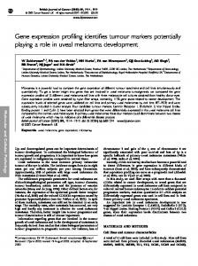
Figure 5. Post hoc validation of selected genes in the T + DES VP dysplasia panel by real-time q-PCR. The data were normalized to the levels of Rpl19. Bars indicated standard deviations (SD) of three to five animals in each treatment group. *P < .05, **P < .01 by one-way ANOVA with Tukey post hoc analysis.
Identification of Estrogen-induced Differentially Expressed Genes That Are Not Related to Dysplasia in Rat LP or VP We also identified two sets of hormone-induced genes that are not related to dysplasia. These genes exhibited differential expression in the VP following T + E2 treatment (14 genes; Figure 1C, left bottom) and in the LP following T + DES treatment (190 genes; Figure 1C, right bottom). They are published in Tables W2 and W3.
Genes Insensitive to the T + E2 and T + DES Treatments Two sets of 156 and 80 genes were identified as insensitive in the LP and/or VP to T + E2 or T + DES treatment, respectively (Figure 6, A–C; Tables W4 and W5). Forty-one genes were found in both hormone-insensitive panels and mapped to an IPA network (Figure 6D) that includes the androgen receptor (Ar), a key regulator of prostate function.
Discussion Exposure of the human prostate to estrogen may increase as men age [2–4] or with increased exposure to dietary estrogens such as DES or zeranol residues in meat, bisphenol A from food containers, or phytoestrogens [9]. The NBL rat provides a relevant model for elucidating mechanisms underlying carcinogenesis caused by such exposures, such as dysplasia and PCa, that consistently developed in the LP or the VP following chronic T–supported treatment with E2 or DES, respectively [15,18–20]. In the present study, we assessed transcriptional profiles elicited by the xenoestrogen DES in comparison with those of the endogenous hormone E2 and observed prostatic lobe–specific differential responses to the two estrogens in NBL rats. Hierarchical clustering revealed that the T + E2 treatment principally affects the LP gene expression profile whereas the T + DES targets that of the VP. Two distinct panels of dysplasia-related genes were identified: the T + E2 LP panel (253 genes) and the T + DES VP
Neoplasia Vol. 10, No. 1, 2008 panel (198 genes), with only 32 overlapping genes. These findings indicate that the two estrogens alter gene expression profiles only in the prostatic lobe that develops dysplasia following each of the hormonal treatments. These findings imply a functional divergence of the two estrogens with respect to their oncogenic actions in the rat prostate. Thus, endocrine disruption of gene expression and dysplasia induction of the two estrogens are categorically different [9], although they may share some biologic convergence [30]. The IPA analysis identified distinct gene interaction networks based on differential expression profiles. It classified genes into functional categories and suggested a possible mechanistic linkage among
Endocrine Disruption in Prostate Carcinogenesis
Tam et al.
37
the differentially expressed genes. We also used this tool to help prioritize genes for confirmation. The T + E2 treatment altered the expression of LP genes primarily in two major networks: one related to cell morphology, cellular growth, proliferation, and movement, and the other related to apoptosis and cell signaling. Exposure to T + DES also affected expression of genes in a similar network associated with apoptosis, cellular development, growth, and proliferation. Intriguingly, it also altered the expression of a set of genes linked to E2, a finding that may explain why, after T + DES treatment, gene expression profiles in the VP shifted closer to those in the treated and untreated LPs, which are under the influence of natural estrogen. By
Figure 6. Heat maps of genes insensitive to (A) T + E2 or (B) T + DES. (C) Venn diagram showing the number of genes insensitive to T + E2 and/or T + DES treatment. Note that 41 genes are found in common in both hormone-insensitive panels and mapped to an IPA network (D) that includes the Ar, a key regulator of prostate function. Gray indicates no change in gene expression levels compared with those in untreated counterparts. See Figure 1 for key to IPA network.
38
Endocrine Disruption in Prostate Carcinogenesis
Tam et al.
first mapping genes to the IPA network before selecting genes for confirmation, we were able to validate most of the expression changes in the microarrays with real-time q-PCR. In this regard, we confirmed three genes (Cebpd, Hoxa1, and Tp53) common in the two dysplasia panels. Importantly, these three genes were upregulated only in the prostatic lobes that developed dysplasia, i.e., the T + E2–treated LP and the T + DES–treated VP. Other lobe-specific genes that were confirmed were Smad7, Klb1, and Nxph3 in the T + E2–treated LP and Mafb, Src, Bcar1, Gpx3, Plau, and Thra in T + DES–treated VP. Our findings suggest that these gene expression changes induced by T + E2/DES treatment could be causative factors of dysplasia development. However, it remains possible that these gene expression changes are simply part of the neoplastic transformation process. Future studies employing laser capture–microdissected samples to establish the temporal relationship between change in gene expression pattern and evolution of dysplasia may shed new light on the cause–effect link between the two. Among all these validated genes, Cebpd, Hoxa1, and Tp53 are the ones upregulated in common in the two dysplasia panels. CCAAT/ enhancer binding proteins (C/EBP) are a highly conserved family of basic leucine zipper transcription factors; six members (C/EBP-alpha to -zeta) have been identified to date. This protein family plays a critical role in cell proliferation, apoptosis, and inflammation, depending on the cell type and specific physiological stress [31]. The precise functional role of C/EBP-delta, in particular, in normal prostate biology and PCa remains poorly understood. Cebpd was shown to be an androgen-repressed gene in the normal rat prostate [32,33], supporting its role as an apoptotic mediator and/or a negative regulator of cell proliferation. In a human androgen–dependent PCa xenograft, androgen withdrawal, however, resulted in a decline in the expression of C/EBP-delta [32]. Most published studies focus on the androgenic regulation of C/EBP-delta; this is, therefore, the first report of the association of estrogen-mediated upregulation of C/EBP-delta gene expression with the early phase of prostate carcinogenesis. A recent study showed overexpression of C/EBP-beta, another member of the family, in proliferative inflammatory atrophy in human prostate specimens, suggesting its role in the inflammation-associated carcinogenesis in the prostate [34]. In microarray data, we observed downregulation of C/EBP-alpha (Cebpa) only in T + E2–treated dysplastic LPs, corroborating its putative suppressive function in epithelial tumorigenesis [35]. Together, these findings raise the possibility that an orchestrated deregulation of C/EBP-family transcripts is involved in the pathogenesis of PCa in humans and rodents. Homeobox gene (HOX ) is a family of transcription factors related to growth and development. In rodents and humans, 39 members of the HOX gene family have been assigned to four gene clusters (A–D) and are numbered according to their expression along the anterior– posterior axis. Hox13 was shown to be involved in cell differentiation during normal prostate development [36], whereas upregulations of HOXC have been observed in PCa cells [37,38]. Knockdown of HOXC6 gene expression led to apoptosis of PCa cells [39], and overexpression of the HOXC8 gene correlated with the loss of tumor differentiation in human PCa [37]. Increased expression of the HOXA1 transcript was detected in human clinical cervical cancer samples and in several cervical cell lines compared with expression in normal tissues [40,41]. Our data suggest that ectopic expression of Hoxa1 is related to the evolution of dysplasia in both LP and VP and that the detailed roles of Hoxa1 in PCa require further study.
Neoplasia Vol. 10, No. 1, 2008 Tp53 is a well-known tumor-suppressive transcription factor and acts as a gatekeeper in signaling pathways involved in monitoring cellular stress such as DNA damage and in the determination of congruous responses (DNA repair, cell growth arrest, or apoptosis) to specific physiological stress. Previous studies demonstrated that mutation of Tp53 is not necessarily as late an event in prostate carcinogenesis as previously reported; it is also frequently found in high-grade prostate intraepithelial neoplasia, a precursor lesion of PCa [42,43]. In this regard, we observed upregulation of Tp53 gene expression in dysplastic rat prostate glands treated with sex hormone, implying a role in early prostate tumorigenesis, but whether these glands express wildtype or mutated Tp53 is not known. Alternatively, the induction of Tp53 expression may be a compensatory response to cellular stress imposed by hormonal changes. We recently demonstrated that T + E2 treatment induced oxidative and nitrosative stress, accompanied by DNA and protein damages, specifically in the LP, which is susceptible to dysplasia/cancer induction [17]. Smad7, an inhibitory Smad, is rapidly induced by TGF-β, thereby creating a negative feedback loop to a variety of TGF-β signaling responses, including proliferation, differentiation, apoptosis, inflammation, tissue remodeling, angiogenesis, and cell adhesion [44]. Disruption of the TGF-β signaling cascade by aberrant overexpression of this negative modulator leads to increased tumorigenicity in colon cancer cells by blocking TGF-β–mediated growth inhibition and apoptosis [45]. In contrast, Smad7 has been shown to mediate apoptosis induced by TGF-β in PCa cells [46], perhaps through crosstalking with other cellular signaling cascades such as the p38 mitogen-activated protein kinase [47] and β-catenin/Wnt [48] pathways. In our study, upregulation of Smad7 in the dysplastic LP (also in the T + DES VP microarray data) suggests perturbations of TGF-β feedback regulation and thus the loss of balance between cell proliferation and apoptosis in the early phase of prostate carcinogenesis. A particularly interesting gene network found in the T + DES– treated VP (Figure 4, B1 and B2) is related to E2 signaling. Members include Src, Bcar1, and Plau. Src, a nonreceptor protein tyrosine kinase, triggers the nongenomic estrogen signaling leading to activation of ERK1/2 and cell proliferation in prostate epithelial cells [49,50]. It remains to be determined if the downregulation of Src by DES represents a classic negative feedback loop. The Bcar1 interacts and modulates Src activity [51]. An increase in Bcar1 gene expression, as observed in the rat dysplastic VP, has also been reported in human PCa [52]. The coordinated dysregulation of both Src (downregulation) and Bcar1 (upregulation) may reflect a disruption of the Srcdependent signaling pathway during the early development of PCa, a notion further supported by the altered expression of Plau, a downstream target of Src [53]. Interestingly, both Bcar1 and Plau are involved in cell survival and migration [54,55]. Our group recently demonstrated significant oxidative stress and inflammation in the T + E2–treated NBL rat LP [17,56]. The microarray data show a relatively small fraction of genes related to redox homeostasis and immune response, possibly due to inherent limitations of the type and coverage size (about 3800 genes) of the array chips we selected. However, T + E2 treatment was found to upregulate Klrb1 in the LP. This gene, also known as NKR-P1A/CD161 in humans, encodes a natural killer (NK) cell C-type lectin–like receptor that regulates NK cell functions. KLRB1 is expressed not only in NK cells but also in immune cell types, such as T cells, monocytes, and dendritic cells [57]. Thus, it is likely that increased expression of Klrb1 in LP with T + E2 treatment is due to the infiltration of immune cells [17].
Neoplasia Vol. 10, No. 1, 2008 The observation of a dramatic induction of Gpx3 in the VP dysplasia panel, as shown by microarray (>17-fold) and q-PCR (>70fold), is consistent with our previous report of a marked elevation of GPX enzyme activity and lipid peroxidation in this lobe after T + DES treatment [56]. Hence, the activation in this glutathioneassociated detoxification enzyme might be a response to hormoneinduced oxidative stress in the gland or represent a cytoprotective mechanism, adapted by dysplastic cells, against oxidative insults. An elevation of GPX3 expression, but not of GPX1 and GPX2, was found in precancerous lesions of NKX3.1 mutant mouse prostate [58]. Taken together, these findings suggest that GPX3 is specifically involved in early prostatic transformation that has a mechanistic link to oxidative stress. In summary, we showed that genes such as Cebpd, Tp53, and Hoxa1 are at the core of disrupted biologic networks related to hormoneinduced dysplasia. Unbiased gene profiling clearly demonstrated differential susceptibility of the LP and VP to natural and xenoestrogen with regard to alterations in gene expression and induction of dysplasia. Our data suggest more functional divergence between DES and E2 in the disruption of prostatic functions than that previously suspected. Methodologically, the combined utility of expression profiling and gene network mapping provides an instrumental platform for transcriptome studies.
Endocrine Disruption in Prostate Carcinogenesis
[15]
[16]
[17]
[18]
[19]
[20]
[21] [22]
[23]
References [1] Taplin ME and Ho SM (2001). Clinical review 134: the endocrinology of prostate cancer. J Clin Endocrinol Metab 86, 3467–3477. [2] Ho SM, Leung YK, and Chung I (2006). Estrogens and antiestrogens as etiological factors and therapeutics for prostate cancer. Ann NY Acad Sci 1089, 177–193. [3] Carruba G (2006). Estrogens and mechanisms of prostate cancer progression. Ann NY Acad Sci 1089, 201–217. [4] Bosland MC (2006). Sex steroids and prostate carcinogenesis: integrated, multifactorial working hypothesis. Ann NY Acad Sci 1089, 168–176. [5] Leav I, Lau KM, Adams JY, McNeal JE, Taplin ME, Wang J, Singh H, and Ho SM (2001). Comparative studies of the estrogen receptors β and α and the androgen receptor in normal human prostate glands, dysplasia, and in primary and metastatic carcinoma. Am J Pathol 159, 79–92. [6] Zhu X, Leav I, Leung YK, Wu M, Liu Q, Gao Y, McNeal JE, and Ho SM (2004). Dynamic regulation of estrogen receptor-β expression by DNA methylation during prostate cancer development and metastasis. Am J Pathol 164, 2003–2012. [7] Ellem SJ and Risbridger GP (2006). Aromatase and prostate cancer. Minerva Endocrinol 31, 1–12. [8] Singleton DW and Khan SA (2003). Xenoestrogen exposure and mechanisms of endocrine disruption. Front Biosci 8, s110–s118. [9] Leffers H, Naesby M, Vendelbo B, Skakkebaek NE, and Jorgensen M (2001). Oestrogenic potencies of zeranol, oestradiol, diethylstilboestrol, bisphenol-A and genistein: implications for exposure assessment of potential endocrine disrupters. Hum Reprod 16, 1037–1045. [10] Epstein SS (1990). The chemical jungle: today’s beef industry. Int J Health Serv 20, 277–280. [11] Herbst AL, Ulfelder H, and Poskanzer DC (1971). Adenocarcinoma of the vagina. Association of maternal stilbestrol therapy with tumor appearance in young women. N Engl J Med 284, 878–881. [12] Troisi R, Hatch EE, Titus-Ernstoff L, Hyer M, Palmer JR, Robboy SJ, Strohsnitter WC, Kaufman R, Herbst AL, and Hoover RN (2007). Cancer risk in women prenatally exposed to diethylstilbestrol. Int J Cancer 121, 356–360. [13] Strohsnitter WC, Noller KL, Hoover RN, Robboy SJ, Palmer JR, Titus-Ernstoff L, Kaufman RH, Adam E, Herbst AL, and Hatch EE (2001). Cancer risk in men exposed in utero to diethylstilbestrol. J Natl Cancer Inst 93, 545–551. [14] Ho SM, Tang WY, Belmonte de Frausto J, and Prins GS (2006). Developmental exposure to estradiol and bisphenol A increases susceptibility to prostate car-
[24]
[25]
[26] [27]
[28] [29]
[30]
[31] [32]
[33]
[34]
[35]
Tam et al.
39
cinogenesis and epigenetically regulates phosphodiesterase type 4 variant 4. Cancer Res 66, 5624–5632. Leav I, Ho SM, Ofner P, Merk FB, Kwan PW, and Damassa D (1988). Biochemical alterations in sex hormone–induced hyperplasia and dysplasia of the dorsolateral prostates of Noble rats. J Natl Cancer Inst 80, 1045–1053. Leav I, Merk FB, Kwan PW, and Ho SM (1989). Androgen-supported estrogenenhanced epithelial proliferation in the prostates of intact Noble rats. Prostate 15, 23–40. Tam NN, Leav I, and Ho SM (2007). Sex hormones induce direct epithelial and inflammation-mediated oxidative/nitrosative stress that favors prostatic carcinogenesis in the Noble rat. Am J Pathol 171, 1334–1341. Thompson CJ, Tam NN, Joyce JM, Leav I, and Ho SM (2002). Gene expression profiling of testosterone and estradiol-17β–induced prostatic dysplasia in Noble rats and response to the antiestrogen ICI 182,780. Endocrinology 143, 2093–2105. Bosland MC, Ford H, and Horton L (1995). Induction at high incidence of ductal prostate adenocarcinomas in NBL/Cr and Sprague-Dawley Hsd:SD rats treated with a combination of testosterone and estradiol-17β or diethylstilbestrol. Carcinogenesis 16, 1311–1317. Ofner P, Bosland MC, and Vena RL (1992). Differential effects of diethylstilbestrol and estradiol-17β in combination with testosterone on rat prostate lobes. Toxicol Appl Pharmacol 112, 300–309. Smyth GK (2004). Linear models and empirical Bayes methods for assessing differential expression in microarray experiments. Stat Appl Genet Mol Biol 3 Article 3. Guo J, Sartor M, Karyala S, Medvedovic M, Kann S, Puga A, Ryan P, and Tomlinson CR (2004). Expression of genes in the TGF-β signaling pathway is significantly deregulated in smooth muscle cells from aorta of aryl hydrocarbon receptor knockout mice. Toxicol Appl Pharmacol 194, 79–89. Karyala S, Guo J, Sartor M, Medvedovic M, Kann S, Puga A, Ryan P, and Tomlinson CR (2004). Different global gene expression profiles in benzo[a] pyrene- and dioxin-treated vascular smooth muscle cells of AHR-knockout and wild-type mice. Cardiovasc Toxicol 4, 47–73. Sartor M, Schwanekamp J, Halbleib D, Mohamed I, Karyala S, Medvedovic M, and Tomlinson CR (2004). Microarray results improve significantly as hybridization approaches equilibrium. Biotechniques 36, 790–796. Sartor MA, Tomlinson CR, Wesselkamper SC, Sivaganesan S, Leikauf GD, and Medvedovic M (2006). Intensity-based hierarchical Bayes method improves testing for differentially expressed genes in microarray experiments. BMC Bioinformatics 7, 538. Benjamini YHY (1995). Controlling the false discovery rate: a practical and powerful approach to multiple testing. J R Stat Soc 57, 289–300. Daly-Burns B, Alam TN, Mackay A, Clark J, Shepherd CJ, Rizzo S, Tatoud R, O’Hare MJ, Masters JR, and Hudson DL (2007). A conditionally immortalized cell line model for the study of human prostatic epithelial cell differentiation. Differentiation 75, 35–48. Rozen S and Skaletsky HJ (1998). Primer3. Available at: http://www-genome. wi.mit.edu/genome_software/other/primer3.html. Livak KJ and Schmittgen TD (2001). Analysis of relative gene expression data using real-time quantitative PCR and the 2(−Delta Delta C(T)) method. Methods 25, 402–408. Buterin T, Koch C, and Naegeli H (2006). Convergent transcriptional profiles induced by endogenous estrogen and distinct xenoestrogens in breast cancer cells. Carcinogenesis 27, 1567–1578. Ramji DP and Foka P (2002). CCAAT/enhancer–binding proteins: structure, function and regulation. Biochem J 365, 561–575. Yang G, Gregory CW, Shang Q, O’Brien DA, and Zhang YL (2001). Differential expression of CCAAT/enhancer–binding protein-delta (C/EBPdelta) in rat androgendependent tissues and human prostate cancer. J Androl 22, 471–480. Desai KV, Michalowska AM, Kondaiah P, Ward JM, Shih JH, and Green JE (2004). Gene expression profiling identifies a unique androgen-mediated inflammatory/immune signature and a PTEN (phosphatase and tensin homolog deleted on chromosome 10)–mediated apoptotic response specific to the rat ventral prostate. Mol Endocrinol 18, 2895–2907. Wang GL, Shi X, Salisbury E, Sun Y, Albrecht JH, Smith RG, and Timchenko NA (2007). Growth hormone corrects proliferation and transcription of phosphoenolpyruvate carboxykinase in livers of old mice via elimination of CCAAT/ enhancer–binding protein α–Brm complex. J Biol Chem 282, 1468–1478. Loomis KD, Zhu S, Yoon K, Johnson PF, and Smart RC (2007). Genetic ablation of CCAAT/enhancer binding protein α in epidermis reveals its role in suppression of epithelial tumorigenesis. Cancer Res 67, 6768–6776.
40
Endocrine Disruption in Prostate Carcinogenesis
Tam et al.
[36] Huang L, Pu Y, Hepps D, Danielpour D, and Prins GS (2007). Posterior Hox gene expression and differential androgen regulation in the developing and adult rat prostate lobes. Endocrinology 148, 1235–1245. [37] Waltregny D, Alami Y, Clausse N, de Leval J, and Castronovo V (2002). Overexpression of the homeobox gene HOXC8 in human prostate cancer correlates with loss of tumor differentiation. Prostate 50, 162–169. [38] Miller GJ, Miller HL, van Bokhoven A, Lambert JR, Werahera PN, Schirripa O, Lucia MS, and Nordeen SK (2003). Aberrant HOXC expression accompanies the malignant phenotype in human prostate. Cancer Res 63, 5879–5888. [39] Ramachandran S, Liu P, Young AN, Yin-Goen Q, Lim SD, Laycock N, Amin MB, Carney JK, Marshall FF, Petros JA, et al. (2005). Loss of HOXC6 expression induces apoptosis in prostate cancer cells. Oncogene 24, 188–198. [40] Shim C, Zhang W, Rhee CH, and Lee JH (1998). Profiling of differentially expressed genes in human primary cervical cancer by complementary DNA expression array. Clin Cancer Res 4, 3045–3050. [41] Hung YC, Ueda M, Terai Y, Kumagai K, Ueki K, Kanda K, Yamaguchi H, Akise D, and Ueki M (2003). Homeobox gene expression and mutation in cervical carcinoma cells. Cancer Sci 94, 437–441. [42] Al-Maghrabi J, Vorobyova L, Chapman W, Jewett M, Zielenska M, and Squire JA (2001). p53 Alteration and chromosomal instability in prostatic high-grade intraepithelial neoplasia and concurrent carcinoma: analysis by immunohistochemistry, interphase in situ hybridization, and sequencing of laser-captured microdissected specimens. Mod Pathol 14, 1252–1262. [43] Downing SR, Russell PJ, and Jackson P (2003). Alterations of p53 are common in early stage prostate cancer. Can J Urol 10, 1924–1933. [44] Hayashi H, Abdollah S, Qiu Y, Cai J, Xu YY, Grinnell BW, Richardson MA, Topper JN, Gimbrone MA Jr, Wrana JL, et al. (1997). The MAD-related protein Smad7 associates with the TGFβ receptor and functions as an antagonist of TGFβ signaling. Cell 89, 1165–1173. [45] Halder SK, Beauchamp RD, and Datta PK (2005). Smad7 induces tumorigenicity by blocking TGF-β–induced growth inhibition and apoptosis. Exp Cell Res 307, 231–246. [46] Landstrom M, Heldin NE, Bu S, Hermansson A, Itoh S, ten Dijke P, and Heldin CH (2000). Smad7 mediates apoptosis induced by transforming growth factor β in prostatic carcinoma cells. Curr Biol 10, 535–538. [47] Edlund S, Bu S, Schuster N, Aspenstrom P, Heuchel R, Heldin NE, ten Dijke P, Heldin CH, and Landstrom M (2003). Transforming growth factor-β1 (TGFβ)–induced apoptosis of prostate cancer cells involves Smad7-dependent activa-
Neoplasia Vol. 10, No. 1, 2008
[48]
[49]
[50]
[51]
[52]
[53]
[54]
[55]
[56]
[57]
[58]
tion of p38 by TGF-β–activated kinase 1 and mitogen-activated protein kinase kinase 3. Mol Biol Cell 14, 529–544. Edlund S, Lee SY, Grimsby S, Zhang S, Aspenstrom P, Heldin CH, and Landstrom M (2005). Interaction between Smad7 and β-catenin: importance for transforming growth factor β–induced apoptosis. Mol Cell Biol 25, 1475–1488. Migliaccio A, Castoria G, Di Domenico M, de Falco A, Bilancio A, Lombardi M, Barone MV, Ametrano D, Zannini MS, Abbondanza C, et al. (2000). Steroid-induced androgen receptor–oestradiol receptor β–Src complex triggers prostate cancer cell proliferation. EMBO J 19, 5406–5417. Chieffi P, Kisslinger A, Sinisi AA, Abbondanza C, and Tramontano D (2003). 17β-Estradiol–induced activation of ERK1/2 through endogenous androgen receptor–estradiol receptor α–Src complex in human prostate cells. Int J Oncol 23, 797–801. Burnham MR, Bruce-Staskal PJ, Harte MT, Weidow CL, Ma A, Weed SA, and Bouton AH (2000). Regulation of c-SRC activity and function by the adapter protein CAS. Mol Cell Biol 20, 5865–5878. Fromont G, Vallancien G, Validire P, Levillain P, and Cussenot O (2007). BCAR1 expression in prostate cancer: association with 16q23 LOH status, tumor progression and EGFR/KAI1 staining. Prostate 67, 268–273. Kleiner S, Faisal A, and Nagamine Y (2007). Induction of uPA gene expression by the blockage of E-cadherin via Src- and Shc-dependent Erk signaling. FEBS J 274, 227–240. Gaylis FD, Keer HN, Wilson MJ, Kwaan HC, Sinha AA, and Kozlowski JM (1989). Plasminogen activators in human prostate cancer cell lines and tumors: correlation with the aggressive phenotype. J Urol 142, 193–198. Zhang X, Fei Z, Bu X, Zhen H, Zhang Z, Gu J, and Chen Y (2000). Expression and significance of urokinase type plasminogen activator gene in human brain gliomas. J Surg Oncol 74, 90–94. Tam NN, Ghatak S, and Ho SM (2003). Sex hormone–induced alterations in the activities of antioxidant enzymes and lipid peroxidation status in the prostate of Noble rats. Prostate 55, 1–8. Poggi A, Costa P, Zocchi MR, and Moretta L (1997). Phenotypic and functional analysis of CD4+ NKRP1A+ human T lymphocytes. Direct evidence that the NKRP1A molecule is involved in transendothelial migration. Eur J Immunol 27, 2345–2350. Ouyang X, DeWeese TL, Nelson WG, and Abate-Shen C (2005). Loss-offunction of Nkx3.1 promotes increased oxidative damage in prostate carcinogenesis. Cancer Res 65, 6773–6779.