Critical Reviews in Biochemistry and Molecular Biology, 36(2):107–260 (2001)
Critical Reviews in Biochemistry and Molecular Biology Downloaded from informahealthcare.com by 107.161.17.122 on 05/20/14 For personal use only.
Generation, Control, and Processing of Cellular Calcium Signals Ernesto Carafoli,1 Luigia Santella,2 Donata Branca,1 and Marisa Brini1 1
Department of Biochemistry, University of Padova, 35121 Padova, and 2Stazione Zoologica A. Dohrn, 80121 Napoli, Italy Referee: Guiseppe Inesi, M.D., Ph.D., Professor and Chairman, School of Medicine, Dept. of Biochemistry and Molecular Biology, Univeristy of Maryland, Baltimore. * Corresponding author: Ernesto Carafoli, University of Padova, Department of Biochemistry, Viale G. Colombo 3, 35121 Padova, Italy. Tel. +39 049 827 6137. Fax +39 049 827 6125. e-mail:
[email protected]
TABLE OF CONTENTS I.
Introduction
............................................................ 110
II. Why Calcium? ............................................................111 III. Calcium Binding Proteins .......................................... 113 A. Ca2+-Modulated Proteins ...................................... 113 1. EF-Hand Proteins ............................................ 113 2. The Calmodulin-Binding Domains in Target Proteins, Including the Ca2+-Independent IQ Binding Motifs ............................................................ 119 3. Non-EF Hand Ca2+-Binding-Proteins ............. 120 a. C2 Domains ................................................. 120 b. Annexins ..................................................... 121 c. Gelsolin ....................................................... 122 4. Ca2+-Binding Proteins Within Cell Organelles ...... 124 B. Membrane-Intrinsic Ca2+ Binding Proteins: Ca2+ Transport Across Cell Membranes ....................... 126 1. Membrane Transport of Ca2+ in Prokaryotes and Yeast Cells ....................................................... 126 1040-9238/01/$.50 © 2001 by CRC Press LLC
107
Critical Reviews in Biochemistry and Molecular Biology Downloaded from informahealthcare.com by 107.161.17.122 on 05/20/14 For personal use only.
2. Membrane Transport of Ca2+ in Higher Eukaryotes ....................................................... 128 a. The Plasma Membrane ............................... 128 i. Plasma Membrane Ca2+ Channels ......... 129 The Voltage-Operated Ca2+ Channels .... 129 The Receptor-Operated Ca2+ Channels .... 133 The Store-Operated Ca2+ Channels..... 136 ii. The Plasma Membrane Ca2+ Pumps (PMCAs) ................................................. 137 iii. The Plasma Membrane Na+/Ca2+ Exchanger ............................................... 141 b. The Endo(sarco)plasmic Reticulum ........... 145 i. Ca2+ Channels in the Endo(sarco)plasmic Reticulum ............................................... 145 Inositol tris Phosphate and Its Channels.............................................. 146 The Ryanodine (cADPribose)-Gated Channels.............................................. 150 The Newest Ca2+ Releasing Second Messenger: Nicotinic Acid Adenine Dinucleotide Phosphate (NAADP+) ... 154 ii. The Endo(sarco)plasmic Reticulum Ca2+ Pumps (SERCAs) ................................... 154 c. The Mitochondria........................................ 158 i. History and Properties of the Ca2+ Transport Process ................................... 158 ii. The Role of Mitochondrial Ca2+ Transport in Cytosolic Ca2+ Homeostasis .............. 161 d. Plant Cell Plasma Membrane and Endomembranes .......................................... 163 e. The Nucleus ................................................ 163 i. Nuclear Cytosolic Ca2+ Gradients .......... 164 ii. The Ca2+ Store of the Nuclear Envelope and its Mobilization ............................... 165
108
IV. Elementary Ca2+ Signals, Ca2+ Waves, and Oscillations ............................................................ 168
Critical Reviews in Biochemistry and Molecular Biology Downloaded from informahealthcare.com by 107.161.17.122 on 05/20/14 For personal use only.
V.
Effects of Ca2+ Signaling on Cellular Processes ....... 170 A. Fertilization ........................................................... 171 B. Ca2+-Directed Phosphorylation and Dephosphorylation of Proteins ..................................................... 174 1. Ca2+-Regulated Protein Phosphorylation ........ 174 2. Ca2+-Regulated Protein Dephosphorylation .... 179 C. Excitation-Contraction Coupling .......................... 183 D. Excitation-Secretion Coupling ............................. 184 E. Excitation-Transcription Coupling ....................... 186 F. Memory and Learning .......................................... 189 G. Nitrogen Monoxide in Ca2+ Signaling .................. 190 H Ca2+-Dependent Intracellular Proteolysis ............. 192 I. Ca2+ in Toxic and Apoptotic Cell Death .............. 197
VI. Conclusions
............................................................ 200
Acknowledgments ............................................................ 201 References
............................................................ 202
ABSTRACT: In the course of evolution, Ca2+ has emerged as the most versatile intracellular messenger. Its concentration within cells is controlled by reversible binding to specific classes of proteins that act as Ca2+ sensors to decode its information before passing it on to targets. The decoding operation is based on specific conformational changes in the sensor proteins. Other proteins intrinsic to membranes simply control Ca2+ concentration without processing its message, by transporting it across membrane boundaries. They are located in the plasma membrane and in the membranes of the organelles (the endo(sarco)plasmic reticulum, the mitochondria, the nuclear envelope), which play distinctive roles in the cellular homeostasis of Ca2+. Ca2+ is an ambivalent signaling agent. It carries information to virtually all processes important to cell life (e.g., it couples excitation to contraction, secretion, gene transcription, and controls enzyme activity through protein phosphorylation-dephosphorylation), but also transmits signals that promote the programmed demise of cells. When escaping control, Ca2+ also precipitates toxic cell death. KEY WORDS: calcium binding proteins, calcium transport, protein kinases and phosphatases, fertilization, memory and learning, cell death. 109
Critical Reviews in Biochemistry and Molecular Biology Downloaded from informahealthcare.com by 107.161.17.122 on 05/20/14 For personal use only.
I. INTRODUCTION Research on the ability of Ca2+ to carry information, and thus to act as a regulator of intracellular processes, has now pervaded most areas of cell biochemistry, physiology, and even pathology, accumulating knowledge at a pace that in the last few years has become explosive. Traditionally, the origin of the Ca2+ concept is traced back to two papers on the influence of blood components on frog heart contraction published by Sydney Ringer nearly 120 years ago. In the second of these papers (Ringer, 1883), Ringer had replaced the London tapwater he had used to make the saline with distilled water and found that the beating of the isolated heart became progressively weaker and stopped altogether after 20 min. To maintain contraction, it was necessary to add a calcium salt to the saline, which became henceforth known as “Ringer’s solution”. Even if serendipitous, the finding was exceptionally important. However, it somehow failed to attract the visibility it deserved. For decades, calcium continued being considered as merely one of the (ionic) factors that may influence the contraction of the heart muscle. Evidently, the observation of Ringer had come ahead of its time, when it was too early to imagine that calcium could do something else, in addition to contributing to the stability of bones and teeth. Thus, even if a couple of contributions that could have directed attention to calcium as a biological messenger had actually appeared in the 1930s (i.e., Chambers and Hale, 1932; Weise, 1934), it was only in 1940 that Heilbrunn
110
(Heilbrunn, 1940) clearly proposed calcium as an activator of intracellular processes, including muscle contraction. Even Heilbrunn, however, considered Ringer’s calcium but one of the factors that had influenced contraction. Key to Heilbrunn’s proposal were experiments in which frog muscle fibers were isolated and exposed to calcium salts: application of the salt to the surface of the fibers elicited no effect, but when the salt was applied instead to the cut ends the adjacent portion of the fiber contracted. Heilbrunn correctly concluded that calcium had diffused through the cut ends to reach the contractile structures inside the fiber, supporting the conclusion a few years later by directly microinjecting calcium solutions into the muscle fiber (Heilbrunn and Wiercinski, 1947). At about the same time, Kamada and Kinosita (1943) published in a Japanese journal results similar to Heilbrunn’s that only became widely known after World War II, showing that local contractures followed the injection of calcium solutions into muscle fibers. Then there was the important discovery made by Bailey in 1942 that the ATPase activity of myosin was activated by Ca2+, but not by Mg2+. It is a tribute to Bailey’s intuition that he concluded that it was the liberation of Ca2+ in the vicinity of the ATPase that controlled contraction. With hindsight, these results should have made immediately obvious that Ca2+ was a key regulator of (heart) muscle contraction. Still, acceptance of the concept was not straightforward. In 1949, no lesser a figure than A.V. Hill published a persuasive commentary negating any role for Ca2+ in muscle contraction. However, times were evidently
Critical Reviews in Biochemistry and Molecular Biology Downloaded from informahealthcare.com by 107.161.17.122 on 05/20/14 For personal use only.
becoming ripe, and in the 1950s the idea that Ca2+ was an intracellular regulator of muscle contraction eventually gained acceptance. One should quote in this context a seminal review article by Sandow that appeared in 1950 (Sandow, 1950) in which the author proposed that Ca2+ released from cortical or membrane regions of the muscle fibers activated actomyosin ATPase, thus mediating the process he named “excitation-contraction coupling”. It is interesting that the idea became generally accepted based on negative results, i.e., on the finding that it was the absence of Ca2+, induced either by its chelation by EDTA (Bozler, 1954) or by the “Marsh factor” (Marsh, 1951), a vesicular fraction later shown to be capable of removing Ca2+ (see below), that relaxed muscle fibers. The next step was the extension of the concept of Ca2+ as an intracellular regulator to cells different from muscle. This took another 20 years or so. Seminal contributions in the period 1972 to 1975 were those by Miledi (1973), who induced the release of neurotransmitters by directly injecting Ca2+ into the presynaptic terminal of squid axons, by Kanno et al. (1973) who promoted exocytosis by injecting Ca2+ into mast cells, and by Timourian et al. (1972), who showed that Ca2+ was the determinant for cleavage furrow in oocytes. A comprehensive study by Rose and Lowenstein (1975) on salivary gland cells, describing the distribution of Ca2+ in the cytoplasm and the restriction placed on its mobility by energized sequestering, also deserves to be mentioned.
II. WHY CALCIUM? In unicellular organisms, each cell is capable of performing all tasks neces-
sary to the vital cycle. Although unicellular organisms may regulate their activity in response to extracellular stimuli, their relationship to other cells is essentially based on the competition for nutrients. Instead, multicellular organisms partition the labor among cells that have become specialized to perform specific tasks, i.e., cells cooperate to ensure the correct functioning of the organism as a whole. This demands the exchange of signals to modulate and coordinate activity. In the course of evolution, multicellular organisms have thus developed complex ways of generating and processing information, based on messengers that carry the primary signals to cells (the first messengers) or decode their information inside them (the second messengers). While the first messengers are numerous, the second messengers known today are only about a dozen: as a rule, they are committed, i.e., they only influence a single target function. The exception is calcium, which has great versatility and controls a very large list of cellular processes. The evolutionary choice of Ca2+ as a universal and versatile intracellular messenger has evidently been dictated by its chemical properties (see Williams, 1999 for a comprehensive review of the topic). The total concentration of Ca2+ within eukaryotic cells can be very high, reaching the mM range. However, Ca2+ easily forms complexes with low-molecularweight anions that are less soluble in the physiological environment than those of Mg2+, the other biologically important and abundant divalent cation. In principle, formation of these complexes would be a means to substantially reduce the total concentration of free Ca2+, an essential requirement for any chemical chosen to act as a messenger. It is indeed self-evident that a messenger must be maintained within
111
Critical Reviews in Biochemistry and Molecular Biology Downloaded from informahealthcare.com by 107.161.17.122 on 05/20/14 For personal use only.
cells at very low free concentrations, to avoid prohibitive energy expenditures to achieve the necessary changes in its concentration in the ambient surrounding the signalling targets. The low-molecularweight compounds that have propensity to complex Ca2+ (e.g., phosphates, ATP, aminoacids, acidic phospholipids of membranes) do so with low affinity, and also easily complex other ions (e.g., Mg2+) that may be more concentrated in the intracellular environment. Other means thus are necessary to reduce the intracellular free Ca2+ concentration to the µM to sub µM level. To this aim, evolution has developed complex (protein) molecules able to exploit the special chemistry of Ca2+ by offering to it specific binding sites. At variance with the low-affinity binding mentioned above, these sites bind Ca2+ preferentially even in the presence of much higher concentrations of other metals, and do it reversibly and with the appropriate high affinity. In doing so they efficiently reduce the ionic concentration of Ca2+ within cells to the sub-µM range demanded by the signaling function. The ability of these sites to efficiently bind Ca2+ even in the greatly reduced ionic concentration range resulting from the low-affinity binding to the compounds mentioned above, and to do so in the presence of much larger concentrations of other cations, e.g., Mg2+, is made possible by the coordination flexibility of Ca2+ (see Carafoli and Penniston, 1985). The combination of charge and dimension of Ca2+ makes it an ideal ligand for irregularly shaped binding cavities, where the distance to the coordinating oxygens may vary greatly. This could be conveniently compared to Mg2+, whose ionic radius is much smaller than that of Ca2+ and whose polarizing power is thus much greater. As a result, the permitted variability in
112
bond length to the coordinating oxygens is much more limited in the case of Mg2+, which thus tends to demand perfectly octahedral binding cavities. Such regularly shaped binding cavities do not come about in proteins, where binding sites of irregular geometry are instead the rule. With appropriate adaptations this type of reasoning could be extended to other ions also abundantly present in cells, e.g., K+ and Na+. The conclusion that emerges is that the advantages of Ca2+ as a ligand for complex (protein) molecules made it a particularly attractive candidate, perhaps even an obligate one, as a messenger that would carry information to the intracellular ambient. There are also other advantages: eukaryotic cells are surrounded by a milieu in which the total concentration of free Ca2+ is orders of magnitude higher than in their interior: mM vs. sub-µM. This creates a large “ Ca2+ pressure” that would tend to force Ca2+ inside cells. However, external Ca2+ has no free admission to the cytoplasm because the plasma membrane is impermeable to it and does not permit its passive diffusion down its steep concentration gradient. It only permits the controlled entry of a limited amount of Ca2+ through carefully regulated proteinaceous channels (to be discussed below). The Ca2+ pressure creates a very dynamic situation, in which the reservoir of messenger available outside cells is virtually unlimited, and in which the regulated opening of the plasma membrane “gates” (i.e., the Ca2+ channels) promotes significant temporary swings in its concentration in the ambient surrounding the cellular targets. The coin, however, also has another side: should the plasma barrier to Ca2+ somehow fail (as is frequently the case in pathology) the Ca2+ pressure would promote the uncontrolled inundation of the cytoplasm
Critical Reviews in Biochemistry and Molecular Biology Downloaded from informahealthcare.com by 107.161.17.122 on 05/20/14 For personal use only.
with Ca2+, bringing its regulated signaling to an end, and effectively transforming Ca2+ into a conveyor of doom. One final point must be mentioned before closing this brief discussion of the advantages of Ca2+: the energetic metabolism of cells is phosphate-oriented: ATP is the universal energetic currency, and it is self-evident, given the propensity of Ca2+ phosphate salts to precipitate, that high concentrations of the ATP cleavage product phosphate in the cell ambient are only possible if means were available to keep the Ca2+ concentration very low. Evolution has evidently exploited the favorable properties of Ca2+ as a ligand to develop means to efficiently lower its concentration to a point where there would be no detrimental consequences for the phosphate-oriented energetic metabolism.
III. CALCIUM BINDING PROTEINS A multitude of proteins bind Ca2+ inside and outside cells, but only a fraction of them do it with the specificity, affinity, and reversibility demanded by the task of controlling its intracellular concentration and of modulating its signals. Because this review deals with the regulation and role of intracellular Ca2+, it will only consider intracellular Ca2+ binding proteins, of which several classes exist. They could be subdivided into two broad categories. There are proteins whose task is solely that of reversibly binding Ca2+ to modulate its concentration in the environment, for instance, by transporting it across membranes. Then there are proteins that bind Ca2+ for a different purpose, i.e., to decode its information. While by reversibly binding
Ca2+ with optimal efficiency in an ambient containing µM to sub-µM concentrations, they do contribute to the control of its concentration, their real task is that of transmitting suitably processed Ca2+ signals to the desired targets. Thus, they can be defined as Ca2+ sensors or, perhaps more appropriately, as Ca2+modulated proteins. Prior to discussing Ca2+-binding proteins, it may be worth emphasizing that Ca2+, in interacting with (enzyme) proteins, acts essentially as an allosteric regulator, never participating as a tightly bound species in catalysis at the active site. This is now common knowledge in biochemistry, and it should have become clear from the discussions above. An interesting exception to this rule is that of diisopropylfluorophosphatase (DFPase) from Loligo vulgaris, whose recently solved crystal structure (Scharff et al., 2001) shows two Ca2+ atoms. One appears to have a conventional allosteric role, promoting the interaction of domains of the enzyme. The other Ca2+, however, appears to be tightly bound at the active site, where it may directly participate in the catalytic process. An additional point of interest of DFPase is the very unusual presence of a histidine nitrogen among the Ca2+-coordinating atoms.
A. Ca2+-Modulated Proteins
1. EF-Hand Proteins The concept of Ca2+-modulated proteins was born with the solution of the crystal structure of parvalbumin (Kretsinger, 1972; Moews and Kretsinger, 1975). This relatively unimportant protein, which was
113
Critical Reviews in Biochemistry and Molecular Biology Downloaded from informahealthcare.com by 107.161.17.122 on 05/20/14 For personal use only.
originally thought to be specific of muscle and whose function is still obscure, binds two Ca2+ atoms per molecule. Its structure has become the prototype for a large family of proteins that have now been subdivided in no less than 66 subfamilies (Nakayama et al., 2000). Taken together, they number nearly 600 members, including some that are exceptionally important, for example, calmodulin. As of today, crystal structures are available for 15 of the 66 subfamilies. These proteins, which are characterized by a Ca2+ binding motif called the EF-hand, bind Ca2+, process its information, and pass it on to (enzyme) targets. According to general consensus, the transduction of the Ca2+ signal by EFhand proteins consists in a series of conformational changes that occur after Ca2+ has become bound. However, it is important to mention that no significant conformational changes after Ca2+ binding have been described for at least two of the EF-hand proteins, e.g., parvalbumin itself and calbindin (the protein that mediates the action of vitamin D), which are thus likely to act instead only as temporal Ca2+ buffers. Not all cellular proteins modulated by Ca2+ belong to the EF-hand family. Some non-EF-hand proteins decipher the Ca2+ signal directly, triggering a number of cellular responses. A prominent example, which is discussed in detail below, is that of protein kinase C, which binds Ca2+ and becomes activated to phosphorylate a variety of important cellular proteins. However, the EF-hand proteins are unquestionably the most important Ca2+-modulated proteins, and are now generally recognized as a paradigm for them. The basic structural module of these proteins, derived from the parvalbumin prototype, is a motif consisting of two helices of about 10 amino acids, interrupted by a 12 amino
114
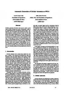
acid loop where Ca2+ becomes coordinated. The helix-loop-helix-motif is repeated from 2 to 12 times. For instance, parvalbumin has motifs A-B, C-D, and E-F (the EF-hand motif has given the name to the entire class of proteins). Normally (albeit not invariably), the EFhand motifs occur in pairs adjacent in the sequence. Two such consecutive motifs are symbolized by a pair of right hands (Figure 1): helix C (and helix E) in parvalbumin run from the tip to the base of the forefinger. The flexed middle finger corresponds to the calcium coordinating CD and EF loops. Helix D (and helix F) run from the base to the tip of the thumb. One important corollary of these structural aspects is that sequence variability in the helices is tolerated, whereas the variability in the residues of the loop is much more restricted. The loops begin with an invariant aspartic acid (position 1), and usually end with a glutamic acid (position 12). Ca2+ is coordinated by seven oxygen atoms of a distorted pentagonal bipyramid. The side chains of four conserved residues in the loop, located approximately at the vertices of an octahedron, provide the oxygens. They occupy positions 1, 3, 5, and 12, the side chain of residue 12 providing two coordinating oxygens. The two coordinating oxygens at the remaining vertices of the octahedron are the carbonyl oxygen of a variety of residues at position 7 and the side chain oxygen of a glutamic acid in position 9, or a water oxygen (as in calmodulin). There are variations to the usual Ca2+ coordination scheme outlined above. First of all, Ca2+ binding is not obligatorily a distinguishing characteristics of EF-hand motifs, as EF-hand motifs in some proteins do not bind Ca2+, sometimes depending on the species of origin. Then, there are loops that use several carbonyl
115
FIGURE 1. The EF-hand Ca2+ -binding motif. The motif is formed by two–helices, oriented at about 90° with respect to one another, flanking a 12 amino acid loop where Ca2+ is coordinated to seven oxygen ligands. The motif is described in fill in the text. (Adapted from Kretsinger [1972], and Persechini et al. [1989].)
Critical Reviews in Biochemistry and Molecular Biology Downloaded from informahealthcare.com by 107.161.17.122 on 05/20/14 For personal use only.
Critical Reviews in Biochemistry and Molecular Biology Downloaded from informahealthcare.com by 107.161.17.122 on 05/20/14 For personal use only.
oxygens instead of the usual oxygens of carboxylic side chains. Other proteins may even use side chains oxygens from neighboring helices instead of the oxygens of the loop. Finally, loops may have extra residues inserted that may coordinate Ca2+ and thus alter the coordination scheme of the conventional loops. Before concluding this discussion on the binding of Ca2+ to EF-hand motifs in proteins, a few other points must be mentioned. One is the location of these proteins. Although as a rule EF-hand proteins reside in the cytosol (and in the nucleoplasm), reticulocalbin is localized in the lumen of the endoplasmic reticulum (Ozawz, 1995; Tachikui et al., 1997). Cab45 (Scherer et al., 1996) and nucleobindin (CALNUC) are localized in the Golgi apparatus, and glycerophosphate dehydrogenase is located on the outer face of the inner mitochondrial membrane. Another EF-hand protein that has been localized in mitochondria is Aralar, a calcium binding member of the mitochondrial inner membrane carriers family, characterized by a C-terminal half that is analogous to other mitochondrial solute carriers and a N-terminal half containing several EF-hands (Del Arco and Satrústegui, 1998; Del Arco et al., 2000). EF-hand proteins may even be extracellular (e.g., osteonectin and QR1) and thus be active in an ambient where Ca2+ remains constant at the mM level. This rules out a Ca2+-modulated function, because under these conditions these proteins will presumably always be saturated with Ca2+. In discussing the location of EF-hand proteins, one could also mention a group of proteins collectively called intracellular neuronal calcium sensors (NCS) (Braunewell and Gundelfinger, 1999; Burgoyne and Weiss, 2001), which in-
116
cludes five subfamilies: the recoverins and guanylyl-cyclase activating proteins, which are primarily expressed in retinal photoreceptor cells and have established roles in the regulation of photo-transduction; the frequenins, visinin-like and Kv-channel-interacting proteins (KChIPs), which are widely expressed in central neurons. Although the functions of the last three families are not clearly defined, it has been shown that they interact with multiple target proteins, and with nucleic acids as well. Interestingly, the nucleotide sequence of one of the NCS proteins (KChIP3) is nearly identical to that of DREAM, the downstream regulatory element antagonist modulator, which is a Ca2+-dependent repressor of transcription (Carrión et al., 1999, and see the section on gene transcription). KChIP3 itself is identical with calsenilin, a protein interacting with presenilin, whose mutations result in familial Alzheimer disease (Buxbaum et al., 1998). One interesting feature of most NCS is N-terminal acylation: several members of the family are N-terminally myristoylated. Binding of Ca2+ to recoverin, and presumably to other NCS proteins, changes their conformation, exposing the myristoyl residue and hydrophobic portions of the molecule, making them available for membrane (or target protein) interaction. The Ca2+myristoyl switch could be a mechanism that affects the compartmentation of signaling cascades in neurons and/or the transmission of Ca 2+ signals to their membranes (Braunewell and Gundelfinger, 1999; Burgoyne and Weiss, 2001). Interestingly, two of the NCS proteins, recoverin and GCA1, have been involved recently in degenerative diseases of the retina. Mutations in the GCAP gene have been associated with
Critical Reviews in Biochemistry and Molecular Biology Downloaded from informahealthcare.com by 107.161.17.122 on 05/20/14 For personal use only.
autosomal dominant cone distrophy (Sokal et al., 1998; Dizhoor et al., 1998). One of the defects [GCAP1(Y99c)] has been related to constitutive activation of guanylyl cyclase that is not properly inactivated by dark levels of Ca 2+ , eventually leading to degeneration of cone cells. The other condition [GCAP1(P50L)] (Sokal et al., 2000) is a milder form of autosomal dominant cone dystrophy in which the mutation reduces ability of the Ca 2+-binding G-CAP. Recoverin has been identified as the autoantigen in a degenerative disease of the retina called cancer associated rethinopathy (CAR), in which patients loose vision due to degeneration of photoreceptors (Polans et al., 1991; Polans et al., 1995). Recent work has shown that recoverin is expressed in tumors of CAR patients, indicating that the disease is linked to the expression of a neuronal specific antigen in nonneuronal (i.e., tumor) cells (Polans et al., 1995). Recoverin is normally sequestered in photoreceptor cells; therefore, its expression in a distant tumor cell triggers the abnormal immunological response. Finally, there is the issue of EFhand proteins in bacteria. Although bacteria do extrude Ca2+ to maintain it at a sub-µM level in the cytosol (values similar to those found in eukaryotic cells have been measured in them, Rosen, 1987), they apparently do not use Ca2+ as a messenger. Nevertheless, one EFhand protein has been detected in a prokaryote, Saccharopolyspora. A last important point is that of the permanent attachment of EF-hand proteins to a target, which has now been documented in several cases. A more striking case along this line is that of proteins with various enzymatic activities, which are hetero*
chimeras containing EF-hand regions. This does not refer to the case of the numerous proteins whose sequence contains one or more EF-hands, but to that of several proteins (among them some kinases, a protease, a phosphatase, and others) in which an EF-hand protein is a built-in part of the sequence. Evidently, the gene encoding EF-hands has this case fused with the gene encoding other functional domains. After having complexed Ca2+, EFhand proteins must transmit its information to targets. Although the latter are generally enzymes, in several cases they are not. EF-hand proteins have been shown to bind to structural cytoskeletal proteins or even to DNA. Most EF-hand proteins are committed, i.e., they interact with a single target, but calmodulin, which is one of the most conserved eukaryotic proteins (Copley et al., 1999), can bind and regulate more than 100 proteins, enzymes and otherwise. Most of what is now understood on the molecular mechanism by which EFhand proteins process the Ca2+ signal and transmit it to targets has come from studies of calmodulin, undoubtedly the most thoroughly studied of all EF-hand proteins. Unlike parvalbumin, which is a compact globular molecule, calmodulin is an elongated molecule, with a 25 residue, solvent-exposed central α–helix that separates the two terminal domains where a total of 4 Ca2+ ions are bound. Although in the crystal structure (Babu et al., 1985) the central helix is rigid and extended (Plate 1*), small angle X-ray scattering work (Heidorn and Trewhella, 1988) has shown that the two terminal lobes of calmodulin are closer to each other than in the crystals, suggesting that the central segment of the molecule is
Plate 1 appears following page 166.
117
Critical Reviews in Biochemistry and Molecular Biology Downloaded from informahealthcare.com by 107.161.17.122 on 05/20/14 For personal use only.
not completely α–helical in solution. This has been supported by NMR relaxation studies (Barbato et al., 1992) that have demonstrated that residues 78-81 of the central linker domain were highly mobile in solution and not in an α–helical conformation. The first threedimensional structure of Ca2+-loaded calmodulin complexed with a target peptide (a 26-residue calmodulin-binding domain from myosin light chain kinase [MLCK] [Ikura et al., 1992]) determined by multidimensional NMR spectroscopy has conclusively documented that calmodulin indeed collapses along the central helix, bringing the two terminal lobes together and engulfing the MLCK peptide (Plate 2a*). The collapse of calmodulin around its binding domain has now been documented for a number of enzyme targets different from MLCK (Meador et al., 1993; Osawa et al., 1999). It is the second of the two conformational changes involved in the processing of the Ca2+ signal by the molecule. The first occurs when calmodulin complexes Ca2+ (Plate 2b,c). In both apo-calmodulin and Ca2+loaded calmodulin two Ca2+-bindings motifs are joined together by a ß-sheet between the two Ca2+ binding loops; however, the helix packing differs in the two states. In apo-calmodulin the two Ca 2+ binding domains are in a “closed” conformation (Finn et al., 1995; Kuboniwa et al., 1995; Zhang et al; 1995), in which the entering and exiting helices of each domain are positioned nearly anti-parallel to each other. After binding Ca2+, each domain undergoes a transition to an “open” conformation, in which the two helices become more perpendicular. Other helix movements also occur when Ca2+ becomes bound, *
Plate 2 appears following page 166.
118
resulting in the exposure of hydrophobic residues on the surface of the molecule. The hydrophobic surface of each of the two terminal domains of calmodulin is rich in methionines, which interact with hydrophobic residues in the binding domain of target enzymes. The flexible nature of the Met side chain and the presence of the polarizable sulfur atom facilitate the interaction, but electrostatic interactions also occur. As discussed in more detail below, all calmodulin-binding domains contain several positively charged residues (James et al., 1995) that bind deeply in the hydrophobic cavities of the C- and N-terminal lobes of calmodulin. The affinity by which calmodulin interacts with its binding domain in target enzymes is modulated by its phosphorylation by protein kinases, a mechanism that recently has come to the forefront of calmodulin research. Two kinase types are involved in the process, casein kinase II (Meggio et al., 1987; Nakajo et al., 1988; Sacks et al., 1992; Quadroni et al., 1994) and a number of tyrosine kinases (Sacks and McDonald, 1988; Benguria et al., 1994; Fukami et al., 1986; Sacks et al., 1992; Joyal and Sacks, 1994; Colca et al., 1987; Corti et al., 1999). Although the phosphorylation process has been mostly studied in vitro, it has been shown to occur in vivo as well (Quadroni et al., 1994; Joyal and Sacks, 1994). The phosphorylation sites by casein kinase II (Quadroni et al., 1994) have been identified at Thr79, Ser81, and Ser101, and found to be the same in vitro and in vivo. Those by tyrosine kinases have been identified at Tyr99 and Tyr139 (Joyal et al., 1996, Corti et al., 1999). The effects of phosphorylation on the function of
Critical Reviews in Biochemistry and Molecular Biology Downloaded from informahealthcare.com by 107.161.17.122 on 05/20/14 For personal use only.
calmodulin have been tested on a number of target enzymes. Although controversial aspects still exist (e.g., Sacks et al., 1995), phosphorylation by casein kinase II tends to decrease the binding affinity of calmodulin for enzyme targets (Quadroni et al., 1998). Interestingly, one of the casein II kinase sites (Ser101) is located in the third Ca2+ binding loop of calmodulin, yet its phosphorylation does not influence the binding affinity of calmodulin for Ca2+ (Quadroni et al., 1998). The finding that two of the casein kinase II phosphorylation sites are located at the center of the flexible loop that mediates the collapse of the central helix after target binding provides a possible explanation for the lowered affinity of phosphocalmodulin for the binding domains. Phosphorylation could make this portion of the helix more rigid, hindering its collapse around the binding domains (James et al., 1995). At variance with casein kinase II tyrosine phosphorylation generally increases the activatory properties of calmodulin, Tyr99 essentially mediating the effect. As is the case for Ser101, the phosphorylation of Tyr99, which is located in the third Ca2+ binding loop of calmodulin, has no effect on its Ca2+ binding affinity (Corti et al., 1999).
2. The Calmodulin-Binding Domains in Target Proteins, Including the Ca2+Independent IQ Binding Motifs The majority of calmodulin binding motifs in proteins, which bind it in the canonical Ca2+-dependent way, are unstructured in the absence of calmodulin, and become α–helical after binding it.
The helix is amphipathic: its hydrophobic side interacts with hydrophobic pockets of calmodulin, whereas the hydrophilic face, containing four or more basic residues, interacts electrostatically with acidic residues in calmodulin (reviewed in James et al., 1995; O’Neil and DeGrado, 1990; Crivici and Ikura, 1995). This is the general pattern of interaction. However, when analyzing in more detail the comparative properties of these calmodulin-binding domains (nearly 200 have now been identified) additional properties have become evident, which has led to their subdivision in classes. Although the classification schemes have varied somewhat, they are all based on the positions of interacting bulky hydrophobic residues in the sequence of the domains, which usually consist of 15 to 30 amino acids. Thus, the classifications have defined the 1-8-14 and the 1-5-10 classes (Crivici and Ikura, 1995; Afshar et al., 1994; Rhoads and Friedberg, 1997). The first and last hydrophobic residues (i.e., W582 and F595 in skeletal muscle MLCK) interact with the hydrophobic cavities of the C- and N-terminal domains in calmodulin, respectively. Thus, in the complex the binding domain assumes an antiparallel orientation with respect to calmodulin. More recently, a third class of calmodulin binding domains has been identified, containing the IQ motif (Rhoads and Friedberg, 1997). This class is of particular interest, because it binds calmodulin in a predominantly Ca2+-independent way, thus rationalizing relatively old, and somewhat unexpected, observations of Ca2+-independent interactions of calmodulin with some targets, e.g., neuromodulin (GAP43/P-57), neurogranin, the heavy chains of unconventional myosins (Cheney and Mooseker, 1992; Alexander et al., 1987;
119
Critical Reviews in Biochemistry and Molecular Biology Downloaded from informahealthcare.com by 107.161.17.122 on 05/20/14 For personal use only.
Baudier et al., 1991; Espreafico et al., 1992). The first observation of this type was made on neuromodulin, whose affinity for calmodulin decreases in the presence of Ca2+, allowing its purification by affinity chromatography (Alexander et al., 1987). The case of unconventional myosins is of particular interest, as their light chains may actually be calmodulin itself (Cheney and Moosekers, 1992). The IQ motif is a consensus sequence IQXXXRGXXXR, which may be incomplete, i.e., the second hydrophobic residue may be missing, and also occurs in many proteins that have not been shown to bind calmodulin (e.g., a subfamily of Ca2+independent protein kinase C (δ, ε, η, and θ), connexin 45, sodium channel proteins, the multidrug resistance protein, L-type Ca2+ channel proteins). It may not be the only motif that binds calmodulin in a Ca2+-independent manner. The macrophage NO-synthase, and the γ–subunit of phosphorylase kinase, which bind calmodulin in a Ca2+-independent way (Xie et al., 1992; Dasgupta et al., 1989; Anagli et al., 1995) do not contain IQ motifs.
teins), but are evidently still capable of deciphering Ca2+ signals. As a rule they bind Ca2+ with lower affinity than the EF-hand proteins. One of these proteins has been briefly alluded to above (protein kinase C, PKC), others will be briefly described below. Those present in the lumen of the endoplasmic reticulum are of special interest. They may have additional functions in addition to regulating Ca2+ storage within the endoplasmic reticulum. The calcium binding sites of nonEF-hand proteins, although generally sharing a high content of acidic amino acids, display a wide range of structural (and functional) features. The bestcharacterized modules are the C2 domains (typical of PKC, phospholipase A2 [cPLA2], phospholipase C [PLC], synaptotagmins and, as very recently discovered, of the catalytic subunit of calpain), the endonexin fold, which is characteristic of the annexin family, and the Ca2+ binding motif in gelsolin, a Ca2+ switch that modulates actin cytoskeleton.
a. C2 Domains 2+
3. Non-EF-Hand Ca -Binding Proteins Numerous Ca2+-binding proteins not containing EF-hand motifs are soluble in the cytosol, contained in the lumen of organelles or loosely associated to membranes. Although some may bind Ca2+ only for buffering purposes, most are also regulated by it, i.e., their function is affected by Ca2+. They have no role in transmitting the Ca2+ information to targets (although in some cases Ca2+ can trigger their interaction with other pro-
120
Although C 2 domains were first identified in PKC (Nishizuka, 1988); most of the available data about their three-dimensional structure were obtained from studies on synaptotagmin I (Sutton et al., 1995), a transmembrane protein of synaptic vesicles believed to act as the main Ca2+-sensor in calciumdependent exocytosis. Its cytoplasmic region comprises 2 C2 domains. The C2 structure consists of a compact β-sandwich of two four-stranded β-sheets. Three of the seven loops connecting
Critical Reviews in Biochemistry and Molecular Biology Downloaded from informahealthcare.com by 107.161.17.122 on 05/20/14 For personal use only.
the β-strands, localized at the top of the β-sandwich, bind a cluster of Ca2+ ions (at least three in the C2A domain of synaptotagmin I), primarily to oxygens of aspartate side chains (Plate 3a). The coordination sphere of bound Ca2+ is incomplete, thus explaining the low affinity of these sites for Ca2+ (Kd > 1.0 mM). Phospholipids probably occupy unsatisfied coordination sites on the bound Ca2+, increasing the affinity of the domain for Ca2+ by three orders of magnitude. Due to this arrangement, C2 domains are often indicated as Ca2+dependent lipid binding domains: the C2 domains of PLC, PKC and cPLA2 are indeed believed to be responsible for the calcium-dependent recruitment of these enzymes to phospholipids of membranes. Sequence homology and structural similarity among C2 domains of different proteins are high in the β-sandwich region, whereas the loops are much less conserved. Thus, the β-sandwich probably constitutes a scaffold supporting the calcium-binding loops, whose variability may be related to the functional specificity of the domain. The three-dimensional structures determined so far have not revealed major conformational changes induced by Ca2+ binding to C2 domains; this raises the problem of how calcium may modulate their function. Studies on synaptotagmin I suggest that a significant change in the electrostatic potential of the domain caused by Ca 2+ binding may be responsible for the Ca 2+-dependent interaction with syntaxin (Shao et al., 1997). In this case the C2 domain would act as a Ca2+-dependent electrostatic switch, without requiring significant conformational changes. *
b. Annexins The annexin family comprises a dozen of structurally similar proteins ubiquitously present in eukaryotic cells, with the exceptions of mammalian erythrocytes and yeasts. A property of all annexins is the ability to bind to negatively charged phospholipids in a Ca2+dependent manner, and hence to associate reversibly to membranes. This would thus be the modulating function of Ca2+. A wide range of functions of annexins have been documented in vitro, including the promotion of vesicle aggregation and fusion, the inhibition of phospholipase A2, anticoagulation, and ion channel activities. These properties strongly suggest the involvement of annexins in membrane traffic processes such as endo- and exocytosis. Despite these findings, the biological roles of these proteins in vivo remain undefined. The molecular structures of annexin V and of other members of the family have been elucidated in detail both in the soluble and membrane-bound forms (Brisson et al., 1991; Mosser et al., 1991; Burger et al., 1996; Huber et al., 1992). All annexins contain four highly conserved repeats of 70 amino acids, the exception being annexin VI, which contains eight such repeats (probably due to gene duplication). Each of the repeats folds into a compact domain consisting of five α-helices of 7 to 16 amino acids wound in a right-handed superhelix. The four domains are arranged in a cyclic array; strong hydrophobic interactions between domains II and III and I and IV, respectively, generate two tight modules that interact more weakly through polar or charged residues. The
Plate 3 appears following page 166.
121
Critical Reviews in Biochemistry and Molecular Biology Downloaded from informahealthcare.com by 107.161.17.122 on 05/20/14 For personal use only.
molecule has a planar, slightly curved shape with a convex and a concave face (Plate 3b) and a central hydrophilic pore that has been suggested to act as Ca2+ channel (Rojas et al., 1990). The Ca2+-binding sites are located on the convex side of the protein, which is the membrane-binding side. The calcium- and phospholipid-binding sites correspond to a 17 amino acid consensus sequence contained in each of the four repeats called the endonexin fold (Geisow et al., 1986; Kretsinger and Creutz, 1986). Ca2+ is coordinated to three carbonyl oxygen atoms at the N-terminus of the fold, and to the carboxylate group of an acidic side chain 38 residues down the sequence; water molecules complete the pentagonal bipyramidal coordination sphere. Three of the four repeats in the annexin molecule bind Ca2+ with moderate affinity (Kd in the micro- to millimolar range, except for annexin VI, which has a Kd of ~ 1 µM); the third repeat does not appear to bind Ca2+ at low concentrations due to deviations from the canonical sequence and structure. As Plate 3b shows, high concentrations of Ca2+ induce a conformational change of domain III resulting in the exposure of a naturally buried tryptophan residue to the molecular surface, and in the creation of a new, canonical Ca2+-binding site within the domain (Berendes et al., 1993; Sopkova et al., 1993). A direct contact between the exposed tryptophan and the ester-bond region of the membrane has been suggested (Meers, 1990; Meers and Mealy, 1993). While the homologous repeats constitute the conserved core of annexins, each member of the family is characterized by an N-terminus of different length (12 to 196 residues) and sequence. The N-terminus is located, together with the
122
C-terminus, on the convex (cytoplasmic) side of the molecule; it represents a regulatory region and probably confers specific biological functions to each annexin. The N-terminus may interact with proteins of the S-100 family (annexins II and XI) (Gerke and Weber, 1985; Tokumitsu et al., 1992) or be posttranslationally modified by phosphorylation, glycosylation, N-myristoylation, cross-linking, acylation, or proteolysis. One interesting development in the area of annexins is their involvement in two human diseases that have been termed annexinopathies. One concerns annexin II, whose anticoagulation role is linked to its ability to promote fibrinolysis on the apical surfaces of vascular endothelial cells by promoting the binding of plasminogen and tissue plasminogen activator (Hajjar et al., 1994). Annexin II has found to be overexpressed in a subset of patients with acute promyelocytic leukemia (Menell et al., 1999). The other disease instead concerns annexin V, which is underexpressed in placental trophoblasts of patients with antiphospholipid syndrome, an autoimmuno condition characterized by the presence of antibodies against anionic phospholipid-protein complexes (Rand et al., 1994). Underexpression of annexin V has also been observed in trophoblasts of preeclamptic placenta (Rand et al., 1997).
c. Gelsolin Gelsolin is a multifunctional protein that binds, severs, and caps actin filaments in a Ca2+-dependent manner (Yin and Stossel, 1979); alternatively, it can nucleate actin polymerization by
Critical Reviews in Biochemistry and Molecular Biology Downloaded from informahealthcare.com by 107.161.17.122 on 05/20/14 For personal use only.
binding two actin monomers. Its importance in functions related to the actin cytoskeleton, its involvement in the signaling pathways linked to phosphoinositides (see below), and the considerable understanding of the structural aspects of its modulation by Ca2+ justify a somewhat more detailed discussion. Two forms of gelsolin, one intracellular and the other (containing an additional leader sequence) extracellular, are produced by the same gene by alternative transcription initiation and selective RNA processing (Kwiatowski et al., 1986). Intracellular gelsolin is involved in cell motility and in motility-related events as a regulator of actin function, e.g., platelet activation, cytokinesis, ion channel modulation, and even apoptosis. Extracellular (plasma) gelsolin acts as an actin-scavenging system to prevent the polymerization of actin released after cell death and the consequent increase in blood viscosity. Point mutations in the gelsolin gene cause a form of familial amyloidosis (Finnish type). Unmasking of a cleavage site for a trypsin-like protease leads to the extracellular deposition of an amyloidogenic gelsolin fragment (Maury et al., 1990; Paunio et al., 1998). Gelsolin contains six 120 to 150 amino acid repeats (G1 to G6) that are found in several actin-binding proteins (Plate 4a, b*); sequence homologies indicate that these repeats have arisen from triplication of an original gene, followed by gene duplication. The six domains are organized into two clusters of similar architecture (G1 to G3 and G4 to G6) connected by a stretch of 53 amino acids: this linker may be cleaved by caspase-3 in vitro and in vivo generating gelsolin N- and C-halves (Kothakota et *
al., 1997). The isolated N-half contains binding sites for actin monomers (G1) and filaments (G2) and is capable of severing and capping actin even in the absence of Ca2+. The isolated C-half, which is devoid of severing and capping activity, requires instead µM Ca2+ to bind actin monomers. Because actin binding and severing by full-length gelsolin requires µM Ca2+, the C-half appears to act on the N-half as a calcium-modulated inhibitory domain. Moreover, the C-half enhances severing by the N-half, probably through cooperative binding to the actin filament, while the N-half contains a binding site for polyphosphoinositides (PIP2, see below), which induce the dissociation of the actin-gelsolin complex (McLaughlin et al., 1993). The fundamental folding motif of the homologous domains of gelsolin has been deduced from the crystal structure of the G1-actin complex. G1 is organized in a central four-stranded β-sheet motif flanked on one face by a four-turn α-helix almost parallel to the strands in the sheet, and on the other face by a shorter α-helix roughly perpendicular to the strands. The shorter helix and the two central strands of the sheet include most of the conserved apolar residues of the repeats. The longer helix binds to a cleft between actin subdomains 1 and 3 through an apolar patch surrounded by polar atoms that form 13 interprotein hydrogen bonds. The actin-complexed G1 domain comprises two Ca2+-binding sites. The type 1 site represents one of the few reported cases of Ca2+ coordination between interacting proteins involving, besides one Asp residue and two carbonyl groups of the gelsolin main peptide chain, Glu167 from actin. By contrast, the type 2 Ca2+-binding site is in-
Plate 4 appears following page 166.
123
Critical Reviews in Biochemistry and Molecular Biology Downloaded from informahealthcare.com by 107.161.17.122 on 05/20/14 For personal use only.
tramolecular: Ca2+ is coordinated by one Asp, one Glu, and a carbonyl oxygen of gelsolin. It has been suggested that this site is involved in phosphoinositide binding, phosphoinositides supplying the lacking ligand for the coordination of Ca2+ (Yu et al., 1992). As predicted by the structural similarities between the two halves of gelsolin, two Ca2+-binding sites, analogous to those of the G1 domain, have been identified in the G4 domain. These four Ca2+-binding sites have low Ca2+ affinity (Kd in the low µM range); however, a fifth Ca2+-binding site, with a Kd of 0.2 µM, is also located in the G6 repeat (Pope et al., 1995). Unfortunately, this site, which may be very important functionally, could not be identified in the electron density maps. Recent work has unraveled the structural basis for the regulation of gelsolin activity by Ca2+, mainly through a comparison of the crystal structures of Ca2+free non-activated gelsolin (Burtnik et al., 1997) and of the C-half bound to two actin monomers (Robinson et al., 1999). In the absence of Ca2+ the six domains of full-length gelsolin pack closely together in a very compact globular structure. The N- and C-halves of the molecule are held together by a C-terminal helix interacting with, and thus blocking, the actin-binding helix of G2. Moreover, the first and third domain within each half (G1 and G3, G4 and G6) are joined into a continuous 10strand β-sheet, masking the actin-binding sites on G1 and G4. Finally, both Asp residues involved in the type 1 Ca2+binding sites of G1 and G4 form salt bridges with Lys residues. The activation of gelsolin by Ca2+ thus requires the separation of the two halves of the molecule and the disengagement of the domains within each half.
124
After binding of Ca2+ to the C-half of gelsolin (most probably to G6) the Cterminal helix is removed from G2, exposing the actin-binding site on G2 and allowing the two halves of the molecule to separate, and hence to bind to parallel actin strands. Binding of Ca2+ to type 1 binding sites dissociate the continuous β-sheets that join G1 to G3 and G4 to G6, allowing G1 and G4 to disengage from G3 and G6, respectively, and to expose their actin binding sites. The occupation of these sites by actin monomers would then create the intermolecular type 2 Ca2+-binding sites. The separation of the domains within each half of the gelsolin molecule involves extensive structural rearrangements: G3 and G6 rotate about 90° both horizontally and vertically and move approximately 40 Å from their original positions (Plate 4). However, further structural changes, not yet experimentally explored, must occur to allow severing of actin by gelsolin, in particular to permit G2 to bind to the actin filament at the interface between two adjacent monomers.
4. Ca2+-Binding Proteins Within Cell Organelles Important proteins that bind Ca2+ to domains differing from EF hands are the calcium-storage proteins located within subcellular organelles, i.e., calsequestrin, calreticulin, chromogranins, etc. They typically bind Ca2+ with low affinity (Kd in the high µM to mM range) and high capacity (20 to 100 mol Ca2+/mole of protein) to regions enriched in acidic amino acids. Their main task is to decrease the free Ca2+ concentration in the lumen of organelles that accumulate it, facilitating its further
Critical Reviews in Biochemistry and Molecular Biology Downloaded from informahealthcare.com by 107.161.17.122 on 05/20/14 For personal use only.
uptake and avoiding potentially negative effects, e.g., the inhibition of the sarco(endo)plasmic reticulum Ca 2+pump SERCA pump at high Ca2+ concentrations or the formation of calcium precipitates. In addition to this “passive” function, many calcium-storage proteins also participate in the control of calcium homeostasis and are involved in Ca2+dependent cellular processes. Calsequestrin, the major Ca2+ storage protein in the lumen of the sarcoplasmic reticulum (SR) of striated muscles (MacLennan and Wong, 1971), has been shown to bind to the internal face of the junctional SR membrane (Franzini-Armstrong et al., 1987; Collins et al., 1990), interacting indirectly, through bridging proteins, with the ryanodine receptor (Ikemoto et al., 1989). It has been proposed that the significant structural changes observed after binding of Ca2+ to calsequestrin (increase of α-helical content, decrease of hydophobicity [Ostwald and MacLennan, 1974]) affect the ryanodine receptor Ca2+-channel, stimulating the release of Ca2+ from the SR lumen (Szegedi et al., 1999). The functional connection between calsequestrin and the receptor has been ascribed tentatively to triadin, a 95-kDa protein that is thought to bind the ryanodine receptor (Caswell et al., 1991; Guo and Campbell, 1995; MacPherson and Campbell, 1993), and junctin, a 26-kDa protein with sequence homology to triadin (Yamaguchi and Kasai, 1998). Both proteins are anchored to the SR membrane by a single transmembrane domain and bind calsequestrin to their luminal domains, probably through regions rich in basic amino acids. An analogous interaction has been suggested between the Ca2+-storage proteins chromogranin A and B and the
Ins-1,4,5-P3 receptor in the secretory granules of neuroendocrine cells (Yoo, 1994; Yoo and Jeon, 2000; Yoo et al., 2000). The coupling between calciumstorage proteins and Ca2+ channels may be a general molecular model for the increase of the efficiency of Ca2+ storage and of the release from intracellular stores. Calreticulin, the main Ca2+-storage protein within the endoplasmic reticulum (ER) lumen, is considered the nonmuscle analogue of calsequestrin (Baksh and Michalak, 1991; Michalak et al., 1992). The two proteins, however, have very little sequence homology, except for similar acidic C-terminal regions that are responsible for the high-capacity, low-affinity binding of Ca2+. The N-terminal domain of calreticulin, which is extremely conserved in eukaryotes, is predicted to contain eight antiparallel βstrands; it binds Zn2+ with low affinity (Kd ~300 µM) and high capacity (14 mol Zn2+/mol of protein), but does not have Zn-finger consensus sequences (Baksh et al., 1995a). The N-domain has also been shown to interact with various chaperones of the ER, such as disulfide isomerase (PDI) and ER protein 57 (ERp57) (Baksh et al., 1995b, Zapun et al., 1998). The central P-domain of calreticulin includes a prolinerich sequence and a high-affinity, lowcapacity Ca2+-binding site (Kd = 1 µM, Bmax = 1 mol Ca2+/mol of protein) (Baksh and Michalak, 1991). This domain shows extensive sequence homology to the lumenal domain of calnexin, a Ca2+-binding chaperone of the ER membrane (Tjolker et al., 1994; Bergeron et al., 1994), and to other Ca2+-binding chaperones (e.g., calmegin and nucleobindin (CALNUC) (Li et al., 1998), and is responsible for the second major function of calreticulin, which is a calcium-de-
125
Critical Reviews in Biochemistry and Molecular Biology Downloaded from informahealthcare.com by 107.161.17.122 on 05/20/14 For personal use only.
pendent lectin-like chaperone activity. Calreticulin, like calnexin, associates transiently with a variety of molecules and promotes their proper folding and assembly: they include ion channels, surface receptors, integrins, and transporters. The interaction between the unfolded glycoproteins and calreticulin (or calnexin) is Ca2+ dependent, requiring the binding of Ca2+ to the high-affinity site of the P-domain (Vassilakos et al., 1998). Moreover, in the case of calreticulin calcium binding to the lowaffinity sites of the C-domain modulates the interaction with PDI and ERp57, and thus appears to be involved in the recruitment of additional proteins participating in the folding process (Corbett et al., 1999).
B. Membrane-Intrinsic Ca2+ Binding Proteins: Ca2+ Transport Aacross Cell Membranes All Ca2+ binding proteins discussed in the sections above do in the end contribute to the buffering of Ca2+, even if this is not their primary raison d’être in the cell. On the other hand, as discussed in detail in an earlier comprehensive review on cellular Ca 2+ homeostasis (Carafoli, 1987), the regulation of the free Ca2+ concentration in cells is the sole function of Ca2+ binding proteins that are intrinsic to membranes, and that transport Ca2+ through them. These proteins are located in the plasma membrane and in the membranes of cell organelles and interact with Ca2+ with widely different affinities: their Kds vary from the mM to the sub-µM range. Some of these proteins, i.e., those organized as channels, interact loosely with Ca2+, and act merely
126
as paths that engage Ca2+ on one side of the membrane to discharge it to the opposite side. They do so in response to gating events operated by potential changes, by the interaction of messengers with specific domains of the channel protein, or by the emptying of intracellular Ca2+ stores. Other proteins interact with Ca2+ with high affinity (i.e., the Ca2+ ATPases [pumps]) and transport it to the opposite side of the membrane against steep concentration gradients using energy liberated by enzymatically cleaved ATP. Still other proteins bind Ca2+ with intermediate affinity at one membrane side and discharge it to the other side in exchange for other ions (Na+, H+) that are transported in the opposite direction. It would be inappropriate to the leitmotiv of this review to discuss these proteins as separate entities isolated from their native membrane environment. This would divorce them and their function from the overall perspective of cellular Ca2+ regulation, which is essentially a matter of the interplay among the various Ca2+ moving membrane systems of the cell. The proteins are described in some detail, both structurally and mechanistically, but emphasis is on the properties that are more relevant to the process of cellular Ca2+ homeostasis, and on their role in the overall Ca2+ regulating function of the membrane systems in which they reside.
1. Membrane Transport of Ca2+ in Prokaryotes and Yeast Cells Although no role for intracellular Ca2+ has been described in bacteria, they do maintain a very low intracellular Ca2+ concentration, and do so by using a number of extru-
Critical Reviews in Biochemistry and Molecular Biology Downloaded from informahealthcare.com by 107.161.17.122 on 05/20/14 For personal use only.
sion systems. Numerous bacterial types use secondary transport modes, i.e., they couple the extrusion of Ca2+ to the import of other ions, using the previously formed electrochemical gradients of these ions as a driving force (Rosen, 1987). In most bacteria the partner ion is H+, but in a minority of cases it is Na+ instead. A Ca2+-phosphate cotransport system that catalyzes the electrophoretic exchange of neutral Ca2+-phosphate for H+ has also been described in Escherichia coli (Ambudkar et al., 1984). The E. coli Ca2+/ H+ antiporter has been solubilized in detergents and reconstituted in liposomes (Nakamura et al., 1986), but no structural information on it is available as yet. In addition to a Ca2+/H+ exchange, Azotobacter vinelandii has also been claimed to contain an electrophoretic Ca 2+ uniporter mechanism (Zimniak and Barnes, 1980), which would operate in the direction of Ca2+ uptake. The concerted operation of the uniporter and of the Ca2+/H+ exchanger would produce the cycling of Ca2+ across the bacterial membrane. The independent regulation of the two legs of the cycle, if occurring, would modulate the level of Ca2+ within the cell, a potentially significant function because Azotobacter vinelandii is one of the few bacteria that have a Ca2+ requirement for growth. Ca2+/Na+ exchangers have so far been described in fewer bacteria, e.g., Halobacterium halobium (Belliveau and Lanyi, 1978). Some bacteria use primary transport systems, i.e., ATP-driven pumps, to extrude Ca2+. Work in the precloning days has documented an ATP-dependent Ca 2+ transport in Streptococci (Kobayashi et al., 1978; Houng et al., 1986; Ambudkar et al., 1986) and reports on Streptococcus faecalis (now Enterococcus hirae) (Solioz and
Carafoli, 1980) and other Streptococci (Ambudkar et al., 1986) have described the solubilization and liposomal reconstitution of the Ca2+ pump. More recent work has described an ATP-driven Ca2+ transporting system in Flavobacterium odoratum (Desrosiers et al., 1996), and in another prokaryote (the Cyanobacterium synechocystis sp. PCC 6803; Geisler et al., 1993) a gene for a putative Ca2+ ATPase has been identified. The gene has up to 30% overall identity with the cDNA of the SERCA3 pump, contains a canonical phosphorylation site (see below), and a domain that has a high degree of homology to the conserved ATP binding site of the SERCA pump. The case of the Flavobacterium enzyme is more problematic, as the protein is able to form a Ca2+-dependent phosphoenzyme, yet shows none of the sequence features that are typical of P-type ATPases (Pfeiffer et al., 1996). Even the site of aspartyl-phosphate formation, which is considered an obligatory feature of P-type pumps, shows very poor homology to that of the SERCA enzyme. In addition, the molecular mass of the Flavobacterium enzyme is much smaller (60 kDa) than that of all other Ptype ATPases, which averages 100 kDa. If the Flavobacterium enzyme is a bonafide Ca2+-ATPase (i.e., it couples the hydrolysis of ATP and the formation of the phosphorylated intermediate to the transport of Ca2+), it is a highly unusual one. The reason for the existence of putative Ca2+ pumps instead of other Ca2+ extrusion systems in the above prokaryotes is not clear. On the other hand, as mentioned above, the very reason why prokaryotes should bother to maintain a low level of cytosolic Ca2+ is also unclear. A better documented and more easily rationalized case is that of yeast (Sac-
127
Critical Reviews in Biochemistry and Molecular Biology Downloaded from informahealthcare.com by 107.161.17.122 on 05/20/14 For personal use only.
charomyces cerevisiae), where two genes have been identified (Rudolph et al., 1989; Cunningham and Fink, 1994a, 1994b) that encode proteins that have about 30% similarity to the SERCA pump (the PMR1 protein) or 40% similarity to the Ca2+ pump of the plasma membrane (PMC) of higher eukaryotic cells (the PMC1 protein). The PMC1 gene product has essential differences with respect to the PMCA pump, because it lacks the calmodulin binding domain, which is one of its distinctive features and is located in the membrane of intracellular vacuoles, not in the plasma membrane. The PMR1 gene product, by contrast, is located in the membranes of the Golgi complex (Antebi and Fink, 1992). At variance with prokaryotes, the raison d’être of Ca2+ pumps in yeast can be understood, because the deletion of both the PMR1 and the PMC1 genes makes yeast cell not viable. Probably, the Ca2+ overload would otherwise permanently activate a limited number of Ca2+-dependent enzymes that have been documented in yeast (Cunningham and Fink, 1994a). The necessity of controlling cytosolic Ca2+ clearly applies to yeast cells as well, but apparently is satisfied in ways that do not demand the variety of systems developed by higher eukaryotic cells.
2. Membrane Transport of Ca2+ in Higher Eukaryotes Three membrane systems move most of the Ca2+ back and forth in the cells of higher eukaryotes: the plasma membrane, the inner mitochondrial membrane, and the endoplasmic reticulum. Although they play different roles in the regulation of the ionic Ca2+ concentra-
128
tion in cells, their concerted operation eventually maintains the Ca2+ homeostasis on which the well-being of cells depends. The nuclear envelope, which is an extension of the endoplasmic reticulum, also has a role in Ca2+ homeostasis (mostly in the nucleoplasm). Its peculiar transport properties, which are also linked to the presence of the envelope pores, demand a different type of discussion.
a. The Plasma Membrane The plasma membrane contains several types of channels that permit the penetration of Ca2+ into the cell down its concentration gradient, and two systems for Ca2+ ejection, a low-affinity, highcapacity Na +/Ca2+-exchanger, and a higher-affinity, low-capacity pump. The types of channels present (and/or predominant) vary with the cell type. So does the relative proportion of the Ca2+ pumps and the Na+/Ca2+ exchangers. While the former are ubiquitously present, the exchangers are particularly abundant in excitable tissues, e.g., heart. Basically, the regulated opening of the Ca2+ channels by either voltage gating or the interaction with ligands or the emptying of intracellular Ca2+ stores, allows a limited amount of Ca2+ to enter the cell. Once inside, Ca2+ fulfills its signaling function, which in some cell types may even translate into its massive liberation from internal stores. An equivalent amount of Ca2+ must then be reexported to avoid Ca2+ overload, and this is accomplished by the pumps and the exchangers. The former have high Ca2+ affinity (Kd500 residues) linking transmembrane domains 5 and 6 (Nicoll et al., 1990). A more recent topography model (Nicoll et al., 1999) based on the accessibility to sulfydryl reagents of cysteines inserted into the molecule by site-directed mutagenesis has convincingly revised the original scheme. The model now predicts that the exchanger protein would instead have only 9 transmembrane domains. Because cysteines in the loops connecting transmembrane domains 6 and 7 were found to be accessible from the cytosol, the sixth hydrophobic segment was shifted to the latter compartment, whereas putative transmembrane domain 9 was proposed to insert half-
way through the membrane, continuing with a cytoplasmic loop into original transmembrane domain 10. Internal S-S bridges by neighboring cysteines have rationalized the commonly made observation of a 160-kDa band in SDS polyacrylamide gels of the exchanger (Santacruz-Tolosa et al., 2000). The internally locked protein moves in the gel with abnormally decreased mobility. A band of about 60 kDa is also routinely observed in SDS gels of the exchanger. The band contains a proteolytically truncated C-terminal fragment of the molecule (Iwata et al., 1995) as well as a shortened splice version of the protein. The exchanger protein has a single glycosylation site (Asn 9), which adds about 10 kDa to its mass. The countertransport function of the protein exchanger is associated with the transmembrane segments, but important regulatory functions reside instead in the large intracellular loop. Surprisingly, the latter is not essential for the exchange activity, as exchanger mutants in which about 85% of the loop was deleted still had significant transport activity (Matsuoka et al., 1993). A region of 20 amino acids at the very beginning of the large loop resembles a calmodulin-binding site. Although the exchanger is not known to bind calmodulin, or to be regulated by it, this region, termed XIP (Li et al., 1991), may play an autoregulatory role: a synthetic XIP peptide is indeed a potent inhibitor of the exchange activity. The XIP region also appears to be involved in the Na+-dependent inactivation process, a form of regulation in which the exchanger current decays within 1 s of the application of Na+ at the intracellular side of the giant patch (Hilgemann, 1989). Because the Km for Na+ inactivation is similar to that for
143
Critical Reviews in Biochemistry and Molecular Biology Downloaded from informahealthcare.com by 107.161.17.122 on 05/20/14 For personal use only.
transport (20 to 30 mM), it appears that the exchanger may be in a state of partial inactivation under physiological conditions. The main loop also contains a regulatory Ca2+ binding site, distinct from the Ca2+ transport site, in a stretch of the loop spanning residues 371 to 525, which contains two clusters of acidic amino acids (Levitsky et al., 1994). The affinity of the site for Ca2+ in excised giant patches corresponds to a Km of about 2 µM, but this value could be much lower in intact cells (~20 to 50 nM; Miura and Kimura, 1989). The highaffinity regulatory site must be occupied by Ca2+ for full exchanger activity: if Ca2+ drops in the cell, or is removed from the patch clamp bath, the exchanger becomes inactivated, as shown by a slow decay of the exchanger current in the giant patch experiments. The high-affinity Ca2+ regulatory site has anomalous properties in an exchanger type cloned from Drosophila (Schwarz and Benzer, 1997; Ruknudin et al., 1997). In this exchanger type activity is fully expressed in EGTA, but is nearly completely inhibited when Ca2+ is raised to 1 µM (Hryshko et al., 1996). A problem that is still open is the regulation of the exchanger by ATP. That the exchanger was stimulated by cytoplasmic ATP had been known for a long time (see, for instance, Blaustein and Santiago, 1977). The effect of ATP (see Hilgemann, 1997, for a recent review) has been attributed variously to kinase-mediated phosphorylations (Caroni and Carafoli, 1981; Iwamoto et al., 1996a), changes in the lipid environment of the exchanger by the action of phospholipid translocases (Hilgemann and Collins, 1992), modulation of the interaction of the exchanger with cytoskeletal proteins, e.g., ankyrin (Li et al., 1993), a direct effect of ATP on
144
the exchanger (so far only detected in amphibian hearts, Iwata et al., 1996; Schuba et al., 1998). Perhaps the most interesting development in the area of ATP-linked regulation is the recent demonstration that ATP stimulates the exchanger in heart sarcolemma by increasing the level of phosphatidylinositol-4,5-bisphosphate (PIP2, Hilgemann and Ball, 1996). The finding is interesting also because it links the exchanger to the general area of cellular Ca2+ signaling and to the molecules that modulate it. The first exchanger to be cloned was that of heart (NCX1), which is expressed in significant levels in other tissues as well. Two additional exchangers have also been cloned: NCX2 (Li et al., 1994), and NCX3 (Nicoll et al., 1996), which are only expressed in detectable amounts in brain and skeletal muscle. Splicing variants have also been described, particularly for NCX1. Because no information is as yet available on the distinctive functional properties of the various exchanger types, the significance of the specific tissue distribution of the three gene products (and of their splice variants) is obscure. The structure of one of the NCX genes (NCX1) has been solved recently (Scheller et al., 1998). Three tissue-specific promoters are used, suggesting that the expression of NCX1 in various tissues could respond differently to environmental stimuli. One interesting recent development is the finding that Ca2+ regulates the transcription of the exchangers in developing cerebellar granule neurons in an isoform-specific way (Li et al., 2000). While the expression of NCX1 remains unaffected during development and eventual apoptotic death, that of NCX3 is instead significantly upregulated over a period of days, whereas that of NCX2 is suppressed
Critical Reviews in Biochemistry and Molecular Biology Downloaded from informahealthcare.com by 107.161.17.122 on 05/20/14 For personal use only.
within 1 h of the exposure of the cell interior to elevated Ca2+ concentrations. As was the case for one of the isoforms of the plasma membrane Ca2+ pump (PMCA4, see above), also the downregulation of the NCX2 transcription is mediated by calcineurin. One special exchanger type is that of retinal photoreceptors, which plays an important role in the vision process. The retinal protein has little sequence identity to the conventional Na+/Ca2+ exchangers, and, most importantly, it countertransports 4 Na+ ions for 1 Ca2+ and one K+ (Km for K+, about 1 mM) (Schnetkamp and Szerencsei, 1993). Finally, a comment on inhibitors. High-affinity, specific inhibitors of the Na+/Ca2+exchanger are not yet available. A number of amiloride derivatives have been proposed, but their specificity for the Na+/Ca2+ exchanger is poor, because other transporters, and even channels, are affected in the same concentration range (Bielefeld et al., 1986; Kaczorowski et al., 1989). A recently described thiourea derivative, KB-R7943 (Watano et al., 1996; Iwamoto et al., 1996b), seems more promising.
the ryanodine-cADP-ribose (cADPr) receptor) for Ca2+ release. The Ca2+ concentration in the ER has been estimated to be in the millimolar range using targeted aequorin (Montero et al., 1995), compartmentalized Ca2+ indicator dyes (Golovina and Blaustein, 1997), and electron energy loss imaging (Pezzati et al., 1997). Until recently, interest in the ER Ca2+ store was limited to its function in the releasing of Ca2+ to the cytosol and in the regulation of cellular events in that compartment. Interest has now expanded and diversified to include functions confined to the ER lumen as well. In addition to processes taking place in the ER itself (such as the modulation of the SERCA pump, of the InsP3 receptor and of lumenal chaperones that ensure the correct folding of newly synthesized proteins), ER Ca2+ is now also recognized as important for cell compartments other than the cytosol, from the plasma membrane to the nucleus (e.g., the trafficking of membranes between the ER and the Golgi complex, nucleo-cytoplasmic transport, the plasma membrane Ca2+ permeability, and the process of apoptotic cell death).
b. The Endo(sarco)plasmic reticulum
i. Ca2+ Channels in the Endo(sarco)plasmic Reticulum Two families of calcium release channels have now been characterized extensively, the ryanodine receptors (RyRs) and the inositol 1,4,5-trisphosphate receptors (InsP3Rs). RyRs are the major Ca2+ release channels in striated muscles, the InsP3Rs have been studied in a larger number of cells, including smooth muscle. The two channels are coexpressed in numerous cell types, from neurons to smooth muscle cells. The emerging picture is that different combinations of Ca2+ release chan-
The endoplasmic reticulum (ER) and its muscle counterpart, the sarcoplasmic reticulum (SR), are the main Ca2+ storage compartments of eukaryotic cells. Their Ca2+ uptake and release functions are controlled by a number of proteins, of which pumps (SERCA-ATPases) are used for the accumulation, lumenal Ca2+binding proteins (calsequestrin and calreticulin) for Ca2+ storage and ligandgated channels (the InsP3 receptor, and
145
Critical Reviews in Biochemistry and Molecular Biology Downloaded from informahealthcare.com by 107.161.17.122 on 05/20/14 For personal use only.
nels within cells may be important in the regulation of specific functions. Evidently, the two channel types have been conserved during evolution, as a significant degree of homology characterizes the sequences of the domain next to the C-terminus, which spans the membrane and contributes to the assembly of the channel proper. RyRs and InsP3Rs also share other structural properties. Both are tetramers composed of identical subunits, and both are unusually large (290 kDa for InsP3Rs and 550 kDa for RyRs). Thus, in the tetrameric state, the two channels are several times larger than the voltage-gated Ca2+ channels, and their Ca2+ conductance is about 10 times greater: 100 pS when compared with about 10 pS for the voltage-gated channels. The mechanism of activation of the two channels is different: in muscle, RyRs of the terminal SR cisternae are activated by the depolarization of the vicinal plasma membrane either through a mechanical coupling mechanism (i.e., charge transfer) involving L-type calcium channels, or through Ca2+ itself penetrating through the same L-type channels (as in the case of heart cells). In the case of the InsP3R, activation occurs by the interaction with InsP3. The RyRs, however, can also be activated by a diffusible second messenger, the nicotinamide adenine dinucleotide (NAD+) metabolite cyclic ADP-ribose (cADPr) (Lee et al., 1989; Galione, 1993; Galione et al., 1991). Inositol tris Phosphate and Its Channels — The InsP3 receptor is the mediator of the cell response to inositol1,4,5-tris phosphate. The history of this fundamental second messenger began with the finding made by M. and L. Hokin over 60 years ago (Hokin and Hokin, 1953) that the interaction of ace-
146
tylcholine with muscarinic receptors on the plasma membrane of pancreas cells stimulated the incorporation of [32P] phosphate into the minor phospholipid phosphatidyl-inositol. It was later found that a small fraction of the phosphatidyl-inositol was phosphorylated in the inositol ring to the 4-phosphate and 4-5 bisphosphate by specific lipid kinases. The finding remained little more than a curiosity for about 20 years, until R.H. Michell (1975) linked it to the activation of calcium signaling in the cells by pointing out that the phosphatidyl-inositol response invariably caused the increase of cytosolic Ca2+, and then 8 years later M. Berridge, having identified with the help of R. Dawson and R. Irvine inositol 1,4,5 tris phosphate (InsP3) as a soluble product of the turnover of inositol lipids, performed a landmark experiment in collaboration with H. Streb, R. Irvine, and I. Schulz using permeabilized pancreatic cells (Streb et al., 1983). The addition of InsP3 to the cell preparation elicited Ca2+ release from a nonmitochondrial pool, clearly establishing 1,4,5 InsP3 as a second messenger that liberated Ca2+ from an internal store, that soon became identified as the endoplasmic reticulum. The finding was soon complemented by the discovery (Kishimoto et al., 1980; Takai et al., 1979) that, in stimulating the turnover of inositol phospholipids to liberate 1,4,5 InsP3, agonists left behind in the membrane diacylglycerol, which specifically activated PKC. Thus, the stimulation of inositol lipids turnover activated simultaneously two parallel signaling pathways, both linked to Ca2+. The chain of events that eventually led to production of 1,4,5-inositol-trisphosphate and diacylglycerol initiates with the action of specific membrane bound lipid kinases that phosphorylate
Critical Reviews in Biochemistry and Molecular Biology Downloaded from informahealthcare.com by 107.161.17.122 on 05/20/14 For personal use only.
the ring of phosphatidyl-inositol in positions 4 and 5. Phosphatidyl-inositol (PtdIns, PI) is a minor phospholipid that is predominantly found in the inner leaflet of the plasma membrane, and its mono (PtdIns-4-P, PIP1) and bis-phosphorylated (PtdIns-4,5-P2, PIP2) forms are but a small proportion of its total. The primary event stimulated by the binding of first messengers to their receptors is the activation of a phosphoinositide-specific phospholipase C (PLC). In addition to acetylcholine, other first messengers also activate phospholipase C, e.g., epinephrin (acting on α1 receptors), vasopressin, thrombin, ATP, PDGF, EGF. Most of them act through G-proteins coupled receptors, in the conventional way in which trimeric G-proteins activate targets (however, the βγ complex has also been shown to activate the β-isoform of PLC; Katz et al., 1992). Some first messengers (EGF, PDGF), however, bind and activate receptors that possess tyrosine kinases activity (Auger et al., 1989). A number of isozymes of PLC have been described and several have been cloned and sequenced. Three PLC types are now recognized, β, γ, and δ, each of which is a discrete gene product (Stahl et al., 1988, Rhee et al., 1989; Suh et al., 1988a; Suh et al., 1988b; Katan et al., 1988; Bennett et al., 1988; Emori et al., 1989). Their sequence similarity is poor except in two domains of 170 amino acids (x) and of 260 amino acids (y). Heterotrimeric G proteins (Gq) activate PLC, but the signal can also be transmitted to PLC independently of G proteins. This is the case of growth factor receptor tyrosine kinases, which stimulate PLC independently of G proteins. The stimulation requires the intrinsic tyrosine-kinase activity of the receptor and leads to the increased phosphorylation of PLC (but not of the β and δ isoforms) on both tyrosine and serine
residues (Wahl et al., 1989a, Wahl et al.,1989b; Meisenhelder et al., 1989; Margolis et al., 1989; Kumjian et al., 1989). All PLC isoforms hydrolyze PI, PIP1, and PIP2, forming diacylglycerol and phosphorylated inositols. The stimulation of PKC by the diacylglycerol and its response to Ca2+ through its C2 domain have been mentioned above. Ins 1,4,5-P3, (InsP3) the only one among the numerous inositol phosphate isomers that is capable of releasing Ca2+ from intracellular stores, interacts with a receptor in the membrane of endoplasmic reticulum that has been purified from rat cerebellum as a glycoprotein of 260 kDa (Supattapone et al., 1988) and then identified using specific antibodies (Ross et al., 1989). The receptor was later found also in the plasma membrane of a number of cell types (Kuno and Gardner, 1987, Khan et al., 1992; Restrepo et al., 1990) and in the nuclear envelope (Nicotera et al., 1990; Mak et al., 1994, Hennager et al., 1995; Gerasimenko et al., 1995; Stehno-Bittel et al., 1995, Santella and Kyozuka, 1997). It has also been suggested that InsP3 receptors may be responsible for Ca2+ release from secretory vesicles (Petersen, 1996) and the Golgi apparatus (Pinton et al., 1998). The receptor has first been cloned from cerebellar homogenates (Furuichi et al., 1989a,b), and three subtypes have been characterized subsequently (see Patel et al., 1999; Furuichi et al., 1999 for recent reviews). The type II receptor is ~70% identical to type I, whereas type III is ~62% identical to either type I or II. The type I receptor is prominently expressed in smooth muscle and brain, particularly in Purkinje neurons. The InsP3R type II, instead, has a very low level of expression (however, its mRNA is abundant in brain, lung, and placenta). The InsP3R type III
147
Critical Reviews in Biochemistry and Molecular Biology Downloaded from informahealthcare.com by 107.161.17.122 on 05/20/14 For personal use only.
has a wider tissue distribution, i.e., it is expressed at high levels in brain and in the gastrointestinal tract. Two additional isoforms, type IV (Ross et al., 1992; De Smedt et al., 1994), and type V (De Smedt et al., 1994) have also been described, and the receptor isoform complexity is further increased by alternative splicing of the primary transcripts. All receptors mobilize Ca2+, but the physiological meaning of their differential expression remains obscure. Perhaps one could mention that the temporal pattern of Ca2+ signals depends critically on the type of expressed InsP3R subtypes (Miyakawa et al., 1999) and differs significantly in the response to agonists, i.e., InsP3 itself, Ca2+, and ATP. In the electron microscope the InsP3R has the appearance of a square of about 25 nm on each side (Maeda et al., 1991). It resembles a fourfold symmetry pinwheel with a channel pore in the center (Chadwick et al., 1990). The best characterized receptor isoform is InsP3 type I. Its amino acid sequence can be divided in three regions (Plate 6*). A large flexible N-terminal domain, which binds InsP3, causing a large conformational change of the domain (Mignery and Südhof, 1990; Miyawaki et al., 1991), a short C-terminal hydrophobic domain that contains the membranespanning sector, and a central regulatory domain of about 1800 amino acids. Deletion and mutational analysis has defined the sequences involved in the binding of InsP3 and in the formation of the Ca2+ channel. It is proposed that the negatively charged InsP3 molecule interacts with a number of positively charged amino acids scattered throughout the putative N-terminal InsP3-binding domain. The binding of InsP3 is steeply cooperative, i.e., it is assumed *
Plate 6 appears following page 166.
148
that InsP3 cooperatively opens the Ca2+ channel (Meyer et al., 1988; Meyer et al., 1990) in the C-terminal region of the receptor. This region, which has sequence homology to the corresponding region of the RyR receptor forms the channel. Six membrane-spanning domains in each monomer are predicted from hydropathy plots, with a large loop (23 residues) between transmembrane domains 5 and 6 that folds into the membrane and is thought to form the pore channel proper together with the equivalent regions of the other three receptor monomers. The binding of Ca2+ ions to this region is thought to occur thanks to the presence of negatively charged amino acids that may concentrate Ca2+ ions around the mouth of the putative pore (Sienaert et al., 1996). The Ca2+ release channel may even be formed by a heterotetramer, because in the same type of cell more than one InsP3R type may coexist. The middle portion of the receptor contains binding sites for various modulators, such as Ca 2+ itself (Mignery et al., 1992), calmodulin (Maeda et al., 1991), and ATP (Maeda et al., 1991; Ferris et al., 1990). It also contains consensus sites for protein kinase A, for the cGMP-dependent protein kinase, for Ca 2+/CaM-dependent protein kinase II (CaMKII), for PKC, and for tyrosine kinases (Furuichi et al., 1999). Tyrosine kinases also phosphorylate the N- and C-terminal portions of the receptor. The InsP3R is operated by InsP3, but is also modulated by Ca2+. Lumenal Ca2+ has been proposed to stimulate the opening of the Ca2+ channel (Irvine, 1990, 1991). The proposal has been supported by several groups (Missiaen et al., 1992a,
Critical Reviews in Biochemistry and Molecular Biology Downloaded from informahealthcare.com by 107.161.17.122 on 05/20/14 For personal use only.
1992b, 1994; Nunn and Taylor, 1992; Oldershaw and Taylor, 1993; Horne and Meyer, 1995), but negated by others (Shuttleworth, 1992; van der Put et al., 1994). Lipid bilayer experiments have, for instance, shown channel inhibition by high Ca2+ (>1 mM) (Bezprozvanny and Ehrlich, 1994). The stimulatory effect of lumenal Ca2+ on the opening of the InsP3R channel only appears to be significant at low lumenal Ca2+ levels, i.e., below 30% of maximal store filling (Parys et al., 1993; Combettes et al., 1996), i.e., the decrease of lumenal Ca2+ below a threshold level could change the sensitivity of the channel to InsP3, modulating Ca2+ release. As for the structural motif in the InsP3R responsive to changes in lumenal Ca2+, the lumenal loop between M5 and the pore region may function as a Ca2+ binding site and sense lumenal Ca2+ (Yoshikawa et al., 1992; Sienaert et al., 1996). The effects of cytosolic Ca2+ on the InsP3R are more variable and may range from desensitization to activation, depending on concentration, cell type and/or experimental conditions. In general, the effects of Ca2+ are biphasic (300 nM inhibitory) (Iino, 1990; Finch et al., 1991). Single-channel studies from cerebellar ER vesicles fused with planar lipid bilayers have shown that the channel has a “bell-shaped” sensitivity to Ca2+ with maximum opening probability at about 200 nM (Bezprozvanny et al., 1991; Mak and Foskett, 1998). This behavior, however, is at variance with that of the type II and type III channels, which are not inhibited by high Ca2+ (Hagar et al., 1998; Ramos-Franco et al., 1998). In summary, InsP3R thus would be regulated by cytosolic Ca2+ at a stimulatory site and an inhibitory site (or at a unique site with changing Ca2+ affinity), whose affinity for Ca2+ would
be high and low, respectively (Dufour et al., 1997; Hajnoczky and Thomas, 1997; Thrower et al., 1998). A putative Ca2+ binding site that could sense the released Ca2+ in a stimulatory or inhibitory way (or both) has indeed been identified near the channel domain of the receptor (Mignery et al., 1992; Sieneart et al., 1996). The effects of cytosolic Ca2+, however, are further complicated by the action of Ca 2+ -dependent kinases and phosphatases (CaM Kinase II, calcineurin), which could also influence the activity of the InsP3R. In addition to Ca2+, a number of other compounds also affect the Ca2+-releasing function of the InsP3R. An interesting agonist is adenophostin (Takahashi et al., 1993), which inhibits the binding of InsP3 to the receptor by interacting with its binding site, but is 100 times more potent than InsP3 as an activator. All InsP3Rs contain consensus sites for ATP binding (GXGXXG). ATP acts allosterically on InsP3R type I, potentiating Ca2+ release (i.e., no ATP hydrolysis is required) (Ehrlich and Watras, 1988; Ferris et al., 1990; Maeda et al., 1991; Bezprozvanny and Ehrlich, 1993). NADH also potentiates Ca2+ release by the InsP3R, very likely by interacting at the same site that also binds ATP (Kaplin et al., 1996). A number of studies have reported controversial effects of calmodulin on InsP3Rs. Very likely, calmodulin acts indirectly on the InsP3R3, by stimulating kinases and/or phosphatases. In general, kinases (PKA, PKC, CaMKII) potentiate InsP3-induced Ca2+ fluxes, whereas the Ca2+-calmodulindependent phosphatase calcineurin depresses them. A potentially very important finding is the recently discovered modulation of the activity of the InsP 3 R by the immunophilin FKBP12. The immuno-
149
Critical Reviews in Biochemistry and Molecular Biology Downloaded from informahealthcare.com by 107.161.17.122 on 05/20/14 For personal use only.
philins have peptidylprolyl-cis-transisomerase activity that is assumed to be involved in protein folding and is inhibited by immunosuppressive drugs. As discussed in a later section, the complex of immunophilins with immunosuppressive drugs inhibits the protein phosphatase activity of calcineurin, the effect being responsible for the suppression of the immunoresponse. The InsP3R of cerebellum copurifies with FKBP12, the immunophilin of the immunosuppressive drug FK506 (Cameron et al., 1995a, Cameron et al., 1995b; Snyder et al., 1998), the copurification being inhibited by FK506. In cerebellar microsomes, FK506 increased by about 10-fold the sensitivity of the receptor to InsP3. It is suggested that FKBP12 stabilizes the InsP3R, which would become leaky when the immunophilin is removed from it by FK506. The binding site for FKBP12 has been mapped at residues 1400 to 1401 or in the modulating domain (Cameron et al., 1997). Heparin is a very potent inhibitor of InsP3 binding (Worley et al., 1987; Ghosh et al., 1988) and has been used extensively in studies characterizing InsP3 binding proteins. Another potent inhibitor, xestospongin (Gafni et al., 1997), has also been described recently. The Ryanodine (cADPribose)Gated Channels — The ryanodine receptor is the other Ca2+-release channel in the membrane of endoplasmic reticulum. It was originally described in the sarcoplasmic reticulum of skeletal and cardiac muscle (Fleischer et al., 1985), but is now known to be widely distributed in nonmuscle cells as well. The name ryanodine receptor originates from the finding that the plant alkaloid ryanodine induced contractures of skeletal muscle (but inhibited cardiac contraction; Jenden and Fairhurst, 1969).
150
Although at the time of the finding Ca2+ release channels had not yet been identified, it gradually became clear that ryanodine acted on a Ca 2+-releasing channel, inhibiting it at concentrations >10 µM, and activating it, i.e., stabilizing it in a subconductance state (Ehrlich et al., 1994), at nM concentrations. The channel was identified utilizing the binding of ryanodine (Fleischer et al., 1985, Imagawa et al., 1987b; Inui et al., 1987; Lai et al., 1988). However, prior to its identification, the Ca2+-release mechanism had already been studied in detail on permeabilized muscle fibers (skinned fibers), isolated SR vesicles, and SR membrane vesicles fused with planar lipid bilayers (Volpe et al., 1986; Fill and Coronado, 1988; Fleischer and Inui, 1989). The binding of ryanodine eventually permitted the isolation and purification of a protein that showed Ca2+ channel activity when incorporated into lipid bilayer membranes (Lai et al., 1988). In 1989 the channel was cloned from rabbit skeletal muscle as a very large protein (5037 residues) (Takeshima et al., 1989). Shortly thereafter, a shorter type, lacking five amino acids (3481to 3485) was also cloned from rabbit and human muscle (Zorzato et al., 1990). The protein forms tetramers, showing a four-leaf clover structure of 27 x 27 nm square, with a central hole of 1 to 2 nm (Saito et al., 1988; Radermacher et al., 1992; Radermacher et al., 1994; Wagenknecht et al., 1997) (Plate 7a*). The receptor has been postulated to contain 4 (Takeshima et al., 1989) or 12 (Zorzato et al., 1990) transmembrane domains (M) clustered toward the C-terminus. Interestingly, the two C-terminal transmembrane domains in the 4-M topology scheme have the same topology as the pore forming M5-M6 region in the InsP3R (Grunwald and Meissner, 1995).
Critical Reviews in Biochemistry and Molecular Biology Downloaded from informahealthcare.com by 107.161.17.122 on 05/20/14 For personal use only.
At least three distinct RyRs have now been identified. RyR1 is expressed predominantly in skeletal muscles, but is also present in other tissues, e.g., brain. RyR2 is expressed in heart and brain, RyR3 predominantly in some regions of the brain (hippocampus, corpus striatum, diencephalon) but also in Jurkat Tlymphocytes. The three receptor types share the general domain organization (Plate 7b), the sequence of RyR2 and RyR3 being 66% and 70% identical to that of RyR1, respectively. A much shorter form of the RyR, corresponding to the 656 C-terminal amino acids, is also expressed in brain (Takeshima et al., 1993). It contains the channel domain, and could be a more simply regulated version of the RyR (Furuichi et al., 1999). All three receptor types are Nglycosylated in the putative channel domain and contain four repeated stretches that occur four times in two doublets, the second doublet containing a phosphorylation site for CaM-kinase II. Three Ca2+ binding sites have been identified on the N-terminal side of the channel domain (Zorzato et al., 1990; Chen et al., 1992). A large N-terminal RyR fragment comprising the Ca2+ release channel segment binds 16 calmodulin molecules with high affinity at 0.1 µM Ca2+, and only 4 at ≤100 µM Ca2+ (Tripathy et al., 1995). Calmodulin activates channel opening at low Ca2+ (≤0.2 µM) but inhibits it at µM Ca2+. ATP also activates the RyR1 channel and membrane-bound protein kinases enhance its opening probability by increasing its sensitivity to Ca2+ and ATP (Hermann-Frank and Varsanyi, 1993). By contrast, phosphorylation by CaM kinase II (in the second repeat doublet) inactivates the channel (Wang and Best, *
1992). As it is the case for the InsP3 receptor, the Ca2+ release channel in the RyR is modulated by the immunophilin FKBP12. FKBP12 binds stoichiometrically to the receptor (Brillantes et al., 1994; Jayaraman et al., 1992; Timerman et al., 1993, 1995) at a site that is relatively distant (>10 nm) from the cytoplasmic entrance of the channel (Wagenknecht et al., 1997). It increases RyR1 activity to full conductance levels, decreases opening time after caffeine activation, and increases mean open time. It is proposed that FKBP12 stabilizes the channel, probably improving the cooperation among the four subunits. It favors either the open or closed state of the channel preventing irregular activity like flickering and partial opening. The immunosuppressant drug FK 506 reverses these effects. Recently, the N-terminal regions of RyRs and InsP3Rs have been reported to contain four domains that are also present as a triplet in yeast proteinmannosyl transferases (Ponting, 2000). These homolougous repeats have been termed MIR (protein O-mannosyltransferase, InsP 3R, and RyR domains). The minimal region of InsP3R that binds InsP3 consists of the half Nterminal portion of the RyR homologous domain and the third and fourth MIR domains. Moreover, it has been suggested that the four MIR domains in the N-terminal regions of InsP3Rs and RyRs possess protein O-mannosyltransferase function. However, because the InsP3R and RyR MIR domains protrude into the cytoplasm rather than in the ER lumen as in the case of yeast protein-mannosyl transferases, this putative enzymatic function of the domains is obscure. Alterna-
Plate 7a appears following page 166.
151
Critical Reviews in Biochemistry and Molecular Biology Downloaded from informahealthcare.com by 107.161.17.122 on 05/20/14 For personal use only.
tively, these domains could have a protein-binding function (Ponting, 2000). Indeed, the InsP3R region encompassing these domains binds the trp3 channel (Kiselyov et al., 1999), and similar InsP3R and RyR regions interact intramolecularly with their C-terminal poreforming portion (Joseph et al., 1995; Yoshikawa et al., 1999; Zorzato et al., 1996). In skeletal muscle, excitation-contraction coupling (ECC) occurs by mechanical coupling involving protein-protein interactions between the L-type Ca2+ channel of the transverse tubules (the dihydropyridine receptors, DHPR) and the ryanodine receptors in the terminal cisternae of the SR membrane. The depolarization of the transverse tubule membrane is sensed by the voltage sensor in the DHPR (see above) and transmitted either directly or through other proteins to the RyR as a signal for the opening of the Ca2+ release channel. A candidate linking protein is triadin, an intrinsic protein in the junctional SR. At variance with the skeletal muscle, Ca2+ release from the SR of cardiac cells demands instead the actual penetration of Ca2+ through the L-type Ca2+ channels of the transverse tubules. The landmark observation by S. Ringer mentioned at the outset of this contribution was actually based on this finding. Information has now become available on the structural determinants of the difference between skeletal and cardiac muscles. When the cardiac α1 subunit of the Ltype channels was expressed in skeletal muscle cells lacking the Ca2+-channel forming α1 subunit, and thus unable of ECC, ECC was recovered, but now required Ca2+ influx (Tanabe et al., 1990). Chimeric studies between skeletal muscle and cardiac L-type channels revealed that the cytoplasmic loop between
152
the second and third transmembrane domain repeats of the channel conferred to the ECC cardiac or skeletal muscle properties. The interaction with the RyR requires two distinct regions of the latter (Grabner et al., 1999). The sequence 1303-1406 in the RyR type 1 has been shown to be critical for ECC (Yamazawa et al., 1997), and a domain in the RyR that mediates retrograde communication to the DHPR has also been identified (Nakai et al., 1996, 1997). ECC in smooth muscles differs from both skeletal and cardiac muscle. In most cases action potentials promote sufficient Ca2+ influx to directly activate contraction. However, in agonist-induced contractions Ca2+ originates instead from intracellular stores, i.e., contraction of smooth muscles occurs even in the absence of extracellular Ca2+. RyRs and InsP3Rs are known to coexist in smooth muscle cells, the latter predominating in the triggering of agonist-induced contraction (Berridge, 1993). Significantly, Iino (1990) found that the opening of InsP3 channels in smooth muscle cells was strongly stimulated by µM Ca2+, thus showing properties of Ca2+-induced Ca2+ release (see below). In the absence of InsP3, however, Ca2+ was unable to promote the opening of the Ca2+ pore. A very important development in the area of the ryanodine receptor/channel, which actually preceded its identification, occurred at the end of the 1960s, when the phenomenon of Ca2+-induced Ca2+ release (CICR) was reported by two independent groups in skeletal muscle sarcoplasmic reticulum (Endo et al., 1970; Ford and Podolski, 1970). Both groups found that Ca2+ in the µM range induced Ca2+ release from sarcoplasmic reticulum vesicles. The phenomenon was later observed in cardiac SR as well (Fabiato and Fabiato, 1972), and its prop-
Critical Reviews in Biochemistry and Molecular Biology Downloaded from informahealthcare.com by 107.161.17.122 on 05/20/14 For personal use only.
erties were then unraveled by numerous studies showing that the structure that mediated CICR was identical with the ryanodine-sensitive Ca2+ release channel. Important tools for the study of CICR were inhibitors like local anaesthetics, Mg2+, ruthenium red, and activators like ATP, other adenine compounds, and, especially, caffeine and other xanthines (Endo, 1985). According to general consensus, CICR is unlikely to be the physiological Ca 2+ releasing process in skeletal muscles, mainly because the concentration of Ca2+ necessary to induce Ca2+ release is too high to operate as a physiological Ca2+ release mechanism. In addition, local anesthetics, which inhibit the contracture of fibers induced by caffeine, do not inhibit contracture induced by plasma membrane depolarization (Heistracher and Hunt, 1969). Similarly, adenine, which under certain conditions acts as an inhibitor, rather than an activator of CICR, inhibits caffeine contracture, but not the contracture induced by plasma membrane depolarization. Finally, buffering of Ca2+ to prevent its rise in the cytosol fails to inhibit physiological Ca2+ release (Baylor and Hollingworth, 1988). Thus, the physiological opening of the Ca2+ release channel in the RyR appears to be triggered by protein-protein interactions between the voltage sensing T-tubules L-type channels and the RyR itself (Chandler et al., 1976) rather than by Ca2+ influx. As mentioned above, the intervention of third proteins (e.g., triadin) in the interaction is a distinct possibility (see above). At variance with skeletal muscle, CICR appears instead to have physiological significance in heart cells. Although problems still exist, e.g., also in heart the concentration of Ca2+ neces-
sary to induce CICR is very high, and the magnitude of Ca2+ influx into the cell does not necessarily correlate with the amount of Ca2+ released (Cannel et al., 1987), there is now general consensus that CICR is the physiological Ca2+releasing mechanism in heart. The situation in smooth muscles is more complex, chiefly due to the coexistence in that tissue of functionally active RyRs and InsP3Rs. However, the InsP3R in smooth muscle is modulated by Ca2+, in the presence of a fixed InsP3 concentration, to confer to it typical CICR properties (Iino, 1990). It emerges from the discussion above that Ca2+ is the modulator of the RyR in cardiac (and smooth) muscles. However, RyR has now been documented in a large number of nonmuscle cells. In these cells the natural second messenger for the RyR appears to be a metabolite of NAD + , cyclic-ADPribose (Lee and Aarhus, 1991). That pyridine nucleotides could induce Ca2+ release from homogenates or microsomes of sea urchin eggs had been discovered more than 10 years ago (Clapper et al., 1987). Ca2+ release occurred after a lag after the addition of NAD+, the lag being evidently linked to the generation of a metabolite from NAD+ that liberated Ca2+ from internal stores. The NAD+ metabolite was then identified as cyclic ADP-ribose (Lee et al., 1989; Lee et al., 1994; Galione et al., 1991), a cyclized derivative having an N-glycosyl linkage between the anomeric carbon of the terminal ribose unit of NAD+ and the N6amino group of the adenine moiety. ADP-ribose cyclase synthesizes cADPr by splitting off the nicotinamide unit from NAD+ and by cyclizing ADP-ribose. The cyclase was first purified as a soluble protein from Aplysia ovotestis, and its crystalline structure was solved
153
Critical Reviews in Biochemistry and Molecular Biology Downloaded from informahealthcare.com by 107.161.17.122 on 05/20/14 For personal use only.
recently (Hellmich and Strumwasser, 1991; Prasad et al., 1996). A membranebound homologue of the Aplysia cyclase, which can also hydrolyze cADPr, is the lymphocyte antigen CD38 (States et al., 1992; Aarhus et al., 1995). CD38 is an ectoenzyme (De Flora et al., 1998), which recently has also been documented in the nuclear envelope (Adebanjo et al., 1999). A very useful tool in the studies of cADPr is its 8-NH2 derivative, which inhibits the effects of cADPr by competing with it for the binding to the receptor. The Newest Ca2+-Releasing Second Messenger: Nicotinic Acid Adenine Dinucleotide Phosphate (NAADP+) — Both the soluble and membrane-bound form of the ADP-ribose cyclase also catalyze the exchange of the nicotinamide group of NADP+ with nicotinic acid to produce nicotinic acid adenine dinucleotide phosphate (NAADP+) (see below). This new derivative of pyridine nucleotides has joined the family of Ca2+releasing second messengers recently (Lee and Aarhus, 1995; Chini et al., 1995; Genazzani and Galione, 1997). Even if the Ca2+ store it depletes is apparently different from the endoplasmic reticulum, it is discussed in this context because of its similarity to cADPr, and because CD38, the enzyme that synthesizes cADPr, apparently also synthesizes NAADP+ (see above). That NAADP+ releases Ca2+ by a mechanism different from that of InsP3 and cADPr, and/or from a (membrane) pool different from that mobilized by the two other messengers is demonstrated by a number of convincing findings: (1) heparin and 8-NH2-cADPr, antagonists of InsP3 and cADPr, do not block the Ca2+ releasing effect of NAADP+ (Lee and Aarhus, 1995); (2) desensitization of the InsP3
154
and cADPr (ryanodine) sensitive channels does not block the effect of NAADP+ (Lee and Aarhus, 1995; Chini et al., 1995); (3) the releasing effect of NAADP+ is additive to that by InsP3 or cADPr (Genazzani and Galione, 1996); (4) depletion of the ER Ca2+ store by thapsigargin abolishes the releasing effect by InsP3 and cADPr, but not that by NAADP + (Genazzani and Galione, 1996). Finally, and perhaps most conclusively, fractionation experiments on Percoll gradients have separated membranes sensitive to InsP3 and cADPr from those sensitive to NAADP+ (Lee and Aarhus, 1995). One could also mention the observation that the release induced by NAADP+ is antagonized by L-type Ca2+ channel blockers, whereas that by InsP3 and cADPr is not (Genazzani et al., 1997). The nature of the Ca2+ pool mobilized by NAADP+, which has so far been found to act on a limited number of cell types, thus is still unknown. Significantly, various experiments have suggested an interplay between the Ca2+ mobilization induced by NAADP+ and those induced by InsP3 and cADPr sensitive channels (Santella et al., 2000; Cancela et al., 1999). In pancreatic acinar cells the co-addition of heparin and an antagonist of the cADPr-sensitive channels substantially inhibited the response induced by NAADP+ (Cancela et al., 1999).
ii. The Endo(sarco)plasmic Reticulum Ca2+ Pumps (SERCAs) That a particulate fraction from rabbit skeletal muscle – not known at that time to contain membrane vesicles – induced relaxation of muscle fibers was shown by Marsh in 1951-1952 (Marsh, 1951; Marsh, 1952). Although not explicitly stated, the finding that ATP was
Critical Reviews in Biochemistry and Molecular Biology Downloaded from informahealthcare.com by 107.161.17.122 on 05/20/14 For personal use only.
necessary for the relaxation of the fibers, while the addition of Ca2+ induced their rapid contraction, somehow indicated the involvement of an ATP-driven Ca2+ removal process in the relaxation event. Final demonstration that an ATPdriven system sequestered Ca2+ into structures that had in meanwhile become known as the sarcoplasmic reticulum (following the rediscovery of the landmark paper published by Veratti in 1902), i.e., that a transporting ATPase was at work was provided by Ebashi in 1960 to 1962 (Ebashi, 1960; Ebashi, 1961; Ebashi and Lipmann, 1962). In 1961, Hasselbach and Makinose (1961) published experiments that led them to explicitly state that an ATP-driven pump drove the uptake of Ca2+ into the muscle ”Erschlaffungsgrana”, i.e., into the vesicles of sarcoplasmic reticulum. The pump rapidly became a popular object of work and was purified from skeletal muscle in 1970 as a 100-kDa protein (MacLennan, 1970). It was also detected, albeit in much smaller amounts, in cell types different from muscle and is now considered an obligatory component of the endoplasmic reticulum membrane. The unique abundance of the protein in the sarcoplasmic reticulum (70% or more of the membrane protein) reflects the specialization of that membrane system in the transport of Ca2+, which is instead but one of the numerous functions of the endoplasmic reticulum. Well before the cDNA of the pump was cloned, extensive knowledge on its properties had been acquired. The essential features, which briefly repeat those described above for the PMCA pump, are those typical of P-type pumps, including the formation of a phosphorylated intermediate on an Asp residue *
(Allen and Green, 1976; MacLennan et al., 1985). At variance with the PMCA pump, the SERCA pump transports 2 Ca2+ per ATP hydrolyzed, and is specifically inhibited by a tumor promoter, thapsigargin (Thastrup et al., 1990), by cyclopiazonic acid, and by 2,5-di(tbutyl)hydroquinone (Inesi and Sagara, 1994). Another difference from the PMCA pump is the regulation by the small hydrophobic protein phospholamban (Tada et al., 1975). Cloning of the pump’s cDNA in 1985 (MacLennan et al., 1985) showed a membrane organization similar to that of the PMCA pump, i.e., 10 transmembrane domains, a somewhat shorter N-terminal protrusion and two main cytosolic units, the second containing the catalytic site. The striking difference with respect to the PMCA pump was the absence of the long C-terminal tail that contains the calmodulin binding domain. A large body of information on the functional and structural properties of the pump was generated by site-directed mutagenesis work (Clarke et al., 1989; Clarke et al., 1990). Particularly important was the demonstration of the inability of the pump to transport Ca2+ when six selected residues were mutagenized in transmembrane domains (M) 4, 5, 6, and 8. These six residues, 4 of them acidic, are conserved in equivalent positions in all P-type pumps (however, see below), and have been proposed to create the 2 Ca2+ binding sites of the protein. Site I would comprise E771 (M5) and T799 (M6), site II E309 (M4), and N796 (M6). E908 (M8) would also partially contribute to site I, whereas D800 (M6) would bridge the two sites (MacLennan et al., 1997) (Plate 8a*). The
Plate 8 appears following page 166.
155
Critical Reviews in Biochemistry and Molecular Biology Downloaded from informahealthcare.com by 107.161.17.122 on 05/20/14 For personal use only.
striking difference in the PMCA pump, which only transports one Ca2+ per enzyme cycle (Niggli et al., 1982) is the absence of the conserved acidic residue in transmembrane domain 5. As could be expected, mutagenesis work on the four remaining conserved residues in M4, M6, and M8 suppressed the ability of the mutated PMCA pump to form a Ca2+-dependent phosphoenzyme intermediate (Guerini et al., 2000b). Apparently, only one Ca2+ binding site, formed by conserved residues in M4 and M6, is present in the that pump (Plate 8b). However, very interestingly, the insertion of the “missing” E-residue in M5 conferred to the PMCA pump a number of properties that resembled those of the SERCA pump. Apparently, the M5 insertion created a second (albeit incomplete) Ca2+ binding site in the PMCA pump (Plate 8c). The understanding of the molecular mechanism of the SERCA pump has been greatly advanced by the recent solution of its crystal structure at 2.6 Å resolution (Toyoshima et al., 2000) (Plate 9). As predicted by the work discussed above, the published structure indeed has two Ca2+ ions bound to the transmembrane domain. They lie side by side near the center of the 4 transmembrane helices (M4-6 and 8) that had been proposed to form the Ca2+ binding sites (Clarke et al., 1989; Clarke et al., 1990; MacLennan et al., 1997). While site I is formed in the space between M5 and M6 (with a contribution by M8) by side chain oxygens of 5 previously identified residues, site II is formed essentially on M4 by carbonyl oxygens of three previously unidentified ligands and by side chain oxygens of three more residues (one in M4, two in M6) that had been *
Plate 9 appears following page 166.
156
proposed based on the mutagenesis work. An important feature of the transmembrane region is the disruption of the M4 and M6 helices to accommodate the Ca2+ binding cavities. The structure also shows three large cytosolic domains termed N (nucleotide binding), P (phosphorylation), and A (the actuator domain, which was formerly called the transducer or beta domain). The phosphorylation site (Asp351) in the P domain is more than 25 Å away from the bound nucleotide (the ATP homologue TNP-AMP) in the N-domain, which is proposed to be mobile in the presence of Ca2+. At least three large conformational changes are predicted to occur during ATP-energized Ca 2+ translocation (MacLennan and Green, 2000). The N and P domain move close to allow phosphorylation of D351, and the A domain move close in the gap with the P and N domains, first occluding Ca2+ in the transmembrane sites, and then disrupting them to change their accessibility to the cytosolic and lumenal sides. The SERCA pump is encoded by three genes (Grover and Khan, 1992), whose products are differently distributed in animal tissues, SERCA1 being expressed in large amounts in fast-twitch muscles and in lower amounts in slow twitch muscles (Brandl et al., 1987). Two alternatively spliced isoforms of SERCA1 have been described. SERCA1a is expressed predominantly in adult muscles, whereas large amounts of transcripts for SERCA1b, which is seven residues longer than the SERCA1a isoform, have been detected in neonatal tissues. SERCA2 has been detected in a large number of tissues besides muscle. One alternatively spliced product, SERCA2b, is of particular interest be-
Critical Reviews in Biochemistry and Molecular Biology Downloaded from informahealthcare.com by 107.161.17.122 on 05/20/14 For personal use only.
cause it replaces the last four residues of the SERCA2a isoform with a 49 amino acid sequence (Lytton and MacLennan, 1988) that contains an additional transmembrane domain (the 11th helix) (Campbell et al., 1992) that forces the C-terminus of the isoform in the lumen of the endoplasmic reticulum. The SERCA2a isoform is confined to muscle (Lytton and MacLennan, 1988; Eggermont et al., 1989), whereas the transcripts of the 2b isoform have been found in smooth muscle and in a variety of nonmuscle tissues. The SERCA2 gene is upregulated during muscle differentiation (Zarain-Herzberg et al., 1990) and in particular the SERCA2a pump is only transcribed when muscle cells differentiate into myotubes (De Smedt et al., 1991; Grover and Khan, 1992). SERCA3 has been cloned from a kidney library (Burk et al., 1989) and is prominently present in platelets (Bobe et al., 1994; Wuytack et al., 1994). In addition to differentiation, thyroid hormones and pressure overload in cardiac cells also upregulate the SERCA2 gene (Nagai et al., 1989). Although much less is known on the transcriptional regulation of SERCA1 (and nothing at all on that of SERCA3) thyroid hormone effects similar to those on the SERCA2 gene have been described for the SERCA1 transcripts as well (Simonides et al., 1990). Apart from transcriptional regulation, the most important regulatory mechanism of the SERCA pump is based on the interaction with phospholamban (see above). This type of regulation only occurs in slow-twitch, cardiac, and smooth muscle, as phospholamban is only expressed in these muscle types. This small (52 residues) hydrophobic protein is the substrate of two protein kinases, protein kinase A and a calmodulin-dependent kinase. Its three-
dimensional structure has been solved recently by NMR methods in the unphosphorylated state (Lamberth et al., 2000). It consists of 2 α–helical regions connected by a β–turn, the first α–helix being cytosolic, the second transmembrane. Of the two contiguous phosphorylation sites (Ser 16 and Thr17), the second is the first of the β–turn. A Pro in the trans conformation (Pro21) lies at the beginning of the C-terminal α–helix. Dephosphorylated phospholamban binds to the pump at two sites, one of them close to the site of phosphoenzyme formation. The site was identified in cross-linking experiments using a variant of phospholamban with a phosphoaffinity label attached to Lys 3 (James et al., 1989c). The other site of interaction, which was identified in mutational experiments (Kimura et al., 1997; Asahi et al., 1999) involves hydrophobic interactions between the membrane-intrinsic portion of phospholamban and the transmembrane domains of the pump, especially helix 6. The binding of phospholamban decreases the affinity of the pump for Ca2+, resulting in its inhibition. Phosphorylation by the two kinases removes phospholamban (presumably its cytosolic portion) from its sites of interaction, freeing the pump from the inhibition. The inhibition/deinhibition cycle is strikingly similar to that described above for the PMCA pump and its calmodulin binding domain, the chief differences being, (1) the extrinsic nature of phospholamban as opposed to the calmodulin binding domain of the PMCA pump, which is an integral part of the molecule, and, (2) the mechanism of the removal, which is evidently different in the two cases. However, the similarity in the two mechanisms is worth emphasizing, and is made even
157
Critical Reviews in Biochemistry and Molecular Biology Downloaded from informahealthcare.com by 107.161.17.122 on 05/20/14 For personal use only.
more convincing by the finding (Hofmann et al., 1994b) that the inhibitory interaction of the calmodulin binding domain of the PMCA pump with its intramolecular receptor sites is also abolished by a phosphorylation step, in this case mediated by protein kinase C, which phosphorylates a Thr residue within the calmodulin binding domain (Wang et al., 1991).
c. The Mitochondria i. History and Properties of the Ca2+ Transport Process Mitochondrial Ca2+ transport has a peculiar history. The process was discovered (or rather postulated) in the early 1950s, well before any other transmembrane Ca2+ transfer process was described. It became directly documented at the beginning of the 1960s and for a while enjoyed great popularity. Then, the experimental demonstration that its Ca2+ affinity was poor, and especially the emergence of endoplasmic reticulum as the chief dynamic Ca2+ regulator in the cell, relegated it to virtual oblivion. The process came back in full force less than 10 years ago, when new probes were developed that showed that micropools of Ca2+ are generated in the vicinity of mitochondria whose concentration is high enough to fully activate their low-affinity uptake system. That isolated mitochondria could take up large amounts of Ca2+ from the suspending medium had been first observed by Slater and Cleland in 1953. However, the uptake occurred at 0°C and thus was concluded to be passive, and not dependent on metabolic energy. Two years later, Chance made a very
158
interesting observation (Chance, 1955). Unlike all other uncouplers of oxidative phoshorylation, Ca2+ stimulated state 4 respiration in isolated mitochondria only temporarily. Taken together with the finding that the duration of the phase of stimulated respiration was proportional to the amount of Ca2+ added, the observation led him to conclude that Ca2+ was somehow “consumed” during the uncoupling phase. Four years later, an obscure Finnish journal published a very remarkable finding (Saris, 1959). The addition of Ca2+ to isolated mitochondria acidified the external medium, leading the author to conclude that H+ and Ca2+ gradients were produced by the uptake of Ca2+ by mitochondria. Eventually, in 1961 Vasington and Murphy measured directly the uptake of Ca2+ by isolated kidney mitochondria coupled to oxidative phosphorylation, and extended the observation 1 year later (Vasington and Murphy, 1962) by showing that the uptake required respiration and inorganic phosphate was inhibited by uncouplers, but was insensitive to the inhibitor of ATP synthesis oligomycin. During the uptake of Ca2+, no phosphorylation of ADP took place. As De Luca and Engstrom (1961) had shown that no respiratory energy was necessary for the uptake of Ca2+ provided that ATP was present, it became obvious that the process of energized Ca2+ uptake was an alternative to ADP phosphorylation in the usage of respiratory chain energy. The requirement for phosphate was soon rationalized by Lehninger et al. (1963). They found that inorganic phosphate was accumulated together with Ca2+, a very important observation because it offered a plausible mechanism for the observed storage of massive amounts of Ca2+ within the mitochondria. Ca2+ and phosphate would precipitate in the matrix,
Critical Reviews in Biochemistry and Molecular Biology Downloaded from informahealthcare.com by 107.161.17.122 on 05/20/14 For personal use only.
leaving its free Ca2+ concentration virtually unchanged. Following the seminal finding of Vasington and Murphy, a number of observation established the properties of the uptake process in both its “massive loading” (i.e., Ca2+ uptake accompanied by phosphate uptake) and “limited loading” (i.e., Ca2+ uptake not accompanied by the simultaneous uptake of phosphate) variants. These early observations, and their interpretation in the prechemiosmotic era, are summarized in a comprehensive review by Lehninger et al. (1967). No detailed discussion of all the properties of the system thus is presented in this review, which focuses on those that are specifically relevant to its subject matter. The uptake reaction exhibits saturation kinetics and is competitively inhibited by Sr2+ (Carafoli, 1965) and other specific inhibitors, e.g., ruthenium red (Moore, 1971). Thus, a carrier-mediated process is evidently responsible for the uptake of Ca2+ (Lehninger and Carafoli, 1969). Such a carrier was later defined, within the framework of the chemiosmotic theory (Mitchell, 1966a,b), as an electrophoretic uniporter, in which Ca2+ crossed the inner mitochondrial membrane with a net charge transfer of 2. The concept of a purely electrophoretic transfer, uncompensated by the accompanying transfer of other ions, was supported by ample experimental evidence (see Scarpa and Azzone, 1970; Rottenberg and Scarpa, 1974). The Ca2+ Km of the uptake system was found to be in excess of 1 µM (Rossi and Lehninger, 1964), and probably as high as 10 to 15 µM (see Scarpa and Graziotti, 1973). Key components of the cytoplasm, the most important being Mg2+, depress the Ca2+ affinity of the uniporter, whereas Mn2+ (Allshire et al., 1985) or other natural agents, e.g., spermine and sper-
midine (Åkerman, 1977), may under certain conditions increase it. Measurements of uptake velocity carried out on several mitochondrial types (Bygrave et al., 1971; Scarpa and Graziotti, 1973) have yielded sigmoidal kinetics, with maximal values varying between 0.1 and 0.6 µmol per mg of prot per min at 25°C, indicating positive cooperativity (with Hill coefficients at about ~2.0). The search for the uniporter has been very intensive, but has yielded essentially no results. A number of fractions were isolated that bound Ca2+ and thus could participate in the transport process (see Sottocasa et al., 1972; Carafoli and Sottocasa, 1974). The uniporter could even be a gated channel. The temperature dependence of mitochondrial influx, which is similar to that of artificial cation channels (Bragadin et al., 1979), would favor the hypothesis of a channel. A second pathway separate from that catalyzed by the Ca2+ uniporter has been described recently. A “rapid” mitochondrial (RaM) Ca2+ uptake pathway (Gunter et al., 1998), which is transiently activated by high [Ca2+]. Mitochondrial Ca2+ transport, when occurring in the absence of inorganic phosphate, was soon found to be dynamically reversible (Drahota et al., 1965). It was originally described as active Ca2+ uptake and passive release, until the advent of the chemiosmotic theory and the measurement of internally negative membrane potentials led to the idea of an energetically downhill uptake mechanism, mediated by the Ca2+ uniporter and efflux mechanisms that are energetically uphill. If Ca2+ were to reach electrochemical equilibrium against the ∆µH across the inner mitochondrial membrane (about 200 to 220 mV negative inside, most of it in the form of a membrane potential differ-
159
Critical Reviews in Biochemistry and Molecular Biology Downloaded from informahealthcare.com by 107.161.17.122 on 05/20/14 For personal use only.
ence; Mitchell, 1966a,b), and considering that cytosolic Ca2+ oscillates between about 0.1 and 1 µM, its concentration in the matrix would rise to about 0.1 to 1 µM, i.e., to values incompatible with both the osmotic properties of mitochondria and, most importantly, with measurements of the mitochondrial Ca2+ concentration (Heaton and Nicholls, 1976). The deviation from equilibrium is due to the fact that the uptake through the electrophoretic uniporter is counteracted by two (electroneutral) Ca2+ efflux pathways: a Na+-independent route, possibly a H+-Ca2+ exchanger, and a Na+dependent pathway, which has been characterized as a Na+-Ca2+ exchanger. The continued operation of the uptake and release limbs of the transport process results in effect in a futile cycle, in which respiratory energy is dissipated. This is the concept of the mitochondrial Ca2+ cycle (Carafoli, 1979). The Na+-linked efflux pathway was first identified in heart mitochondria (Carafoli et al., 1974) and was then characterized in detail in a series of studies by Carafoli, Crompton, and their associates (Crompton et al., 1976, Crompton et al., 1977; Crompton et al., 1978a; Crompton et al., 1978b). The route operates with a sigmoidal dependence on the concentration of extramitochondrial Na+: half maximal Ca2+ release rate occurs at 6 to 8 mM Na+, corresponding to 12 to 18 nmol of Ca2+ per mg of protein per min at 25°C. The pathway is inhibited by a number of compounds of pharmacological interest, among them diltiazem, clonazepam, and CGP37157 (Cox and Matlib, 1993). Both 2:1 (Affolter and Carafoli, 1980) and 3:1 (Wingrove and Gunter, 1986; Jung et al., 1995) Na+ to Ca2+ exchange stoichiometries have been proposed. The Na+ driven pathway is the primary Ca2+ efflux
160
mechanism of most mitochondria, but the primary efflux mechanism of liver, kidney, lung, and smooth muscle mitochondria is largely Na+ independent. The Na+-independent Ca2+ efflux appears to be a Ca2+/H+ exchanger that exchanges at least two H+ for each Ca2+ to make the passive Ca2+/H+ exchanger energetically feasible (Fiskum and Lehninger, 1979; Gunter and Pfeiffer, 1990). The Vmax of the pathway does not exceed 1.2 nmol Ca2+ per mg protein per min. Another pathway leading to the Ca2+ release has been identified by Lehninger et al. (1978). Conditions producing the oxidation of mitochondrial NADH induced Ca2+ release, whereas conditions producing its reduction induced its reuptake. The phenomenon has been further analyzed by Lötscher et al. (1979, 1980), who found that the process was associated to the hydrolysis of NADH and to the release of nicotinamide from mitochondria. Lötscher et al. (1979, 1980) also found that the process was electroneutral and not associated with damages to the mitochondrial membrane, as the membrane potential across the inner membrane remained at its normal value throughout the entire Ca2+ release process. In addition to Ca2+ efflux via specific transporters, the inner mitochondrial membrane may under certain conditions undergo permeability changes (Beatrice et al., 1980), leading to the loss of membrane potential and to the leakage of small ions (Beatrice et al., 1982). These permeability changes are induced by high Ca2+ concentrations. It has been suggested that they are mediated by a high conductance unselective channel, named permeability transition pore (PTP), that exhibits a prominent dependence on matrix Ca2+ and is inhibited by cyclosporinA (CsA). Although
Critical Reviews in Biochemistry and Molecular Biology Downloaded from informahealthcare.com by 107.161.17.122 on 05/20/14 For personal use only.
reversible PTP openings occur both in isolated mitochondria (Huser et al., 1998) and in intact cells (Petronilli et al., 1999), whether they are accompanied by Ca2+ release in situ remains unclear. As mentioned, the idea that mitochondria could play a key role in the control of cytosolic Ca2+ was dealt a serious blow by the finding that the Ca2+ affinity of the uniporter system was so modest. However, the demonstration that mitochondria could accumulate, alongside with Ca2+, inorganic phosphate, and thus precipitate in the matrix large amounts of Ca2+ in the form of hydroxyapatite, led to the idea of another role for this process. The proposal that mitochondria could protect the cytosol against the damage induced by excessive Ca2+: mitochondria would remove excess Ca2+ from the cytosol and store it until the injuring cause responsible for the Ca2+ overload in the cytosol would disappear. At that point, the hydroxyapatite deposits in the matrix would slowly dissolve, and Ca2+ would leave mitochondria at a rate compatible with the transport capability of the plasma membrane Ca2+ejecting systems. Thus, mitochondria would buy precious time for the cell, enabling it to overcome situations of Ca2+ emergency. However, the release would only be temporary. Because the uptake of Ca2+ and the production of ATP are alternative in the usage of respiratory energy, a prolonged phase of Ca2+ uptake eventually will deprive cells of ATP, inhibit the action of the pumps that remove Ca2+ from the cytosol, and thus create a vicious cycle. The role of mitochondria as temporary safeguards against Ca2+ overload in the cytosol is now generally accepted. However, in the 1970s a second important role for their Ca2+ handling ability became recognized. Work by Denton
and his associates showed that the process regulated intramitochondrial oxidative metabolism, because Ca2+-activated pyruvate dehydrogenase phosphate –phosphatase- (PDHP-P) and hence pyruvate dehydrogenase (half-maximal response at about 1 µM Ca2+; Denton et al., 1972; Hansford, 1981). Later on, two other intramitochondrial dehydrogenases, the NAD+-dependent isocitrate dehydrogenase (Denton et al., 1978) and 2-oxoglutarate dehydrogenase (McCormack and Denton, 1979), were also shown to be activated by Ca2+ within the 0.1 to 10 µM concentration range. Because PDHP-P and the two dehydrogenases control the activity of the citric acid cycle, and thus in the end the production of ATP, these findings were obviously important. Whereas nonmitochondrial systems may be the key actors in cellular Ca2+ homeostasis, but it is self-evident that only the mitochondrial transport system can regulate intramitochondrial Ca 2+ . Thus (see Denton and McCormark, 1980), the regulation of the matrix enzymes came to be regarded as the most important function of mitochondrial Ca2+ transport.
ii. The Role of Mitochondrial Ca2+ Transport in Cytosolic Ca2+ Homeostasis The third phase of mitochondrial Ca2+ transport, which has seen its robust revival as an effective means to rapidly regulate cytosolic Ca2+, was ushered in by important technological advances, for example, the trapping of fluorescent indicators within organelles (Miyata et al., 1991; Minta et al., 1989). However, the most significant advance has been the development of recombinant targeted aequorin (Rizzuto et al., 1992), a Ca2+ probe that can be directed to the various
161
Critical Reviews in Biochemistry and Molecular Biology Downloaded from informahealthcare.com by 107.161.17.122 on 05/20/14 For personal use only.
cellular compartments, including mitochondria. The use of this probe has shown that mitochondria in a variety of cell types accumulate Ca2+ very rapidly, but only do so if their uniporter is offered the chance to experience a local Ca2+ ambient whose Ca2+ concentration is much higher than that of the bulk cytosol. This is made possible by the close proximity of intracellular or plasma membrane Ca2+ channels to mitochondria, so that when Ca2+ is released after agonist stimulation via InsP3 or ryanodine receptors (Brini et al., 1997; Duchen, 1999; Montero et al., 2000), or after activation of plasma membrane channels (Babcock et al., 1997), a microdomain of high Ca2+ concentration will be temporarily created around mitochondria that is adequate to fully activate their uptake uniporter. Otherwise, the Ca2+ increases induced by physiological stimuli in the bulk cytosol, which normally do not exceed 1 to 2 µM, would be inadequate to induce a significant activation of the uniporter. These concepts are schematically illustrated in Plate 10*. The use of recombinant aequorin has permitted to analyze in detail the activation of the three matrix enzymes by Ca2+, and to correlate the changes in their activity with the intramitochondrial free Ca2+ concentration. The work has measured simultaneously mitochondrial Ca2+ and NADH production in living cells (Rizzuto et al., 1994) and has been extended to single cells (loaded in this case with fluorescent indicators, Hajnoczky et al., 1995), showing that oscillations in cytosolic Ca2+ were paralleled by changes of mitochondrial Ca2+ and by increased NADH production. Interestingly, the NADH increase persisted longer than the Ca2+ transients, *
Plate 10 appears following page 166.
162
suggesting that mitochondria may translate a high-frequency Ca2+ oscillation in the cytosol in a sustained metabolic stimulation. A correlation between the changes of mitochondrial Ca2+ and ATP production has also been demonstrated recently using recombinant targeted luciferase (Jouaville et al., 1999). The matter of the activation of the matrix enzymes by Ca2+ still has unsettled aspects, because the values of matrix Ca2+ measured with a low-affinity aequorin (Montero et al., 2000) have indicated peak values as high as 500 to 700 µM, whereas the fluorescent indicator rhod-2 has indicated values in the range of 1 to 2 µM (Babcock et al., 1997). Very likely, the two probes tend to privilege the response of different mitochondrial populations, i.e., aequorin would be biased toward highly responding mitochondria, whereas rhod-2 would be biased toward the lower responding population. The matter is of some importance, because the Km (Ca) of the mitochondrial enzymes is in the range 1 to 10 µM. One problem with the mitochondrial Ca 2+ transport process is the amount of Ca2+ accumulated, which is but a small fraction of that flowing through intracellular or plasma membrane channels. Mitochondria thus may essentially act as powerful local Ca2+ buffers, regulating fluxes across Ca2+ channels. They would do so by rapidly dissipating the high [Ca 2+] microdomains generated at the mouth of the InsP3 channels, thus suppressing their positive (or negative) effects on the InsP3R and RyR. A nice example (Landolfi et al., 1998) is that of BHK cells, where mitochondrial Ca2+ uptake reduces the [Ca2+] inhibitory effect on the InsP3 channel.
Critical Reviews in Biochemistry and Molecular Biology Downloaded from informahealthcare.com by 107.161.17.122 on 05/20/14 For personal use only.
d. Plant Cell Plasma Membrane and Endomembranes Ca2+ transport across the membranes of plant cells shares some properties with that in animal cells, but also has distinguishing features. The pumps share similarities with both the PMCA and the SERCA pumps, except that their organellar distribution may differ. For instance, a PMCA-type pump, first described by Gross and Marmé more than 20 years ago (1978), can be located both in the vacuoles (Askerlund, 1996; Hwang et al., 1997) or in the plasma membrane (Hwang et al., 1997; Rasi-Caldogno et al., 1995). A PMCA-type pump with weaker calmodulin affinity and a mass (116 kDa) slightly lower that that of the conventional PMCA ATPase has actually also been suggested to be located in the endoplasmic reticulum in barley cells (Dainese et al., 1997). A comparison of leaves and roots has shown that the plasma membrane pump is dominant in leaves, the internal pump in the roots. Another pump is located mainly in the endoplasmic reticulum, and has properties, including inhibitor sensitivity, that link it to the SERCA-type pumps (Hwang et al., 1997; Dainese et al., 1997). The plant vacuolar membrane also possesses a high-capacity H+/Ca2+ exchange activity (Schumaker and Sze, 1985; Blumwald and Poole, 1986; Blackford et al., 1990), which is crucial to the maintenance of the low cytosolic Ca2+ concentration by catalyzing a pH gradient-energized uptake of Ca2+ into the vacuoles. This is an important function, because plant respond to a large variety of stimuli, e.g., to red light, to touch, to gravity, with an elevation of cytosolic Ca2+. The H+/Ca2+ transporter
has not been identified as yet, but two genes have been described in Arabidopsis thaliana (Hirschi et al., 1996) that suppress a mutant of Saccharomyces cerevisiae that has a defect in vacuolar Ca2+ uptake. Both genes encode proteins having similarity to bacterial H+/Ca2+ antiporters. Gene CAX1 encodes the high-efficiency H+/Ca2+ exchanger, CAX2 the low-efficiency exchanger.
e. The Nucleus The presence of a nucleus is the principal feature that distinguishes eukaryotic from prokaryotic cells. The nuclear envelope (NE), which separates the nucleoplasm from the cytoplasm, is formed by two concentric membranes: the outer membrane is continuous with the ER, and the space between the inner and outer membranes is directly connected with its lumen. The barrier represented by the two nuclear membranes is interrupted by the nuclear pores (nuclear pore complexes, NPCs, Paine et al., 1975; Feldherr et al., 1984) that allow the selective traffic of nucleic acids, proteins, and RNAs. In the traditional view, the nuclear envelope does not represent a barrier to small molecules and is seen rather as a static, sieve-like structure with large aqueous holes. The traditional view of the envelope, however, is difficult to reconcile with results indicating that the pores may under certain conditions restrict even the movement of small molecules, including Ca2+. In principle, it would seem advantageous if the nucleus were capable of independent Ca 2+ regulation. A number of specific nuclear func-
163
Critical Reviews in Biochemistry and Molecular Biology Downloaded from informahealthcare.com by 107.161.17.122 on 05/20/14 For personal use only.
tions, 2+e.g., the transcription of genes are Ca sensitive, but they all respond to Ca2+ on a slower time scale than some reactions in the cytosol, which in some cases demand very rapid Ca2+ oscillations (e.g., muscle contraction). Therefore, it would make sense to shield the nucleoplasm from the rapid and extensive Ca2+ changes frequently occurring in the cytosol. Because the matter of nuclear Ca2+ regulation is complex, and riddled with controversies, it deserves to be discussed in some detail. The nuclear pores are supramolecular assemblies made of over 100 proteins embedded in the double-membrane of the envelope. Three-dimensional reconstructions reveal eight symmetrical units that surround a large central pore (Hinshaw et al., 1992). Due to its structural intricacy, the pore complex has been suggested to be a coupled transporter for ions and macromolecules rather than a water-filled hole. Patch clamp experiments on isolated pronuclei have indeed shown that the envelope contains K+selective channels with multiple conductance states (Mazzanti et al., 1990). The results have been repeatedly confirmed (Dale et al., 1994), showing that in experiments of this type no current flows for long periods of time despite the high density of pores in the patches. Thus, it has thus been speculated that the eight channels that surround the central pore could be the pathway for ions, whereas the central pore would be the pathway for macromolecules (Mazzanti and De Felice, 1995; Santella, 1996). Electron, but especially atomic force microscopy (Stoffler et al., 1998), show that most NPCs have a central plug whose detection depends on the concentration of Ca2+ in the envelope store, and that could be related to the translocation of particles (Greber and Gerace, 1995;
164
Stehno-Bittel et al., 1995a; Pérez-Terzic et al., 1996). Atomic force microscopy and patch clamp results have also shown that extranuclear Ca2+ and ATP alter the conformation of the NPCs (Rakoska et al., 1998; Assandri et al., 1995). It has been proposed recently (Bustamante et al., 2000) that Ca2+ and ATP would act indirectly, i.e., through the space within the NE, on the gating of the pores, possibly by a mechanism mediated by a Ca2+ binding glycoprotein (gp210) that faces the lumen of the NE (Greber and Gerace, 1992).
i. Nuclear Cytosolic Ca2+ Gradients As mentioned, the idea that the nuclear envelope could restrict the passive diffusion of Ca2+ and thus permit the establishment of the nuclear/cytosolic Ca2+ gradients that are routinely detected with fluorescent indicators went against the traditional view and has been criticized vigorously. One argument has been that the fluorescent properties of the dyes can be altered by the nuclear environment: that Ca2+ indicators may behave differently depending on their subcellular localization has indeed been shown (Pérez-Terzic et al., 1997). Sequestration of the dye into intracellular stores has also been claimed to be responsible for the different Ca2+ responses measured in the nucleoplasm (Al Mohanna et al., 1994), and it has been argued that some of the observed nuclear/ cytoplasmic Ca2+ gradients may have been artifactual because no corrections had been applied for local factors like autofluorescence, which could have acted differently in the nuclear and cytosolic compartments (O’Malley, 1994). These criticisms may apply to reports on a number of cell types, which have
Critical Reviews in Biochemistry and Molecular Biology Downloaded from informahealthcare.com by 107.161.17.122 on 05/20/14 For personal use only.
provided evidence in favor of persistent nuclear/cytoplasmic Ca2+ gradients using Ca2+ indicators introduced in the cytoplasm by the usual esterification procedure (Williams et al., 1985; Waybill et al., 1991; Lui et al., 1998). One recent development has been the targeting to the nucleus of aequorin or of high molecular-weight calcium indicators that would thus presumably remain trapped there and exclusively indicate nuclear calcium. These experiments (Brini et al., 1993; Allbritton et al., 1994) have shown that upon stimulation of the cells the kinetics of cytosalic and nuclear [Ca2+] increases were indistinguisable and the changes in nuclear Ca2+ followed with almost no time delay those in the cytosol, suggesting that the nuclear membrane did not represent a major barrier to the diffusion of Ca2+. Others, however, have used the same technique and found that Ca2+ signals evoked by stimulation (i.e., of HeLa cells with histamine) were invariably lower in the nucleus than in the cytoplasm (Badminton et al., 1996; Badminton et al., 1998). Persistent gradients of Ca2+ between the cytoplasmic and nuclear compartments have also been observed repeatedly in prophase-arrested starfish oocytes (Santella and Kyozuka, 1994; Santella and Boshover, 1999). These dormant cells, activated to reinitiate meiosis by hormonal stimulation, contain a very large nucleus that can be easily impaled to introduce into it calcium indicators in vivo. The case of oocytes is important, because in this case no artifacts linked to the esterification loading procedure (Connor, 1993) were responsible for the observed gradient. The unesterified dye was microinjected directly into the cytoplasm or into the nucleus, where it remained trapped. Interestingly, when the dye was injected into the cytosol, the
nuclear region showed an intense glow, which manual removal of the nucleus proved to be due to the perinuclear spaces (Santella and Kyozuka, 1994). Another striking case of nuclear/cytoplasmic Ca2+ gradients is that of rat sensory neurons loaded with fluo-3. Following depolarization of the plasma membrane small increases of cytoplasmic Ca2+ (< 300 nM) propagated rapidly to the nucleoplasm, whereas increases of greater amplitude were delayed for seconds at the nuclear envelope (Al Mohanna et al., 1994). These results, however, have also been criticized, because the accuracy of the calibrations of cytosolic and nuclear calcium may have been inadequate (Bootman et al., 2000). The suggestion has been made that the elevation of nuclear Ca2+ could derive from the diffusion through the NE of elementary Ca2+ release events (Ca2+ puffs, see below) occurring in the membrane stores in the perinuclear space (Bootman et al., 2000). At this point in time general statements valid for all cells and conditions are evidently not possible: the matter of the permeability of the nuclear envelope to Ca2+ is still very controversial. A conservative view could be that the nuclear pores may be either passively permeable to Ca2+, or somehow restrict its passage, depending on conditions (in vivo as well), and/or on cell type. Oocytes are probably to be the most convincing examples of restricted Ca2+ permeability.
ii. The Ca2+ Store of the Nuclear Envelope and its Mobilization Isolated nuclei incubated in the presence of ATP and submicromolar Ca2+ accumulated large amount of it (Nicotera et al., 1989; Gerasimenko et al., 1995), because the nuclear membranes contain
165
Critical Reviews in Biochemistry and Molecular Biology Downloaded from informahealthcare.com by 107.161.17.122 on 05/20/14 For personal use only.
a Ca2+ transporting pump closely related to that of the endoplasmic reticulum (Lanini et al., 1992), Ca2+ is presumably taken up in the space between the outer and inner nuclear membranes that is continuous with the lumen of the endoplasmic reticulum. The accumulated Ca2+ is released by InsP3 (Nicotera et al., 1990; Malviya, 1990; Gerasimenko et al., 1995; Humbert et al., 1996) and by cADPr (Gerasimenko et al., 1995). Both messengers release the accumulated Ca2+ directly into the nucleoplasm. Functionally active channels sensitive to InsP3 and to cADPr have been documented in the nuclear envelope also in vivo: injection of caged InsP3 into the nucleus of Xenopus laevis (Hennager et al., 1995), or starfish oocytes (Santella and Kyozuka, 1997) after blocking the endoplasmic reticulum InsP3 receptors in the cytoplasm with heparin induced an immediate Ca2+ transient after irradiation. Nuclear Ca2+ oscillations induced by InsP3 have been observed in mammalian oocytes (Lefebre et al., 1995), and nuclear Ca2+ transients have been induced by also injecting cADPr directly into the nucleus (Santella and Kyozuka, 1997). Evidently, InsP3 and cADPr (ryanodine) receptors are present and functioning in the inner membrane of the envelope. The matter of the pump is probably more complex: although the experiments above clearly established its presence in the envelope, a report in which the inner and outer envelope membranes were separated detected it exclusively in the outer envelope membrane. In the same report an InsP4-mediated mechanism that drove Ca2+ uptake into the envelope was also detected (Malviya, 1994). The presence of functionally active InsP3 sensitive channels in the inner membrane of the nuclear envelope would demand that InsP 3
166
should somehow become available within the nucleus. In principle, InsP3 formed in the plasma membrane could diffuse to the nucleus through the envelope pores. However, a discrete phospho-inositide signaling pathway, separate from that in the plasma membrane, has been identified recently in the nucleus (Divecha et al., 1993). Most enzymes involved in the phosphoinositide cycle, including the lipid kinases that synthesize PI(4,5)P2, different isoforms of PLC (PLCβ1a and PLCβ1b) (Divecha et al., 1993; Martelli et al., 1991; Martelli et al., 1999a), inositol phosphate-related phosphatases and the DAG kinase have all been documented. What remains to be understood is how the nuclear phosphoinositide machinery responds to primary messengers acting on the plasma membrane, i.e., how nuclear PLC can be activated. Interesting reports have appeared. The levels of PI(4,5)P2 within the nucleus decreased following stimulation of Swiss 3T3 cells by IGF, whereas no change was observed in the overall levels of this lipid in the cell (Divecha et al., 1991). IGF-1 stimulation of 3T3 cells also induced a transient phosphorylation of nuclear PLC-β1 mediated by the mitogen-activated protein (MAP) kinase (Martelli et al., 1999b). PI(4,5)P2 has been claimed to be hydrolized by a nuclear PLC (PLCβ1) that has been shown to be involved in the nuclear Ca2+ oscillations observed during the reinitiation of meiosis in mouse oocytes (Avazeri et al., 1998). However, the decrease of PI(4,5)P2 (Divecha et al., 2000) could have been due to the higher activity of PLCβ1 toward PI(4)P, which is the precursor of PI(4,5)P2. The γ PLC isoform has also been studied. Immunofluorescent staining and confocal microscope analysis have shown that PLC-γ1 increased in the nuclei of regenerating liver cells, suggesting a relationship between the
Critical Reviews in Biochemistry and Molecular Biology Downloaded from informahealthcare.com by 107.161.17.122 on 05/20/14 For personal use only.
S-phase of the cell cycle and the intranuclear localization of this PLC isoform (Neri et al., 1997). An observation that could be relevant to the activation of nuclear PLC is the translocation of the whole heterotrimeric GTP-binding protein Gi to the nucleus in response to growth factor stimulation, because a potential site of interaction of the βγ dimer with the catalytic domain of PLCβ2 has been mapped recently (Kuang et al., 1996; Sankaran et al., 1998) the βγ subunits could conceivably activate nuclear PLCβ1 (Crouch and Simson, 1997). One could quote in this context a somewhat older observation (Chiba et al., 1993) in which the injection of the βγ subunits of trimeric G-proteins in the cytoplasm close to the nuclear envelope of starfish oocytes induced the reinitiation of meiosis earlier than when injected in the cytoplasm near the plasma membrane (however, the puzzling finding was also made that the injection of the βγ dimer directly into the nucleus was also less effective). PLCγ1 is also present in the nuclei of transformed and proliferating cell lines (Diakonova et al., 1997), and PLCγ2 has been localized within the nucleus as well. Liver nuclei have been claimed to contain an active D-3 phosphoinositide cycle that generates PI(3,4,5)P3, PI(3,4)P2, and PI(3)P in an independent phosphoinositide turnover pathway (Lu et al., 1998). Phosphoinositides are substrates for phosphoinositide 3-kinases (PI3-Ks), a family of lipid kinases that phosphorylate the 3’ position of the inositol ring on phosphatidylinositol (Corvera and Czech, 1998). It is noteworthy that the nuclear PI3-K (Zini et al., 1996) has not only been found in the envelope membrane but also in the nuclear matrix were other
phosphoinositide components, including PI(4,5)P2, are also present (Maraldi et al., 1995). Recently, it has been reported that PI3-K translocates to the nucleus following stimulation by growth factors (Neri et al., 1994) and in an interleukin (IL)1-dependent manner (Bavelloni et al., 1999). A potential target of intranuclear PI(3,4,5)P3 could be PLC-γ1 (Rameh et al., 1998). Moreover, it should be remembered that PLCδ1, a member of the δ family of PLC of isozymes, has been shown to localize within the nuclear compartment at elevated calcium concentrations (Katan, 1998). Thus, the nucleus appears to contain all necessary ingredients to perform Ca 2+ regulation in its internal space, including a complex array of phosphoinositide enzymes. The problem of the crosstalk between the plasma membrane and the nuclear envelope, even if some promising leads are beginning to appear, is still a major obstacle to the concept of autonomous nuclear Ca2+ regulation. The other major problem is that of the nuclear pores: even if one accepts that they may exist in both Ca2+ permeable and Ca2+ sealed states, their opening is likely to offset the operation of the envelope transporters, and thus inundate the nucleoplasm with Ca2+, or deprive it of it, depending on the direction of the nuclear/cytoplasmic Ca2+ gradient. The following conciliatory view could be offered as a way out of the problem: the pores could be large capacity Ca2+ buffers for the nucleus as a whole, whereas the envelope Ca2+ store, and the channels/transporters that act on it, would be finetuners of nuclear Ca 2+ , acting on microdomains close to the envelope membrane in response to local needs for Ca2+ signaling regulation (Santella and Carafoli, 1997).
167
Critical Reviews in Biochemistry and Molecular Biology Downloaded from informahealthcare.com by 107.161.17.122 on 05/20/14 For personal use only.
IV. ELEMENTARY Ca2+ SIGNALS, Ca2+ WAVES, AND OSCILLATIONS The opening of the Ca2+ channels in the plasma membrane and in the endo(sarco)plasmic reticulum introduces Ca2+ in the cytosol. Because the channels only remain open a short time, a highly concentrated pulse of Ca2+ remains temporarily localized at the cytosolic mouth of the channels before diffusing away from it. These elementary Ca2+ releasing events have been given various names: the most popular are sparks and puffs. They define the elementary Ca2+ release events by the two types of intracellular channels: the InsP3 receptors (puffs; Yao et al., 1995) and the ryanodine/cADPr receptors (sparks; Cheng al., 1993). Random opening of one (or of a few) channels results in positive feedback by the locally increased Ca2+ on other vicinal channels, causing generative Ca2+ release. However, this requires that the concentration of second messenger in the ambient (InsP3) be relatively high. At relatively low concentrations of the messenger (InsP3) the Ca2+ increase fails to spread because the sensitivity of receptor channels in the vicinity would be too low to be activated by Ca2+ diffusing from the site of initial release. The failure of the Ca2+ puff to spread at low messenger (InsP3) concentration is most likely due to the large distance between “puff sites”. This has been estimated to be about 6 µm (Parker and Yao, 1991), a distance at which the Ca2+ increase produced by the initial puff would decrease enough to be inadequate to trigger the opening of additional channels. Thus, the localized Ca2+ signals at the mouth of the intracellular channels (as well as those
168
generated by the plasma membrane channels) have two essential functions (Berridge et al., 1998): they can either remain localized and transmit the Ca2+ signal to targets in the immediate vicinity, or they can recruit additional Ca2+ channels, triggering a chain of autocatalytic Ca2+ releasing events that result in the generation and spreading of Ca2+ waves across the cell. In essence, this is the phenomenon of Ca2+-induced Ca2+ release, which had been originally described for the ryanodine receptors, but is operational for the InsP3 receptors as well (see the discussion above on the effects of Ca2+ on the InsP3 receptors). By inducing the activation of Ca2+ channels throughout the cell, the original elementary event thus transforms the Ca2+ signal from localized into global, i.e., into a signal that now carries information to processes located all over the cell. Ca 2+ waves have been classified based on the speed of their motion through the cell (Jaffe, 1993, 1995): fast waves, first visualized in fertilizing medaka eggs (Gilkey et al., 1978), travel at a speed of 15 to 30 µm/s at 20°C; slow waves, which have been first seen to accompany cytokinesis in medaka eggs and may drive a number of slow surface contractile processes that accompany development (Fluck et al., 1991), travel at a speed of about 0.3 to 3.0 µm/s at 20°C. Less well-defined ultrafast waves, which correspond to subplasma membrane Ca2+ transients accompanying action potentials, have also been described (Hagiwara, 1981; Jaffe, 1993). The amplitude of the waves varies with the cell system, but can reach impressive values in some cell systems, e.g., up to 30 µM in fertilizing eggs (but considerably less in differentiated [mammalian] cells).
Critical Reviews in Biochemistry and Molecular Biology Downloaded from informahealthcare.com by 107.161.17.122 on 05/20/14 For personal use only.
The matter of Ca2+ waves is intimately connected to that of Ca2+ oscillations: fluctuations in cytosolic Ca2+ have long been known to be produced by the periodic openings of plasma membrane Ca2+ channels, such as induced, for example, by the rhythmic changes of the plasma membrane potential in heart. More recently, a novel type of Ca2+ oscillation, linked instead to the mobilization of Ca2+ from intracellular stores, has been discovered in cells that do not fire action potentials. The initial observation was made on hepatocytes (Woods et al., 1987), and was then extended to a large number of other cells (see Tepikin and Petersen, 1992, for a review). The mechanism of the repetitive spiking has been debated vigorously (see Berridge, 1990; Jakob, 1990; Tsien and Tsien, 1990). In principle, the oscillatory pattern could be due to the diffusion of Ca2+ itself, as discussed above, or to that of another propagator, e.g., InsP3. Abundant evidence supports the first alternative: the most compelling argument is probably the persistence of the waves in cells injected with high concentrations of a nonhydrolyzable analogue of InsP3 (Wakui et al., 1989). If the Ca2+ oscillation were instead produced by InsP3, a wave of highly concentrated InsP3 would also have to oscillate across the cell. Thus, it is now generally accepted that the oscillatory pattern is generated by a process of CICR, combined with its periodic blockade by the depletion of the stores, and with the direct inhibition of the receptor channels in the ER/SR by high Ca2+ (Parker and Ivorra, 1992; Payne et al., 1990). The Ca2+ spike is then eventually terminated by the SERCA pump. The oscillatory pattern of Ca2+ signaling has generated a great deal of interest and has produced a very large number of publications. While most of
the work has centered on the mechanism of the repetitive spikes, comparatively less attention has been devoted to their functional significance. On this aspect, it is becoming increasingly clear that the oscillatory pattern may be a means to permit elevations of Ca2+ to occur, but in a way that would avoid its potentially deleterious permanent increase while still allowing its signaling function to go on. This may be essential for processes that demand periods of signaling larger than a single brief Ca2+ transient: the repetitive spiking thus may be the only way cells have to function properly while at the same time avoiding Ca2+ death. However, the oscillatory pattern does more than this: exciting new contributions have now shown that oscillations, and their amplitude and frequency (that may vary from a few seconds to minutes), have specific effects on gene transcription, a process that in a number of cases is modulated by Ca2+ (see below) (Dolmetsch et al., 1997). Experimental protocols have now been proposed to modulate both the amplitude and the frequency of the oscillations, e.g., by releasing InsP3 from the caged state in the cytosol with modulated pulses of ultraviolet light (Li et al., 1998), or by activating the CCE channels of the plasma membrane by depleting the ER stores with thapsigargin (Dolmetsch et al., 1998). In this second protocol, the stable increase in the permeability of the cells to Ca2+ generates oscillations whose frequency and amplitude can be modulated by rapidly changing the concentration of Ca2+ in the external medium. Li et al. (1998) found that in RBL-2H3 basophilic leukemia cells oscillations of cell Ca2+ induced by the uncaging of InsP3 were more effective in activating the transcription factor NFAT than a sustained
169
Critical Reviews in Biochemistry and Molecular Biology Downloaded from informahealthcare.com by 107.161.17.122 on 05/20/14 For personal use only.
Ca2+ increase. Optimal efficiency required oscillations whose period was about 1 min, oscillations with shorter or longer periods being less effective. Dolmetsch et al. (1998) studied instead the activation of three transcription factors in Jurkat cells: NFAT, Oct/OAP, and NF-κB. They also found that oscillations were more effective in activating them than a sustained Ca2+ increase, provided that the amplitude of the oscillations did not exceed 300 nM. They also found that the three transcription factors responded in the same way to a stable increase of cell Ca2+, but only did so in the presence of the oscillatory pattern when its period did not exceed 400 s. Oscillations of lower frequency only activated NF-κB. These are very exciting results: they are likely to depend on the interplay between the three transcription factors and the Ca2+-dependent phosphatase calcineurin, which mediates their action. Calcineurin is discussed in some detail later on. Here, suffice it to say that the key factor in the response of the transcription factors to oscillations is likely to be the persistent activation of calcineurin, which mediates their transfer to the nucleus by dephosphorylating them. One likely reason for the different response of NF-κB to the frequency of the oscillations is the different speed at which it abandons the nucleus after nuclear kinases have rephosphorylated it. The frequency of the oscillations also modulates the activity of CaM Kinase II, another Ca2+-dependent enzyme that plays an important role in gene transcription. CaM Kinase II is apparently able to recognize the frequency of the oscillations, varying its activity accordingly (De Koninck and Schulman, 1998). Ca2+ waves may also propagate to neighboring cells to integrate their activities within a tissue. The waves pass
170
from one cell to the next through gap junctions. They have been observed in epithelial cells, where the ciliated epithelium that lines the airways is a particularly striking example. The intracellular Ca2+ waves coordinate the beating of the cilia to expel mucus and debris (Boitano et al., 1992). Intracellular waves have also been observed between other cell types (Berridge and Dupont, 1994; Charles et al., 1991), where they allow them to interact functionally over significant distances. Intercellular waves are blocked by InsP3 inhibitors (either of its formation or of its interaction with the receptor). Thus, it is suggested that InsP3 will diffuse through gap junctions to neighboring cells. When its concentration there will reach a threshold value it will initiate a regenerative Ca2+ wave, just as in the parental cell (Boitano et al., 1992).
V. EFFECTS OF Ca2+ SIGNALING ON CELLULAR PROCESSES To compile a comprehensive list of cell processes that are controlled by Ca2+ signals would border on the futile: quite simply, nearly everything cells do during their life is under Ca2+ supervision. As Berridge et al. (1999) have pointed out, Ca2+ control operates during the entire life span of cells: it begins at fertilization, with the triggering of new life, continues with development and differentiation, and ends with cell death. Within this time period, the influence of Ca2+ spreads over the most important activities of differentiated cells. Historically, muscle contraction was the first to be recognized, marking the birth of the Ca2+ concept (see the initial section of
Critical Reviews in Biochemistry and Molecular Biology Downloaded from informahealthcare.com by 107.161.17.122 on 05/20/14 For personal use only.
this review). Fertilization, secretion, metabolic control, and, most notably, nervous functions as important as learning and memory have gradually also become recognized examples of the roles of Ca2+. In the following sections, a selection of Ca2+-modulated processes are discussed, from fertilization to death.
A. Fertilization The discovery that eggs can be activated by raising their free calcium concentration with a needle goes back to the early 1920s (Bataillon, 1911; Loeb, 1921). The essential component of the activation is a local Ca2+ entry because no activation occurred if the medium contained no Ca2+. The Ca2+ entry produced a substantial local increase of the free calcium concentration (Moser, 1939; Yamamoto, 1954a), which was later shown to depolarize the plasma membrane (Jaffe, 1985). Membrane depolarization is also produced by the interaction with the sperm, which inserts cationic channels into the plasma membrane of the egg (Lynn and Chambers, 1984; McCulloh and Chambers, 1992). These and other observations on a large variety of marine eggs led to the calcium theory of activation, which was supported by experiments in which eggs were activated using a Ca2+ ionophore (Chambers et al., 1974). Direct evidence that sperm interaction indeed induced a Ca2+ increase was eventually provided (Ridgway et al., 1977; Jaffe, 1985) by experiments in which a transient rise in the luminescence of injected aequorin was observed in medaka eggs after sperm interaction. The results were then extended to sea urchin eggs (Eisen et al., 1983) and to eggs from a great
number of species, from marine invertebrates to mammals. In cnidarians, echinoderms, fish, and frogs the Ca2+ wave starts from the point of sperm interaction and propagates as a single transient to the antipode of the cell (see Stricker, 1999 for a review on the spatio-temporal pattern of the signals), whereas oscillatory changes in free calcium prevail instead in ascidians and mammalian oocytes (Kyozuka et al., 1998; Jones, 1998). The mechanism(s) by which the sperm-induced Ca2+ signals are produced to initiate the development of the early embryo is obscure. In sea urchin eggs and frog oocytes, phosphoinositide lipid turnover and the generation of InsP3 have also been measured and found to coincide with the calcium wave (Ciapa et al., 1992; Snow et al., 1996; Lee and Shen, 1998). In sea urchin eggs the injection of InsP3 was shown to elicit a cortical granule exocytosis similar to that following sperm egg activation. As the effect was abolished by calcium chelators (Turner et al., 1986), it was plausibly suggested that InsP3 mediated Ca 2+ release at fertilization (Whitaker and Irvine, 1984). Following these early results, the InsP3 involvement in the Ca2+ rise at fertilization has been supported by numerous other findings. InsP3 injection into ascidian oocytes causes Ca 2+ release (Yoshida et al., 1998). Activation of PLCγ of starfish oocytes and sea urchin eggs occurs during fertilization and stimulates the production of InsP3. Also, the injection of a PLCγ SH2 domain to inhibit PLCγ into echinoderm eggs inhibits the release of Ca2+ at fertilization (Carroll et al., 1997; Carroll et al., 1999; Shearer et al., 1999). In addition, fertilization of hamster oocytes is inhibited by antibodies against the InsP3
171
Critical Reviews in Biochemistry and Molecular Biology Downloaded from informahealthcare.com by 107.161.17.122 on 05/20/14 For personal use only.
receptor (Miyazaki et al., 1992). Other data, however, militate against the exclusive role of InsP3. For instance, in mouse oocytes (Melhmann et al., 1998) the activity of PLCγ and PLCβ is not essential to oocyte activation, and it has been shown that the InsP3 receptor antagonist heparin, even if retarding the generation and the propagation of the Ca2+ wave in sea urchin and starfish oocytes fails to completely block it (Mohri et al., 1995; Santella et al., 1999; McDougall et al., 2000). Thus, additional mechanisms may apparently also mobilize Ca2+ from internal stores at fertilization. In sea urchin eggs the cADPr/ryanodine receptor channels, which were actually first described in these eggs (Clapper et al., 1987) contribute to the Ca2+ mobilization (Galione et al., 1993a; Lee and Aarhus, 1995). Because NAADP-sensitive Ca2+ stores have also been characterized in sea urchin eggs, in starfish and ascidians oocytes (Perez-Terzic et al., 1995; Lee and Aarhaus, 1995; Genazzani et al., 1997a; Galione et al., 2000; Santella et al., 2000) the three Ca2+ messengers may all have a function at fertilization (Albrieux et al., 1998). At fertilization, NAADP sensitive stores could initiate the liberation of Ca2+, as a result of the sperm-egg interaction (Santella et al., 2000). Interestingly, cGMP (Ciapa and Epel, 1996) and the NO radical stimulate cADPr synthesis (Willmott, et al., 1996). Because high concentrations of NO synthase are present in sperm after activation by the acrosome reaction, and because the microinjection of NO donors or of recombinant NO synthase reproduces the events of egg activation, the finding may be physiologically significant (Kuo et al., 2000). *
Plate 11 appears following page 166.
172
Three hypotheses have been proposed on the mechanism by which spermatozoa trigger Ca2+ transients (Plate 11*): (1) the sperm conduit hypothesis, (2) the ligand/receptor hypothesis, (3) the activating sperm factors hypothesis. According to the first hypothesis, egg activation would depend on the influx of Ca2+ during the latent period that precedes the depolarization event (Créton and Jaffe, 1995). This would require the presence of Ca2+ in the external milieu. Data on this have been conflicting, e.g., fusion between mouse gametes, and Ca2+ oscillations, also occur in the absence of Ca2+ (Jones et al., 1998). The ligand/receptor hypothesis postulates that the binding of a first messenger (in this case a sperm) to a receptor on the outer surface of the plasma membrane would activate phospholipase C, promote the formation of diacylglycerol and InsP3, and induce Ca2+ release (Berridge, 1993). Two signaling pathways may be involved in the release of Ca2+ (Plate 11a, b). The first (Plate 11a) (Kline et al., 1988) postulates a G-protein linked sperm receptor and was suggested by experiments in which the injection of mRNA for the serotonin or acetylcholine receptors into frog oocytes induced G protein activation after exposure of the oocytes to the two neurotransmitters. The proposal was supported by results on eggs of other species (Shilling, et al., 1994), and by the finding that oocytes of invertebrates, amphibians, and mammals can be activated by the injection of GTPγS (Kline et al., 1991; Machaty et al., 1995). Unfortunately, however, G-protein antibodies (Moore et al., 1994; Williams et al.,
Critical Reviews in Biochemistry and Molecular Biology Downloaded from informahealthcare.com by 107.161.17.122 on 05/20/14 For personal use only.
1998) failed to block sperm-induced egg activation. That a pathway not involving a canonical G-protein signaling cascade, but involving instead tyrosine kinases was involved in sperm-induced egg activation, was originally proposed by Ohlendieck and Lennarz (1995), who suggested as the sperm-receptor candidate a transmembrane 350-kDa protein that would interact with bindin, the major protein of the acrosomal vesicle that becomes exposed on the acrosomal process following the acrosomal reaction (Vacquier and Moy, 1977). The sperm receptor has been claimed to become phosphorylated on tyrosine residues (Abassi and Foltz, 1994). The concept of tyrosine phosphorylation has been expanded by later work showing that receptor tyrosine kinases are also capable of generating InsP3 (Plate 11b). When the mRNA for PDGF was injected into starfish oocytes, the addition of PDFG to the incubation medium induced intracellular Ca2+ increase (Moore et al., 1994; Shilling et al., 1994; Yim et al., 1994). Similar results were obtained in Xenopus oocytes, in which the treatment with EGF induced activation (Yim et al., 1994). Apparently, then, a receptor tyrosine kinase pathway is also able to activate egg phospholipase C (the γ isoform, see the section on InsP3) in a reaction that is rapidly stimulated at fertilization. The involvement of a PLCγ, which is activated when its two src-homology (SH2) domains bind to an activated tyrosine kinase, was supported by experiments in which starfish eggs were injected with a PLCγ SH2 domain fusion protein to inhibit PLCγ. The injection delayed, or completely blocked, depending on the concentration of the injected protein, the Ca2+ release at fertilization (Carroll et al., 1997). The activation of PLCγ was suggested to be related to the action of an upstream Src-family tyrosine kinase
(Giusti et al., 1999a; Giusti et al., 1999b; Giusti et al., 2000), i.e., sperm-egg interaction in starfish would cause egg activation by sequential activation of a Src-family kinase identified as a 57-kDa protein (Abassi et al., 2000) and of PLCγ. The Fyn kinase has also been suggested to participate in the signaling pathway producing the Ca2+ wave at fertilization of sea urchin eggs (Kinsey and Shen, 2000). The sperm factor hypothesis (Plate 11c) is based on a the presence of a diffusible messenger in the cytoplasm of the spermatozoa that enters the oocyte cytoplasm after sperm-oocyte fusion to trigger Ca2+ increase. A large number of reports has shown that sperm extracts can indeed induce Ca2+ oscillations in oocytes, because spermatozoa establish cytoplasmic continuity with them during the latent period (Dale and Santella, 1985). This presumably allows the diffusion of messengers into them (Whitaker and Swann, 1993; Swann, 1996). Among the findings supporting the sperm extract concept, one could quote the following: the microinjection of soluble components from crushed spermatozoa into sea urchin and ascidians oocytes activated them (Dale et al., 1985). The intracytoplasmic injection of sperm extracts in mammalian oocytes induced Ca2+ oscillations (Swann, 1990, 1996; Dale et al., 1996; Wu et al., 1997), and did it also in heterologous oocytes (Wu et al., 1997). The injection of sperm extracts not only induced Ca2+ oscillations but also meiotic division in marine worms and in ascidian oocytes (Wilding et al., 1997; Stricker, 1997; Kyozuka et al., 1998; Runft and Jaffe, 2000). More recently, human spermatozoa have also been microinjected into human oocytes activating them (Tesarik et al., 1994; Dale et al., 1996). Taken together, these data strongly suggest that a soluble sperm
173
Critical Reviews in Biochemistry and Molecular Biology Downloaded from informahealthcare.com by 107.161.17.122 on 05/20/14 For personal use only.
component is responsible for the Ca2+ increase that activates the oocytes at fertilization. The nature of the component has not been established, but spermatozoa contain a number of potential Ca 2+-releasing molecules, including InsP3 itself, cGMP, and NO synthase (Iwasa et al., 1990; Whitaker and Crossley, 1990; Wu et al., 2000). Data from a number of laboratories now point to the role of a protein, at least in mammalian (Swann, 1996), marine worm (Stricker, 1997) and ascidian spermatozoa (Kyozuka et al., 1998). The active component of the hamster sperm has been suggested to be an oligomeric protein of monomer molecular weight 33 kDa that was called oscillin (Parrington et al., 1996). The protein was identified by injecting sperm extracts into mouse eggs and by fractionating the extract to enrich it in the protein, whose human homologue is glucosamine 6-phosphate isomerase (GPI). However, injection of recombinant human GPI into mouse oocytes failed to initiate a Ca2+ response, whereas the injection of soluble sperm extracts initiated it (Wolny et al., 1999). It thus was concluded was that a sperm factor is involved in the oscillatory Ca2+ response during mammalian fertilization, but a protein different from GPI is responsible for it. Irrespective of the mechanism by which the elevation of intracellular Ca2+ at fertilization is produced, its immediate consequence is the exocytosis of cortical granules, which is responsible for the elevation of the fertilization envelope. The physiological role of the latter is to prevent polyspermy. The exocytotic process is so extensive that it may double the surface area of the egg (Jaffe et al., 1978; Schroeder, 1978). Fortunately, a compensatory mechanism exists: when depolarization initiates the
174
depolarization of the plasma membrane from –70 mV to 0 mV (Jaffe and Robinson, 1978) ω-agatoxin-sensitive Ptype channels are opened. The influx of Ca2+ into the egg initiates a burst of compensatory endocytosis that returns the total surface area of the egg to the normal prefertilization level (Vogel et al., 1999).
B. Ca2+-Directed Phosphorylation and Dephosphorylation of Proteins
1. Ca2+-Regulated Protein Phosphorylation Because protein phosphorylation is a mechanism of fundamental importance for the control of cellular activity, it is only obvious that Ca 2+ should have a role in it. The activity of proteins, enzymes or otherwise, is regulated by two types of phosphorylation: on serine/threonine residues and on tyrosine residues. So far, all described Ca 2+ -regulated kinases are of the Ser-Thr type. Ca 2+-dependent kinases are numerous. By contrast, only one Ca 2+ -dependent Ser-Thr protein phosphatase has been described. As a rule, Ca2+ does not modulate protein kinases directly, i.e., by binding to them without intermediation: rather, it activates them via calmodulin. A prominent exception to the rule is PKC, some isoforms of which are instead activated directly by Ca2+ through the C2 domains. This has been mentioned in one of the preceding sections and is not discussed further. Another exception is
Critical Reviews in Biochemistry and Molecular Biology Downloaded from informahealthcare.com by 107.161.17.122 on 05/20/14 For personal use only.
only apparent. This is the case of some plant kinases, termed CDPK, which interact directly with Ca2+ (Roberts and Harmon, 1992; Roberts, 1993), but do so through a built-in calcium-modulated regulatory domain with homology to calmodulin, including four canonical EFhand calcium binding motifs (Harper et al., 1991). The acronym CDPK stands for calmodulin-like domain protein kinases. Calmodulin-dependent kinases can be divided in two groups (for a recent comprehensive review see Schulman and Brown, 1999). Committed kinases, which phosphorylate only one substrate (or a limited number of related substrates), and multifunctional kinases, which phosphorylate instead a large number of unrelated substrates. The committed kinases are myosin light chain kinase (MLCK), whose only substrate is the light chain of myosin, phosphorylase kinase, which phosphorylates glycogen phosphorylase, and calmodulin kinase III, also called elongation factor2 kinase because it phosphorylates elongation factor-2 (Ryazanov et al., 1997). The substrate specificity of the committed CaM kinases ensures that the response they generate is precisely restricted to a single regulatory event. By contrast, the broad substrate specificity of the multifunctional CaM kinases allows responses that may reach simultaneously diverse areas of cell activity, providing the necessary coordination of complex cascades of reactions. The multifunctional CaM kinases include CaM kinases I, II, and IV (a multifunctional CaM kinase V has also been identified recently). MLCK has the structural organization of all members of the CaM kinase family (see below). Its calmodulin-binding domain, which is located immedi-
ately downstream of the catalytic domain, is only 17 residues long. MLCK phosphorylates a Ser residue in the light chain of myosin (MLC), which is a member of the EF-hand family of proteins. However, unconventional myosins may instead contain several calmodulins as light chains. Phosphorylation of MLC has different effects in smooth and striated muscles. In the former it relieves the inhibition of myosin ATPase and thus triggers contraction (Walsh, 1994), in the latter MLC phosphorylation increases the speed and maximal extent of isometric contraction (Sweeney et al., 1993). The effects of MLCK-directed phosphorylation in heart are less clear. MLCs in heart cells are phosphorylated by MLCK (Venema et al., 1993; Clement et al., 1992). Although to a smaller extent than in smooth muscle, also in heart the phosphorylation of MLCs may be involved in the regulation of contractility. However, an interesting potential new role of MLCK in heart has been discovered recently during the early hypertrophic response to antagonists such as angiotensin II or phenylephrine (Aoki et al., 2000). During the development of hypertrophy, cardiac myocytes increase the organization of contractile proteins into sarcomeric units (sarcomere organization). MLCK specifically mediates the process, promoting the phosphorylation of MLCs both in cultured cardiac myocytes and in the adult heart. MLCK also plays a role in nonmuscle cells by phosphorylating the MLCs of unconventional myosins. These myosins are involved in cell motility processes through the regulation of the cytoskeleton organization. Phosphorylase kinase can phosphorylate a number of proteins in vitro, but its only substrate in vivo is the b form of phosphorylase. The kinase is an
175
Critical Reviews in Biochemistry and Molecular Biology Downloaded from informahealthcare.com by 107.161.17.122 on 05/20/14 For personal use only.
hexadecamer formed by four α, four β, four γ, and four δ subunits. The γ subunit contains the active site, whereas the δ subunits are four calmodulin molecules (Cohen, 1978), which are tightly bound to the γ subunit in the absence of Ca2+ (the γ subunit contains two calmodulin binding domains which sandwich an autoinhibitory sequence, Dasgupta et al., 1989). The kinase holoenzyme binds four more calmodulin molecules in the conventional Ca 2+-dependent way to a calmodulin-binding site in the α-subunit (James et al., 1991). However, if calmodulin is incubated with the isolated phosphorylase subunits, binding sites are detected on the β–subunits as well (James et al., 1991). Calmodulin binding stimulates phosphorylase kinase, but the conversion of the enzyme to the fully active state also requires phosphorylation of at least two critical residues in the β–subunit by PKA. Calmodulin kinase III (elongation factor-2 kinase) contains the catalytic subdomains present in the majority of protein kinases, but they are atypical and have only limited homology with other kinases (Redpath et al., 1996). The putative calmodulin binding domain is located C-terminally to the catalytic domain. The kinase phosphorylates the eukaryotic translation elongation factor 2 (EF-2), decreasing its activity and leading to the inhibition of peptide chain elongation. The activity of CaM kinase III is selectively activated in proliferating cells, whereas inhibition of the kinase blocks cells in G(0)/G(1)-S and induces a decrease in viability (Parmer et al., 1999). As expected, the activity of the kinase is inhibited by calmodulin antagonists. Unusually, the widely used nonselective protein kinase inhibitor staurosporine is rather ineffective. However, another kinase inhibitor, rottlerin,
176
specifically suppresses EF-2 phosphorylation at micromolar concentrations (Gschwendt et al., 1994). Consistent with their name, the multifunctional kinases phosphorylate a number of unrelated substrates. CaM kinase IV was initially thought to be a cerebellar enzyme and was accordingly named Gr, where the notation CaM kinase Gr stands for granule cells (Ohmstede et al., 1989). It is now known that the kinase is not confined to cerebellar granule cells, or to brain, although its tissue distribution is certainly more restricted than that of CaM kinase II. Two CaM kinase IV species have been identified in cerebellum: they are the products of alternative splicing of the primary transcript, CaM kinase IVβ has a N-terminal 28 amino acid insert and is restricted to cerebellar granules, CaM kinase IVα is more widely distributed. Among the substrates phosphorylated by CaM kinase IV synapsin I and the cAMP response element binding protein (CREB) are the most important. The phosphorylation of CREB on Ser13 is of primary importance in transcriptional regulation of immediate early genes, and is discussed in more detail later on. CaM kinase I, originally identified in brain extracts as a synapsin I kinase that phosphorylates a site in synapsin I different from that phosphorylated by CaM kinase II (Huttner et al., 1981; Kennedy and Greengard, 1981), also has broad tissue specificity. Preparations of CaM kinase I (Nairn and Greengard, 1987) contain two distinct proteins (DeRemer et al., 1992), termed CaM kinase 1a and 1b, whose properties are very similar to those of a newly isolated brain kinase termed CaM kinase V. Because all three proteins are phosphorylated by a CaM kinase-kinase (that is itself a calmodulin-regulated kinase;
Critical Reviews in Biochemistry and Molecular Biology Downloaded from informahealthcare.com by 107.161.17.122 on 05/20/14 For personal use only.
Tokumitsu et al., 1995; Lee and Edelman, 1994; Lee and Edelman, 1995) their difference may be related to their phosphorylation state (DeRemer et al., 1992b). They could, however, also be related to the alternative splicing of primary transcripts (Naito et al., 1997). CaM kinase I phosphorylates several substrates, among them synapsin I and II, CREB, and, outside the central nervous system, the cystic fibrosis transmembrane conductance regulator (Picciotto and Cohen, 1992). CaM kinase II has a broad tissue distribution (Hanson and Schulman, 1992), although it is expressed in particularly large amounts in the forebrain (Erondu and Kennedy, 1985; Miller and Kennedy, 1986; Burgin et al., 1990). It is encoded by four genes, α, β, γ, and δ. The isoforms originating from each of the four genes are highly homologous in the catalytic domains and in portions of the regulatory and association domains (see below). They differ because of 11 types of inserts assumed to originate from alternative splicing and ranging in length from 9 to 127 residues introduced in four variable positions (Schulman and Brown, 1999). CaM kinase II is a multimer, consisting of 6 to 12 monomers that could be identical or different. The substrates phosphorylated by CaM kinase II include synapsin I, tyrosine and tryptophan hydroxylases, cytoskeletal proteins, plasma membrane ionic channels, enzymes of the carbohydrate metabolism, CREB. Thus, the spectrum of processes regulated by CaMkinase II covers the entire range of cellular activities. However, the enzyme is now considered of special importance to the nervous system, where, according to general consensus, it regulates the process of memory formation and storage (see below). The find that CaM ki-
nase II is particularly abundant in the forebrain, where it accounts for 2% of the hippocampal protein (it accounts for 0.25% of the total brain protein; Erondu and Kennedy, 1985) is in line with its general importance to brain. By comparison, its levels in nonneuronal tissues are on the average about 50 times lower than in brain. In a somewhat simplified representation, the basic structure of all calmodulin-regulated kinases consists of a catalytic domain and an autoregolatory domain C-terminal to it. The catalytic and regulatory domains are collectively designated as domain A. The autoregulatory domain suppresses catalytic activity in the absence of bound calmodulin. A linker sequence B connects the catalytic/association domains to an association domain, subdivided in segments C and D, responsible for the binding to other subunits of the kinase (as in CaM kinase II), or to target proteins. Splicing inserts (see above) occur at several variable positions. Between the catalytic/autoregulatory and the linker domain, between the linker domain and the association domain, between subdomains C and D of the association domain, and C-terminally to subdomain D. One particularly interesting insert (insert III) between subdomains C and D of the association segment creates a nuclear localization sequence (KKRK) that targets the large CaM-kinase II holoenzyme to the nucleus, from which isoforms lacking the insert are instead excluded (Srinivasan et al., 1994; Brocke et al., 1995). Interestingly, phosphorylation of a Ser residue adjacent to the insert by either CaM kinase I or CaM kinase IV blocks the nuclear translocation of the isoform (Heist et al., 1998). Other splicing inserts may target CaM kinase II to
177
Critical Reviews in Biochemistry and Molecular Biology Downloaded from informahealthcare.com by 107.161.17.122 on 05/20/14 For personal use only.
other destinations, for example, to synaptic vesicles (Benfenati et al., 1992) or to proteins that contain SH3 domains (Urquidi and Ashcroft, 1995). The catalytic domain of CaM kinases, based on the crystal structure of other protein kinases, and on the recently solved crystal structure of CaM kinase I (Goldberg et al., 1996), is predicted to be bilobal, with a smaller N-terminal lobe that binds ATP and a large C-terminal lobe that binds the substrates and contains most of the amino acids involved in the catalytic event, which occurs in the cleft between the two lobes. The consensus sites in the substrates are less stringently defined in the multifunctional than in the committed kinases, but invariably require a basic residue (generally an Arg) three positions upstream of the phosphorylated Ser/Thr. The autoinhibitory domain of CaM kinases contains the calmodulin-binding domain, which may be adjacent or overlap with it. The autoinhibitory sequence may be positioned at the active site, acting as a gate to it: the interaction of calmodulin with its binding site will remove it from its site of interaction. The crystal structure of CaM kinase I (Goldberg et al., 1996) has shown that the autoinhibitory domain establishes hydrophobic interactions over an extensive area of both N- and C-terminal lobes, depressing the activity of the kinase by acting as a pseudosubstrate and by other interactions. The molecular mechanism by which calmodulin displaces the autoinhibitory sequence is now well understood and has been discussed in the initial sections of this review. Calmodulin collapses around its binding domain, which is organized as an amphipathic helix (Ikura et al., 1992; Meador et al., 1992; Meador et al., 1993), establishing numerous hydrophobic and
178
electrostatic interactions with it and resulting in the displacement of the autoinhibitory gate from its site of interaction. The affinity of calmodulin for the kinases is variable: it is lowest in CaM kinase II (Kact 20 to 100 nM), and highest in MLCK (Kact 1 nM). However, it can be regulated by phosphorylation of calmodulin (Quadroni et al., 1998), or of the kinases themselves. This may occur through the action of external kinases, or by autophosphorylation. For instance, autophosphorylation of CaM kinase II transforms it into the kinase with the highest CaM affinity (Kact 60 pM; Meyer et al., 1992), whereas the phosphorylation of a Ser immediately downstream of the CaM binding domain by external kinases (e.g., PKA, PKC) decreases the calmodulin binding affinity of MLCK (Stull et al., 1993). The matter of phosphorylation has implications that reach beyond the affinity for calmodulin. CaM kinase-kinase increases the activity of CaM kinases I and IV severalfold by phosphorylating threonines in the activation loop in the catalytic site (Thr 177 and Thr 196, respectively). The effect is due to a substantial decrease of the Km for the substrates (Inoue et al., 1995; Selbert et al., 1995). In addition, as a result of the phosphorylation, the activity of the two kinases becomes partially independent of Ca2+/calmodulin. At variance with CaM kinases I and IV, the multimeric CaM kinase II does not utilize an external specialized kinase, but a formally similar mechanism in which each subunit acts as a kinasekinase, phosphorylating the vicinal neighbor in the holoenzyme. This is an intramolecular, as opposed to intermolecular, kinase cascade. Autophosphorylation of CaM kinase II subunits involves two sites: thr 286 in the autophosphorylation domain and Thr
Critical Reviews in Biochemistry and Molecular Biology Downloaded from informahealthcare.com by 107.161.17.122 on 05/20/14 For personal use only.
305/Thr 306 in the CaM binding domain. Calmodulin promotes the phosphorylation of the first site, while preventing the second from being phosphorylated. Intersubunit phosphorylation occurs when a subunit activated by Ca2+/ CaM is presented with Thr 286 of an adjacent subunit made accessible by the binding of CaM to it. As a result of the phosphorylation of the first site (called autonomy site, Thr 286), the affinity for calmodulin increases very significantly (see above), because its rate of dissociation from the binding domain becomes greatly reduced. At low Ca2+ (100 nM) the time required for CaM to dissociate is increased from 0.2 to 10 to 20 s, and the Km for the substrates also decreases significantly. However, these are not the only effects of Thr 286 phosphorylation. Another important effect is to make the autoinhibitory domain somehow ineffective, thus rendering the enzyme largely independent of calmodulin, i.e., the kinase remains active when calmodulin dissociates from it. Phosphorylation of Thr 305 and Thr 306 (the inhibitory sites) blocks the rebinding of dissociated CaM. The trapping of CaM by the phosphorylated kinase and its transformation into an autonomous form has an important functional consequence, i.e., the kinase acquires the ability to sense the frequency of repetitive Ca2+ spikes. This has been briefly alluded to in a preceding section, and is summarized in Figure 3. Each brief (and thus subsaturating) Ca2+ spike would lead to the binding of a few CaM molecules to the holoenzyme, so that some CaMloaded subunits would become activated. Only occasionally, however, they would have a vicinal CaM-bound subunit whose Thr 286 they could phosphorylate. At the end of the Ca 2+ spike, Ca 2+ would rapidly dissociate from the
nonphosphorylated subunits, but not from the few that are autophosphorylated. If the next spike occurs within 10 to 20 s, CaM would again bind to some free subunits, increasing the chance that subunits that had remained active having retained CaM from the previous spike would now have vicinal subunits to phosphorylate. The probability and rate of autophosphorylation of subunits would thus increase at each Ca2+ spike, the key factor naturally being the frequency of the spikes. When the spiking frequency is sufficiently high, more and more CaM would gradually be trapped by the subunits in the holoenzyme. When, by contrast, the frequency of the spiking intervals exceeds 10 to 20 s (Hanson et al., 1994), the long time intervals between individual spikes would allow trapped CaM to dissociate and the autophosphorylated subunits to become dephosphorylated, preventing the increasing accumulation of CaM with successive spikes. Thus, autophosphorylation may allow CaM kinase II to function as a frequency detector that decodes the frequency of Ca2+ signals into discrete increases of kinase activity. The scenario has been validated recently by subjecting immobilized preparations of the kinase to rapid perfusions with Ca2+/ calmodulin (De Koninck and Schulman, 1998), and by observing that the activity response was modulated by the amplitude and duration of the Ca2+ exposures (but also by the subunit composition and previous state of activation of the enzyme).
2. Ca2+-Regulated Protein Dephosphorylation In contrast to the multiplicity of Ca2+controlled protein kinases, only two
179
180
FIGURE 3. Activation of individual subunits in the CaM kinase II holoenzyme during successive Ca2+ spikes. The kinase is depicted as an hexamer, the phosphorylated monomers (phosphate is on Thr 286) are black. The grey subunits are those that have bound calmodulin. The first spike is visualized to induce binding of calmodulin to three subunits, but only one of them becomes autophosphorylated because it is one that has a proximal subunit with bound calmodulin. During the interval between spikes this subunit retains calmodulins and remains active. The second spike is visualized to promote the addition of three calmodulin, so that now four subunits have bound calmodulin and are active. This increases the probability of a neighboring subunit that has bound calmodulin and can thus become autophosphorylated. The process goes on with successive Ca2+ spikes until the kinase is fully saturated with calmodulin and maximally active. (Reprinted from Schulman and Braun, 1999.)
Critical Reviews in Biochemistry and Molecular Biology Downloaded from informahealthcare.com by 107.161.17.122 on 05/20/14 For personal use only.
Critical Reviews in Biochemistry and Molecular Biology Downloaded from informahealthcare.com by 107.161.17.122 on 05/20/14 For personal use only.
Ca2+-dependent Ser/Thr protein phosphatases have so far been characterized. One is a dedicated enzyme responsible for the dephosphorylation of the insulin-sensitive intramitochondrial pyruvate dehydrogenase (Denton et al., 1972), the other is a calmodulin-regulated enzyme of broad substrate specificity, which was first discovered in brain extracts (Klee et al., 1980). The enzyme, which is very abundant in brain, was termed calcineurin. In the currently adopted nomenclature (Ingebritsen and Cohen, 1983) it corresponds to protein phosphatase 2B. A third Ca2+-controlled protein phosphatase may also exist: a cDNA clone of the Drosophila gene (retinal degeneration C (rdgC)) contains a domain with significant homology to the catalytic domain of protein phosphatases, and a domain with similarity to calmodulin (Steele et al., 1992). A family of human proteins with homology to the Drosophila gene product have been identified (Sherman et al., 1997), and it has been postulated that they may be active in rhodopsin dephosphorylation (Vinos et al., 1997). Calcineurin has been mentioned already in the sections, above, partly because of its unusual molecular properties, but, and especially, because of the crucial importance it has now acquired in processes as fundamental to cell biology as immunosuppression and gene transcription. Because a number of recent reviews (e.g., Klee et al., 1999) offer an in-depth and updated coverage of the enzyme, and because one of its most interesting aspects, i.e., its role in excitation transcription coupling, will be dealt with in a separate section, only a succinct discussion is presented here. Calcineurin is a heterodimeric protein consisting of A and B subunits. The A-subunit contains the catalytic site and,
C-terminal to it, a calmodulin binding domain. The B subunit is absolutely required for the activity of the holoenzyme (Merat et al., 1985). It is a EFhand Ca 2+ binding protein of the calmodulin family, whose structure, recently determined by NMR methods, shows the two conventional Ca2+-binding lobes connected by a flexible helix linker (Anglister et al., 1993; Grzesiek and Bax, 1993). Calcineurin B binds three Ca2+ ions with moderate affinity (Kd in the µM range), and one with high affinity (Kd< 0.1 µM). Thus, the holoenzyme is under dual Ca2+ regulation: by direct binding of it to the B subunit, and by calmodulin binding to the A subunit. Isoform diversity exists for both subunits. A second isoform of the B subunit (B2) has been isolated from testis, which also contains the other (B1) isoform (Nishio et al., 1992). Three different isoforms of the A subunit (α, β, γ) have been recognized (Guerini and Klee, 1989; Ito et al., 1989; Kuno et al., 1989; Kincaid et al., 1990; Muramatsu et al., 1992). They are about 80% identical to each other and contain a peculiar polyproline motif at the N-terminus. Additional isoforms of both subunits originating from alternative splicing of the transcripts have also been described. Catalysis at the active site of the A subunit has been proposed (Wang and Graves, 1991; Martin and Graves, 1994) to be metal assisted and to occur in two steps: the first step is a protonation of the phosphoester bond by a metal-activated water molecule, the second is the cleavage of the bond by a second metal activated water-molecule. The proposal is consistent with the requirement of calcineurin catalysis for divalent metals, which have been identified with Zn2+ and Fe2+ (King and Huang, 1984). In the recently published crystal structures of
181
Critical Reviews in Biochemistry and Molecular Biology Downloaded from informahealthcare.com by 107.161.17.122 on 05/20/14 For personal use only.
calcineurin (Griffith et al., 1995; Kissinger et al., 1995) the catalytic domain is composed by two groups of β–strands covered by α–helices. The catalytic center, containing the two metals, is blocked by a C-terminal autoinhibitory domain. As discussed above for the CaM kinases, and as is the case for most calmodulin-regulated enzymes, calmodulin activates calcineurin by removing the autoinhibitory domain from the active site. This, however, may also have negative consequences, because the displacement of the autoinhibitory domain may expose the catalytic metals to oxygen radicals (Wang et al., 1996), to which calcineurin is very sensitive. The 3D structure shows that calcineurin B interacts hydrophobically with an amphipathic helix that extends out of the A subunit: unlike calmodulin, the subunit remains in an extended configuration. The role of the Ca2+ binding sites in the B subunit is unlikely to be regulatory, as the exposure of the dimer to very low Ca2+ concentration fails to dissociate the two subunits. However, the complete removal of Ca2+ inactivates the enzyme irreversibly, suggesting a structural role for the high affinity of Ca2+ binding sites of subunit B (e.g., in the folding and stabilization of the subunit; Stemmer and Klee, 1994). Importantly, calcineurin B, and the polar side of the amphipathic helix of the A subunit that binds it, interact with the immunophilin of the immunosuppressive drug FK506 (FKBP12). FK506 itself is also bound by both calcineurin B and the A subunit helix that binds it. The interaction with the immunosuppressant is very important: although a number of calcineurin inhibitors exist, the inhibition by the immunosuppressive drugs FK506 and cyclosporin complexed to their respec-
182
tive immunophilins (Liu et al., 1991) has attracted most of the attention. As pointed out above, the immunophilins have other targets in the cell: although the immunosuppressive drugs may interfere with their interaction with the InsP3 and ryanodine receptors and inhibit their propyl cis-trans isomerase activity, it is generally accepted that the inhibition of the enzymatic activity of calcineurin is the basis of their immunosuppressive action. The substrate specificity of calcineurin is rather broad. Among the proteins that are dephosphorylated by it in vivo some are worth mentioning because of their importance to signaling pathways: the two protein phosphatase inhibitors, the type II regulatory subunit of PKA, Gproteins, neurogranin, neuromodulin, the NO-synthase, proteins involved in the endocytic pathway, and the nuclear factor of activated T-cells (NFAT). The other Ca2+-regulated protein phosphatase is the intramitochondrial pyruvate dehydrogenase phosphate phosphatase (PDHP-Pase). The enzyme dephosphorylates the E1 subunit of the PDH complex (Pettit et al., 1972; Denton et al., 1972) counteracting the action of a specific kinase that phosphorylates it: as a result of the dephosphorylation, the PDH complex becomes activated. Ca2+ activates the phosphatase by decreasing its Km for Mg2+, which is essential for its activity (Thomas et al., 1986). The phosphatase is also activated by the treatment of lipogenic tissues with insulin, but the mechanism of insulin activation is still unknown (Denton et al., 1989). PDHP-Pase binds to the E2 subunits of the PDH complex when Ca2+ is present (Lawson et al., 1993), the presence of the E2 subunit being required for the phosphatase to act on the E1 subunit. PDHP-Pase is a dimer composed of a
Critical Reviews in Biochemistry and Molecular Biology Downloaded from informahealthcare.com by 107.161.17.122 on 05/20/14 For personal use only.
97-kDa subunit whose function is unknown and a 50-kDa catalytic subunit (Teague et al., 1982). The 50-kDa subunit has been cloned and shown to contain an EF-hand motif. However, this motif is unlikely to bind Ca2+, first of all because operational EF-hands motifs normally occur in pairs, and, secondly, because the putative Ca2+ binding loop lacks some of the amino acids that are critical to its correct folding (McCormack and Denton, 1999).
C. Excitation-Contraction Coupling Although the term excitation-contraction coupling was introduced by Sandow in 1950, the concept that a stimulus could propagate from the plasma membrane to the contractile elements of muscle had been aired repeatedly before. The novel aspect of Sandow’s proposal was the suggestion that Ca2+ was the element that linked the action potential to the development of contraction, which was consistent with the finding made by K. Bailey (1942) that the ATPase activity of myosin was strongly activated by Ca2+ (albeit at concentrations that were much higher than those that came to be routinely used in later experiments). The next important step, which came only after a long time, was the demonstration that activating Ca2+ came from intracellular storage places, which were later identified with the terminal cisternae of the sarcoplasmic reticulum. Perhaps the first experiment in this direction was performed by Jöbsis and O’Connor in 1966, showing that Ca2+ rose in the sarcoplasm following the application of a stimulus to the plasma membrane. As discussed in de-
tail in the sections above, the process by which Ca2+ is released from the sarcoplasmic reticulum is now fairly well understood, and is not discussed again. However, to understand in full the process of excitation-contraction coupling some additional information on the contractile proteins is necessary. At the time of Bailey’s observations on myosin it was not known that what matters to contraction is not myosin per se, but a complex set of proteins that includes actin, tropomyosin, and troponin. At about the same time of Bailey’s observations, Straub had discovered actin (1942). When combined with purified myosin it produced actomyosin, which contracted beautifully in the presence of ATP. However, the system consisting of pure actin and myosin, even if contracting, had no sensitivity to Ca2+, meaning that it lacked one of the most important features of muscle contraction. It was this negative observation that led to the discovery of two other proteins, tropomyosin and troponin, which were essential for the Ca2+ responsiveness of actomyosin contraction. Tropomyosin was discovered by Bailey in 1948 (Bailey et al., 1948) and later found (Ebashi, 1963) to confer Ca2+ sensitivity to the superprecipitation of actomyosin. However, the preparations of “native” tropomyosin used in these early experiments were soon found to consist of two proteins: a filamentous tropomyosin of about 80 kDa, and a new globular protein of about the same mass, which was termed troponin (Ebashi and Kodama, 1965). The tropomyosin-troponin complex depresses the contractile interaction of actin and myosin. The inhibition is removed by Ca 2+, which binds to the troponin component allowing contraction to occur. Troponin thus is the Ca 2+ sensor in the myofibrils. It
183
Critical Reviews in Biochemistry and Molecular Biology Downloaded from informahealthcare.com by 107.161.17.122 on 05/20/14 For personal use only.
is distributed in the actin filament at regular intervals of about 40 nm along the end to end fibrous tropomyosin molecules, which occupy the grooves of the actin filament. Troponin itself was then found to be a complex of three proteins, troponins C, I, and T (Greaser and Gergely, 1971; Ebashi, 1974). Troponin I is the inhibitory subunit of the complex, as it inhibits the ATPase activity of the actomyosin and the contractile interaction of myosin and actin-tropomyosin, whereas troponin T is the tropomyosin-binding component of the complex that regulates the interaction of tropomyosin and troponins C and I. Troponin C is the Ca2+ binding component of the troponin complex, and thus is central to the subject matter of this review. It is a EF-hand protein of about 17 kDa that has great similarity to calmodulin: Ca2+ binding potentiates its interaction with troponin I (Potter and Gergely, 1974), promoting the dissociation of the inhibitory domain of troponin I, which is located in a region near the middle of the molecule from actintropomyosin. The inhibitory domain of troponin I shifts from actin to troponin C, releasing the ATPase inhibition, and allowing the contractile interaction between actin and myosin to occur. Troponin C has four Ca2+ binding sites, two with high affinity, two with low affinity (Ebashi et al., 1968). The Ca2+ activation of myofibrillar contraction is caused by the binding of Ca2+ to the low-affinity sites. As is the case for calmodulin, troponin C is dumbbell shaped, the two terminal Ca2+-binding loops engulfing the target peptide of troponin I in the presence of Ca2+, in a conformation similar to that of calmodulin-target peptide complexes. Ca2+ that activates troponin comes essentially from the emptying of the
184
sarcoplasmic reticulum store. This has been discussed in detail in the section on intracellular Ca2+ channels, and it applies to excitation-contraction coupling in skeletal and cardiac muscles. In smooth muscles, action potentials could bring in sufficient Ca2+ to activate contraction, although in agonists-induced contraction Ca2+ is liberated essentially from intracellular stores in smooth muscles as well. However, in sharp contrast to skeletal and cardiac muscles, in smooth muscle myosin and actin are unable to contract in response to ATP unless the regulatory light chains of myosin are phosphorylated by MLCK (Hartshorne, 1997). The Ca2+ regulation of smooth muscle contraction thus is not mediated by the troponin-tropomyosin complex, but by the Ca2+-calmodulindependent MLCK.
D. Excitation-Secretion Coupling Secretion of biologically active compounds stored within intracellular vesicles occurs frequently in response to cellular stimulation and is mediated by second messengers. Among them, Ca2+ is of special importance. It was first involved in the process when Hodgkin and Keynes (1957) suggested that its influx into nerve terminals could have had a role in the secretion of acetylcholine. Shortly thereafter, Douglas and Rubin (1961) showed that Ca2+ was important in the secretion of catecholamines from adrenal medulla cells. Since then, the phenomenon has been studied in numerous other cells and extended to a number of compounds, especially neurotransmitters and hormones. The basic phenomenon that underlies the
Critical Reviews in Biochemistry and Molecular Biology Downloaded from informahealthcare.com by 107.161.17.122 on 05/20/14 For personal use only.
process is the fusion of the vesicles that store the compound to be secreted with the plasma membrane, followed by the exocytotic emission of the compound to the extracellular space. Ca2+ acts in the process in conjunction with a set of membrane proteins on the vesicles and on the plasma membrane, which interact to bring the vesicles close to the latter. A detailed description of these proteins and of their molecular mechanism of action would be out of the scope of this contribution. Here, suffice it to say that they were originally discovered as receptors for the N-ethylmaleimide-sensitive factor (NSF) and the soluble NSF attachment protein (SNAP), and thus were named SNAREs (SNAP receptors). They include a number of proteins having different roles in the exocytotic process, e.g., synaptobrevins (or VAMPs) and synthaxins (Söllner et al., 1993). Exocytosis consists of several steps, some of which are Ca2+ dependent (Burgoyne and Morgan, 1995; Thomas et al., 1993; Smith et al., 1998), going from the vesicle recruitment to the plasma membrane, to their docking, to their eventual fusion. Whatever the Ca2+ role, the overall process may sense it with affinities that vary from nerve terminals (Km, about 200 µM; Heidelberger et al., 1994) to endocrine cells (Km, about 20 µM; Proks et al., 1996). Within the same cell, different granule populations may even be secreted with widely different Ca2+ affinities (Nüsse et al., 1998), which are likely to be related to the involvement of distinct Ca2+ sensors: of these several have been postulated, e.g., synaptotagmin, annexins, the S-100 proteins, and even calmodulin (Geppert et al., 1994; Lang et al., 1997; Peters and Mayer, 1998; Schafer and Heizmann, 1996). The relatively poor
Ca2+ affinity of the system opens the problem of whether Ca2+ can ever reach the supra-µM levels necessary to activate it (problems of this type have already been alluded to when discussing other systems in previous sections). The commonly accepted way to overcome the problem is by postulating that in the immediate vicinity of the plasma membrane Ca2+ may briefly rise to very high levels due to the action of the plasma membrane channels. Whereas the role of voltage-operated Ca2+ channels in creating subplasma membrane microdomains of high Ca2+ concentrations, and thus in activating the SNARE exocytotic machinery is now generally accepted, that of the store-operated Ca2+ channels is less clearly defined. The intracellular Ca2+ pool in the endoplasmic reticulum has also been implicated in a secretion process, i.e., in the regulation of the phagosome-liposome fusion during phagocytosis in neutrophils (Jaconi et al., 1990). One problem with the exocytosis of secretory granules is the addition of excess vesicular membrane to the plasma membrane. However, the increase in cell surface area is compensated by a process of endocytotic membrane retrieval that has now been observed in numerous cell types, in some of which it is thought to be Ca 2+ dependent (see Artalejo et al., 1995; Smith and Neher, 1997; Engisch and Nowycky, 1998). A particularly interesting case is that of the cortical granules exocytosis following see urchin egg fertilization. The process, which has been discussed in some detail above, is induced by the release of Ca2+ to the cytoplasm from intracellular stores and is followed by a burst of endocytotic activity that requires the penetration of Ca2+ through P-type channels (Vogel et al., 1999).
185
Critical Reviews in Biochemistry and Molecular Biology Downloaded from informahealthcare.com by 107.161.17.122 on 05/20/14 For personal use only.
E. Excitation-Transcription Coupling Changes in intracellular Ca2+ fluxes have been known to influence gene expression for more than 10 years. The first observation was made on the expression of the prolactin gene in cultures of CH3 cells (White, 1985), where the addition of Ca2+ stimulated the expression of the gene up to 200-fold. Interestingly, the effect was inhibited by anticalmodulin drugs, an observation that, already at that early stage, implicated calmodulin in the process of gene expression regulation. The number of genes whose transcription is affected by Ca2+ has rapidly increased. Although both late response and immediate-early genes have been reported to be sensitive to Ca2+, most of the findings have been made on the latter gene type (see Santella and Carafoli, 1997, for a review). The activation of rapidly expressed genes by Ca2+ was first described for the c-fos protooncogene in PC12 cells about 15 years ago (Greenberg et al., 1986). The response to Ca2+ penetrating into the cells was detected in about 1 min, and became maximal in about 30 min. Although this early study established that activating Ca 2+ came from outside through voltage-gated channels, later studies (Schontal et al., 1991) showed that the same gene could also be activated by Ca2+ released from intracellular stores. Interestingly, also in the case of this gene indications were obtained for the involvement of calmodulin in the activation process (Morgan and Curran, 1986). The matter of the contribution of the various types of plasma membrane Ca2+ channels to the activation of the expression of the c-fos gene has been considered in a series of studies by
186
Greenberg and his associates (see, for instance, Bading et al., 1993), using neuronal cells that contain both voltageoperated Ca2+ channels and channels gated by glutamate (or its agonist NMDA). Because the two channel types have different locations in the plasma membrane (dendrites versus soma) and their mechanisms of activation differ, they mediate influxes of Ca2+ of different magnitude and duration into different cytoplasmic domains. Thus, it would not be surprising if Ca2+-sensitive genes discriminated between fluxes through the two channel types by responding differently to them. Most of the information on Ca2+ and gene transcription has been generated by work on CREB, a transcription factor of the bZIP family (Sheng et al., 1990; Hai et al., 1989) that binds to the cAMP response element (CRE) and to the Ca2+-response element (CARE). A great deal of work has been focused on the activation of transcription by CREB in response to Ca2+ (or to cAMP) showing the apparent involvement of its phosphorylation by CaM kinases II and IV (Sun et al., 1994; Wakin and Aswad, 1994). CREB phosphorylation occurs on Ser 133, predominantly by CaM kinase IV, CaM kinase II (the nuclear isoform mentioned in a preceding section) being less effective in phosphorylating this particular Ser. In addition, CaM kinase II also phosphorylates Ser 142, which is inhibitory. Therefore, because CaM kinase IV only phosphorylates Ser 133 (and does so more efficiently than CaM kinase II), whereas CaM kinase II phosphorylates both the activating and the inhibiting Ser, CaM kianse IV appears to be much more effective in activating CREB than CaM kinase II. The matter of genes responding differently to Ca2+ fluxes coming from dif-
Critical Reviews in Biochemistry and Molecular Biology Downloaded from informahealthcare.com by 107.161.17.122 on 05/20/14 For personal use only.
ferent channels and thus spatially defined and/or of different magnitude and duration has been considered briefly in the section on Ca2+ oscillations. Experiments of Hardingham et al. (1997) on a mouse pituitary cell line in which the nuclear and cytoplasmic Ca2+ pools have been manipulated separately by injecting into them nondiffusible Ca2+ chelators have shown that the two Ca2+ pools have distinct roles in the regulation of CREB. The experiments have later been extended (Chawla et al., 1998) generating results that are relevant to the whole matter of transcriptional activation by CREB. Activation of the Ca2+ signaling pathways in the cytoplasm stimulates the phosphorylation of Ser 133 (however, by a mechanism independent of CaM-kinases), but, despite the phosphorylation, CREB remains transcriptionally inactive in the absence of nuclear Ca2+ increases. Nuclear Ca2+ activates a second regulatory event that leads to transcriptional activation, and that involves the recruitment of a co-activator, the CREB binding protein CBP. CBP contains a transcriptional activation domain that is specifically controlled by cAMP, by nuclear Ca2+, and by CaM kinase IV, which is localized in the nucleus (Jensen et al., 1991). The response of genes to Ca2+ is also shaped by the temporal coordinates of the signal. This was elegantly shown in the experiments by Li et al. (1998) and Dolmetsch et al. (1998) that have been quoted above when discussing Ca2+ oscillations. In both cases Ca2+ oscillations were found to be more effective in activating genes than a sustained Ca2+ increase. Most interestingly, the response of the genes for three proinflammatory transcriptional regulators was dictated by the amplitude and frequency of the oscillations in a gene-specific way.
Evidently, the factor (most likely calcineurin) that mediated the response of these genes was somehow capable of decoding the information contained in the temporal aspects of the oscillatory signal into a gene-specific activatory response. Calcineurin has now become a central actor in the regulation of the gene response to Ca2+. Its presence in the nucleus has been first documented in 1993 (Bosser et al., 1993). Although its effects are invariably related to the dephosphorylation of proteins, it translates the latter into the modulation of gene expression in more than one way. In Tlymphocytes, whose activation is linked to the entry of Ca2+, the transcription factor NFAT (nuclear factor of activated T-cells) regulates the expression of a set of T-cell-specific genes (Clipstone et al., 1992; Clipstone et al., 1994; O’Keefe et al., 1992). Calcineurin associates with phosphorylated NFAT in the cytoplasm and transfers it into the nucleus while dephosphorylating it (Shibasaki et al., 1996). Dephosphorylated NFAT will be trapped into the nucleus until nuclear kinases rephosphorylate it, allowing its exit to the cytoplasm (Beals et al., 1997). This nuclear import mechanism is not confined to T-lymphocytes, e.g., recently it has also been described in hippocampal neurons (Graef et al., 1999), where a variant of NFAT (NFAT-3c) has been demonstrated. A similar mechanism also prevails in Saccharomyces cerevisiae, where the transcription factor cr21p is dephosphorylated by calcineurin and translocated to the nucleus (Stathopoulos-Gerontides and Cyert, 1997). Regulation of genes by calcineurin in budding yeasts, among them those for two Ca2+ pumps and a Ca2+/H+ exchanger, has also been described (Matheos et al., 1997). In this case, the
187
Critical Reviews in Biochemistry and Molecular Biology Downloaded from informahealthcare.com by 107.161.17.122 on 05/20/14 For personal use only.
target of calcineurin is a transcription factor termed TCN1. At variance with NFAT, the regulation of CREB phosphorylation by calcineurin in neurons follows a different pathway. The phosphatase becomes activated by Ca2+ entering into the cell (the nucleus) following brief bursts of synaptic activity and dephosphorylates activated protein phosphatase inhibitor 1, leading to its inhibition. As a result, protein phosphatase type 1 now expresses full activity and dephosphorylates CREB, inhibiting gene expression (Bito et al., 1996; Deisseroth et al., 1996). By contrast, longer lasting stimuli activate instead CREB-mediated gene expression because they maintain the transcription factor in a phosphorylated state for a longer time. This has been proposed to be due to the inactivation of calcineurin by oxygen radicals produced under these conditions (Bito et al., 1996; Wang et al., 1996). The case of CREB thus is another striking example of an effector (in this case calcineurin) being able to decode the information in the temporal aspects of the Ca2+ message into functionally specific signals. Also, the sensitivity of the genes for the proin-flammatory transcription regulators to the frequency/amplitude of Ca2+ oscillations (see above) is likely to be indirectly related to the calcineurin-mediated nuclear import mechanism. The amount of time spent by the three dephosphorylated factors in the nucleus before being rephosphorylated is factor specific. A recent development in the transcriptional regulation by calcineurin is the finding that the downregulation of the transcription of one of the PMCA pumps (Guerini et al., 1999; Guerini et al., 2000a) and of one of the Na/Ca exchangers (Li et al., 2000) in cerebellar granule neurons is dependent on
188
calcineurin. Although no information on the molecular details of the process is as yet available, it is noteworthy that in the case of PMCA4 the downregulation of the transcription requires the de novo synthesis of a (proteinaceous) factor, whereas in the case of NCX2 it does not. At variance with PMCA4 and NCX2, in the same cerebellar neurons calcineurin instead mediates the strong upregulation of the expression of InsP3 receptor type 1 (Genazzani et al., 1999). The transcriptional effects of Ca2+ described so far are all mediated by protein phosphorylation/dephosphorylation events. A recent observation has now shown that Ca2+ can also influence gene transcription directly, without the intermediation of protein kinases and phosphatases. This is the case of the antagonist modulator of the downstream regulatory element (DREAM) in the prodynorfin gene (Carrion et al., 1999), which is involved in neuronal processes as important as memory acquisition and sensitivity to pain (Weisskopf et al., 1993; Naranjo et al., 1991). The expression of the gene is regulated by derepression of the downstream regulatory element (DRE), a DNA sequence that acts as a locationdependent gene silencer (Carrion et al., 1998). DREAM, a tetrameric EF-hand protein that contains four EF-hand Ca2+-binding motifs per monomer, binds to DRE in the absence of Ca2+, repressing transcription. Its affinity for DRE is reduced by the binding of Ca2+, allowing DREAM to dissociate from DRE, and gene transcription to resume. The prodynorphin gene is not the only target of DREAM. The c-fos gene is regulated in a similar way, suggesting a broader role for DREAM in gene regulation. Interestingly (see one of the sections above), DREAM is nearly identical to colsenilin, a protein that binds to presenilin and may be involved in Alzheimer’s disease. Calcium regulation has also been claimed for
other transcription factors, e.g., those of the AML1 family (Corneliussen et al., 1994).
Critical Reviews in Biochemistry and Molecular Biology Downloaded from informahealthcare.com by 107.161.17.122 on 05/20/14 For personal use only.
F. Memory and Learning Processes that change the strength of synapses are generally assumed to underlie memory storage. They are longterm potentiation, LTP, which is the sustained increase of the efficiency of synaptic transmission caused by brief trains of high-frequency stimulation, and long-term depression, LTD, which is the sustained depression of synaptic transmission caused by the brief activation of an excitatory pathway. Although the mechanism by which the change is stored is not conclusively established, the involvement of CaM kinase II is suggested by numerous lines of evidence (see Lisman, 1994, for a review). One important point that must be emphasized at the outset of this discussion is the specific location of the α isoform of CaMkinase II at synapses, where it is the most abundant protein in the postsynaptic density (Kennedy, 1993). This is a structure that is physically connected with domains of the postsynaptic membrane that also contain the channels that mediate synaptic transmission (Craig et al., 1993; Rogers et al., 1991; Blackstone et al., 1992). Although nuclear DNA has also been suggested to be involved in memory storage (Bailey et al., 1992), it would be clearly advantageous to store the changes in synaptic strength directly at the thousand of individually modified synapses that characterize some type of neurons. The molecular basis of the storage process, which in essence should correspond to the molecular basis of memory, could be the structural modification of proteins. In this case CaM ki-
nase II is a particularly attractive candidate because, apart from its specific synaptic location, its autophosphorylation (see the section on protein kinases and phosphatases), which is induced by a brief exposure to Ca2+ converts it to a state of Ca2+-independent activity that could essentially self-perpetuate. This is a particularly important point, because protein modifications are reversible and, in addition, proteins have finite lifetimes. It would be difficult to rationalize longterm memory storage using unstable mechanisms/molecules. The properties of the intersubunit autophosphorylation of the CaM kinase II holoenzyme would overcome these difficulties. Thus, CaMkinase II could plausibly “remember” the synaptic event that had triggered its autophosphorylation. Theoretical analyses have been proposed indicating that the spontaneous return to the inactive state of the holoenzyme phosphorylated on multiple subunits would be extremely unlikely, providing a potential mechanism for the storage of information for very long times (Lisman and Goldring, 1988; Lisman and Goldring, 1989). The induction of LTP in hippocampal slices induces Ca2+-independent CaM-kinase II activity and indeed increases CaM-kinase II phosphorylation (Fukunaga et al., 1993). The same effects are induced by the influx of Ca 2+ into various isolated cells (Ocorr and Schulman, 1991; Fukunaga et al., 1992). The targeted disruption of the CaM kinase II a gene inhibits LTP in hippocampal slices and impairs spatial memory, a form of memory that depends on the hippocampus (Silva et al., 1992a; Silva et al., 1992b) and that, like the memory for facts and events, is explicit. At variance with it, other forms of learning were unaffected in the knockout mice. One problem with gene ablation experiments is
189
Critical Reviews in Biochemistry and Molecular Biology Downloaded from informahealthcare.com by 107.161.17.122 on 05/20/14 For personal use only.
that they would affect indiscriminately the entire brain, affecting in an equal way structures like the hippocampus and other anatomical systems that are involved, for instance, in implicit memory (a memory for perceptual and motor skills). To achieve overexpression of Ca2+-independent CaM kinase II α in restricted regions of the forebrain Mayford et al. (1996) have used a forebrain specific promoter in combination with the tetracycline transactivator (Gossen and Bujard, 1992; Furth et al., 1994). A persistently active Ca2+-independent CaM kinase II α was obtained by replacing Thr 286 with an Asp, a procedure that mimics the Ca2+induced effect of the autophosphorylation (Cho et al., 1998). The deficit in LTP induced by the disruption of the CaM kinase II gene was now explored in detail in the CaM kinase II α Asp 286 in transgenic mice. As a result of the targeted expression of CaM kinase II α Asp 286 the hippocampal LTP in response to 100 Hz stimulation was unaffected, but the response to low-frequency stimulation (10 Hz) shifted in favor of LTD as opposed to LTP. In the range of 5 to 10 Hz, which is the frequency typical of the endogenous hippocampal activity oscillations in animal during spatial exploration, LTP was eliminated, the loss being associated with a severe impairment of spatial memory. When the transgene expression was suppressed, both the LTP and the memory defects were reversed. By contrast, expression of CaM kinase II α Asp 286 specifically in the lateral amigdala and in the corpus striatum led to a deficit in fear conditioning, which is a form of implicit memory. Thus, the Ca2+ signaling pathway that has CaM kinase II as the main actor is critical to the storage of both explicit and implicit memory in a way that is specific for the various brain areas.
190
G. Nitrogen Monoxide in Calcium Signaling Nitrogen monoxide (nitric oxide, the NO-radical) plays a messenger role in a wide range of physiological processes, including neurotransmission, blood clotting, blood flow, arterial pressure, and macrophage defense mechanisms. What makes NO unique among messengers is its ability to diffuse independently of any membrane transport mechanisms, so that the most efficient way to regulate its concentration is by controlling its synthesis. Here, Ca2+ plays a fundamental role. That nitrogen oxides are products of mammalian metabolism had been known long since, and it had also been observed that inflammatory conditions are associated to an increase in nitrate production. However, the turning point in NO research came with the identification (Palmer et al., 1987; Ignarro et al., 1987) of the endothelium-derived relaxing factor (EDRF, Furchgott and Zawadzki, 1980) with nitric oxide. The enzyme that synthesizes NO is an unusual NADPH-dependent monooxygenase that requires L-arginine as a precursor (Palmer et al., 1988) and results in the production of citrulline and the NO radical through a reaction that is inhibited by L -monomethylarginine (Palmer and Moncada, 1989). It was then established that the reaction requires other cofactors as well (heme, FAD, FMN, and tetrahydrobiopterin) (Knowles and Moncada, 1994) and depends on calmodulin. The matter of calmodulin requires additional discussion. Three distinct mammalian isoforms of the synthase have been identified (Knowles and Moncada, 1994): one is the neuronal form (nNOS) that is expressed con-
Critical Reviews in Biochemistry and Molecular Biology Downloaded from informahealthcare.com by 107.161.17.122 on 05/20/14 For personal use only.
stitutively in neurons, binds calmodulin, and is activated by it like all other conventional calmodulin-modulated enzymes (Zhang and Vogel, 1994). Another is expressed constitutively in endothelial cells (eNOS); It is the enzyme historically responsible for the synthesis of EDRF. It responds to calmodulin much as the nNOS does. The third isoform was first identified in macrophages, but was later also found in other tissues, e.g., liver, and was characterized as an enzyme inducible by endotoxin and cytokines (iNOS). This isoform is not dependent on external calmodulin, because it contains it as a very tightly bound subunit. Thus, once the enzyme has been induced it will remain permanently active (at least for a few hours). The calmodulin binding domains of the brain and macrophage synthases have been identified (Vorherr et al., 1993; Anagli et al., 1995). They are not particularly similar, although both have the required hydrophobic/basic composition and contain the amino acids critical for the interaction with calmodulin. However, whereas the binding of calmodulin to an isolated synthetic domain of nNOS was Ca2+ dependent as expected, that of the iNOS occurred even in the presence of EGTA. The historical classification of these three isoforms as eNOS, nNOS, and iNOS may actually be an oversimplification, because it has meanwhile become known that the isoforms can be also expressed in cells different from those designated above, and can all be induced, e.g., eNOS and nNOS by estrogen. One significant difference between iNOS and the other two isoforms is the amount of NO produced. The noninduced brain and endothelium isoforms produce nmolar amounts of it, whereas the induced macrophage en-
zyme (or the enzyme induced in other cells) produces 100 to 1000 greater quantities. This is important, because nitric oxide is attacked by the superoxide radical forming peroxynitrite •NO+O2•- → ONOOwhich easily oxidizes many important biological molecules. Peroxynitrite can also form nitronium ions that readily nitrate tyrosine residues and thus interrupt signaling pathways. Under normal physiological conditions the concentrations of superoxide will be kept very low by superoxide dismutase, so that the chances of its reacting with NO to produce peroxynitrite are extremely reduced, and NO will be able to diffuse out of the endothelium (or of neurons) to reach its targets. However, if the amount of NO produced is very large, and/or the cell produces substantial amounts of superoxide, then peroxynitrite is formed and causes extensive oxidative injury. About 20 years ago, it was found that NO and various NO-releasing agents such as nitroglycerine and sodium nitroprusside activated the soluble form of guanylyl cyclase (Murad et al., 1978) by binding to the iron in the heme of the prosthetic group. The increased cGMP causes a decrease in cellular Ca2+, and does so by more than one mechanism. The cGMP-activated protein kinase phosphorylates phospholamban (Cornwell et al., 1991), leading to a more effective removal of Ca2+ from the cytosol by the SERCA pump (see above). It has also been shown that the kinase phosphorylates (and presumably inhibits) the InsP3 receptor in vascular smooth muscle: Ser1755 is in a site that has the required consensus sequence RRXS (Komalavilas and Lincoln, 1994). Other
191
Critical Reviews in Biochemistry and Molecular Biology Downloaded from informahealthcare.com by 107.161.17.122 on 05/20/14 For personal use only.
mechanisms that have been proposed are the inhibition of the production of InsP3 (Hirata et al., 1990; Rapoport, 1986), the activation of Ca2+ activated K+ channels (Chen and Rembold, 1992; White et al., 1993; Robertson et al., 1993), and the activation of the plasma membrane Ca2+ pump (Vrolix et al., 1988). Smooth muscle relaxed as a consequence of one or more of these mechanisms allows the vessels to dilate, lowering blood pressure. If NO is instead produced by the constitutive synthase in cells different from the endothelium, e.g., as a consequence of the penetration of Ca 2+ into neurons mediated by the glutamate-operated NMDA channels (Southam and Garthwaite, 1993; Bredt and Snyder, 1989), it would diffuse to nearby neurons, including the presynaptic neuron, and play a role in the modulation of synaptic plasticity. The matter still has controversial aspects, but the process could function as follows (Schuman and Madison, 1994). The activation of a presynaptic fiber releases glutamate that will open up NMDA channels in the postsynaptic dendrite, allowing the penetration of Ca2+ into it. The NO synthase will become activated, NO will be produced and diffuse out of the cell. If NO will enter a synapse that is already activated, potentiation will occur, possibly in the form of increased glutamate release (Zhuo et al., 1993). If NO instead diffuses to a poorly activated synapse glutamate release will be decreased and depression ensues (Boulton et al., 1994). Apart from the central nervous system, NO is also the mediator of transmission in peripheral nerves that do not operate through acetylcholine or norepinephrine (Rand and Li, 1995). They are the nitroergic neurons of several peripheral tissues, including those of the car-
192
diovascular, urogenital, respiratory, and digestive systems. For instance, it has been shown that nitroergic transmission mediates the relaxation of the smooth muscles of corpora cavernosa, causing their relaxation and penile erection. The role of nitric oxide produced by the transcriptional induction of iNOS is different from that in blood vessels or in neurons. The effects is cytotoxic and is linked to the far greater amounts of NO produced and to its persistence in the ambient for much longer times. The most important inducers of iNOS are interferon γ, tumor necrosis factor, interleukin 1, interleukin 2, lipopolysaccharide (which is a component of the cell wall of Gram-negative bacteria). The NO produced by the induced iNOS of macrophages diffuses out of them and into target cells where it exerts a cytotoxic effect by acting on iron sulfur centers of important macromolecules/enzymes, including aconitase and complexes I, II, and III of the respiratory chain. As a result, the ability of cells to produce ATP becomes substantially impaired. It has also been proposed that stimulated macrophages produce sufficient NO to inhibit ribonucleotide reductase, an enzyme essential for DNA synthesis. The inhibition of DNA synthesis may be cytotoxic to rapidly dividing cells, e.g., tumor cells.
H. Ca2+-Dependent Intracellular Proteolysis A report appeared a few years ago claiming that the activity of the proteasome may be enhanced by Ca2+ (Kawahara and Yokosawa, 1994). Another report has instead claimed that the activity of the proteasome is inhibited
Critical Reviews in Biochemistry and Molecular Biology Downloaded from informahealthcare.com by 107.161.17.122 on 05/20/14 For personal use only.
by Ca2+. The 29-kDa proteasome regulatory subunit binds Ca2+ to a domain rich in lysine and glutamic acid residues, which is common to other Ca2+ binding proteins (the KEKE motifs) (Realini and Rechsteiner, 1995). The binding of Ca2+ to the KEKE motifs depresses the ability of the regulatory subunit to activate the proteasome. Because these reports have remained more or less isolated in the literature, firm conclusions on a possible role of Ca2+ in the activity of the proteasome are not possible at the moment. Additional experimental information is evidently necessary. A Ca2+-dependent protease has been shown to degrade a family of nuclear matrix proteins, the lamins (Clawson et al, 1992; Tokes and Clawson, 1989; Neamati et al., 1995), and thus is known as the “nuclear scaffold (NS) protease”. However, the most important nonlyso-somal Ca2+-dependent proteases are calpains, the eponyms of a superfamily of structurally related proteins whose members have been detected in a variety of organisms ranging from mammals, to insects, nematodes, and yeasts (Sorimachi et al., 1997; Carafoli and Molinari, 1998). Calpains and calpain-like proteins share homologous cysteine protease domains phylogenetically distinct from those of other cysteine proteases such as papain, cathepsins, caspases, etc. In addition to the “conventional” calpains, which are expressed in all cells and have homologous Ca2+ binding domains, the superfamily includes several tissue-specific and atypical calpains, which have been only poorly characterized. The homology between atypical and conventional calpains concerns mainly the protease domains; in fact, many atypical calpains lack apparent Ca2+ binding motifs (and have not been included in this review),
suggesting that their activities may be Ca2+ independent. However, calpains are the most important intracellular Ca2+dependent proteins. They are a family of proteins whose members may be expressed in all tissues or in a tissue-specific way (Carafoli and Molinari, 1998). They have been detected in all animals, from mammals, to molluscs, to insects. They have also been detected in fungi, but not in plants, yeasts, or bacteria. Ubiquitous calpains are heterodimers composed of an 80-kDa catalytic subunit of which several isoforms are known, and a common 30 kDa smaller subunit whose role is still unclear. Tissue-specific calpains are less well characterized, but one of them, which is specific for skeletal muscle and is known as calpain 3 or p94 (Sorimachi et al., 1989; Sorimachi et al., 1993), recently has attracted considerable interest because the disruption of its gene causes limb girdle muscular dystrophy type 2A. p94 does not bind to a 30-kDa regulatory subunit, but interacts instead with the giant muscle protein connectin/titin (Sorimachi et al., 1995; Kinbara et al., 1998). The domain organization in the two isoforms of dimeric calpains is essentially the same. The small subunit has a glycine-rich N-terminal region (domain V) that is cleaved after calpain activation and whose hydrophobicity suggests a role as a membrane anchor. It is connected through a polyproline linker to a C-terminal Ca2+-binding domain (domain VI), which has a high degree of similarity to the Ca2+-binding domain IV of the catalytic subunit. The crystal structure of homodimeric domain VI has been solved recently in the apo- and in the Ca2+-bound forms (Blanchard et al., 1997; Lin et al., 1997). The structure reveals five EF-hands motifs, of which
193
Critical Reviews in Biochemistry and Molecular Biology Downloaded from informahealthcare.com by 107.161.17.122 on 05/20/14 For personal use only.
three bind Ca2+ at physiological concentrations (EF hand 1, EF hand 2, EF hand 3). EF hand 1 coordinates Ca2+ very unusually, because its loop lacks some prototypic side chains, including that of the N-terminal Asp. It also lacks the conserved Gly, which is replaced by a Met. EF hand 4, whose Ca2+ coordinating ligands are the least conserved, also binds Ca2+ atypically, and more weakly, at the C-terminal end of the loop, in an unusual octa-coordination geometry. EF hand 5, instead, does not bind Ca2+, because a two-residue insertion at the end of its loop perturbs its conformation, i.e., it extends the distance of Ca2+ to the C-terminal coordinating Glu. However, the packing together of EF hand 5 domains is responsible for the formation of domain VI homodimers and could also be involved in the formation of the calpain heterodimer, through the interaction of EF hand 5 of domain VI with the equivalent region of domain IV in the large subunit. A sixth potential EFhand Ca2+ binding motif had been predicted between domains II and III of the catalytic subunit (Andersen et al., 1991), but has now become an integral component of subdomain IIb (see below; Strobl et al., 2000). The catalytic subunit of calpain diverged about 300 millions years ago into µ and m subunits, differing functionally in Ca2+ affinity (Berti and Storer, 1995). µ calpain is activated in vitro by µM Ca2+, m-calpain by mM Ca2+. The crystal structure of a heterodimer composed of the catalytic subunit of m-calpain and the truncated or full-length small subunit has been solved recently in the Ca2+free form (Hosfield et al., 1999; Strobl et al., 2000) (Plate 12*). Very unusually, the active center is disrupted *
Plate 12 appears following page 166.
194
into two halves, located in different (sub)domains (indicated as domains D-I and D-II in Hosfield et al. [1999] or as subdomains IIA and IIB in Strobl et al. [2000]) that are held apart by interactions with spatially adjacent regions of the heterodimer. As a result, the active site cysteine (Cys105) is about 10 Å away from the histidine (His262) and asparagine (Asn286) that complete the catalytic triad in all thiol proteases (Kamphuis et al., 1984), and thus is unable to form a competent catalytic triad. Subdomain IIa is mainly helical and shares with papain the N-terminal loop leading to catalytic Cys105 and three of the helices but includes additional helices and some novel β strands. At a glycine hinge it passes over to the barrel-like subdomain IIb, whose shape is more similar to papain. To achieve superposition of the two calpain subdomains with the corresponding domain of papain, i.e., to fuse the two (sub)domains into a papain-like catalytic domain, the barrel-like subdomain IIb of calpain would have to be rotated and translated (Strobl et al., 2000). A significant constraint to this fusion is exerted by the 19-residue N-terminal α-helix of the catalytic subunit, which is autolyzed after Ca2+ activation. This N-terminal anchor clamps the adjacent (sub)domain comprising the first half of the active center to the small subunit through contacts with a hydrophobic pocket within domain VI and helical dipole-dipole interactions with it. The much lower Ca2+ requirement of autolyzed calpain would result from the liberation of the α-helical anchor from the small subunit after Ca2+ binding to domain VI and subsequent cleavage. The increased freedom of movement of the
Critical Reviews in Biochemistry and Molecular Biology Downloaded from informahealthcare.com by 107.161.17.122 on 05/20/14 For personal use only.
first half of the catalytic site would then allow its approach to the second half. However, even if subunit autolysis lowers the requirement of calpain for Ca2+, the autolyzed enzyme still requires Ca2+, indicating that additional Ca2+-dependent conformational changes are required to assemble a competent catalytic site. Conformational changes induced by the binding of Ca2+ to the EF hand motifs of domain IV could be transmitted to the (sub)domain housing the second half of the catalytic site through domain III that makes several contacts with each domain of the protein and thus would act as a “transducer” (Hosfield et al., 1999; Strobl et al., 2000), as had been suggested previously (Vilei et al., 1997). An extremely acidic loop in domain III, which forms interdomain salt bridges with basic residues in subdomain IIb, could also promote the Ca2+-induced fusion of the two halves of the catalytic site (Strobl et al., 2000). Ca2+ ions would bind to the acidic loop reducing its strong negative character. The lowering of electrostatic interaction would permit subdomain IIb to “turn over” to subdomain IIa, leading in essence to the fusion of the two subdomains. This electrostatic switch mechanism could explain the different sensitivities of calpains to Ca2+, because m-calpain contains a larger number of acidic residues in the loop than µ–calpain, and thus would require more Ca2+. Domain III, however, could also play a role in the membrane association of calpain. The domain is an eight–stranded antiparallel β–sandwich that shares structural characteristics with the C2 domains that have been described in one of the sections above as Ca2+-dependent phospholipid binding folds. Because calpain activation in vivo is assumed also to be induced by the Ca2+-dependent associa-
tion with membranes (Mellgren, 1987; Molinari and Carafoli, 1997), domain III could mediate the interaction. However, the effect of Ca2+ on calpain activation still has obscure facets, one being the persistence of the dimeric organization in the fully active state. Whereas claims have been made that Ca2+ dissociates a fully active 80-kDa subunit (Yoshizawa et al., 1995), others have presented data showing that calpain subunits remain associated during catalysis (Mellgren and Lane, 1988). Although no structural information is as yet available on p94, its domain organization repeats essentially that of the catalytic subunit of ubiquitous calpains. However, p94 contains two peculiar inserts (IS1 and IS2, Sorimachi et al., 1993). Insert IS2, which is located at the end of domain III, resembles a nuclear localization sequence and thus may mediate the transfer of the protein to the nucleus where it has been located frequently. However, insert IS2 is also essential for the binding of p94 to the giant sarcomeric protein connectin/titin (Sorimachi et al., 1995). Clearly, the Ca2+ sensitivity of µ and, especially, m-calpains is a problem for their function in vivo, because the concentrations of Ca2+ necessary to activate them are out of the physiological range. This could be a lesser problem for p94, which had originally been claimed to be Ca2+ independent despite the five EFhand motifs it contains (Kinbara et al., 1998). However, recent work has shown that p94 is not only Ca2+ dependent, but is optimally sensitive to it in the sub-µ molar range (Branca et al., 1999). The matter of the requirement for high Ca2+ would demand endogenous activators that shift it toward substantially lower concentrations. One such protein activator has been described recently
195
Critical Reviews in Biochemistry and Molecular Biology Downloaded from informahealthcare.com by 107.161.17.122 on 05/20/14 For personal use only.
(Melloni et al., 1998), and it had been previuosly proposed that the association of calpain with membrane phospholipids is an effective way to lower its Ca 2+ requirement (Coolican and Hathaway, 1984; Saido et al., 1991; Cottin et al., 1993). However, the most interesting activating mechanism is that provided by the association of calpain with nuclear matrix proteins complexed to DNA (Mellgren, 1991; Mellgren et al., 1993). m-calpain cleaves several nuclear matrix proteins, proteolysis occurring in the presence of as little as 3 µM Ca2+ in the presence of DNA. As the cleaved proteins may bind DNA, the formation of a tripartite complex of matrix proteins, DNA, and calpain is likely to be necessary for proteolysis. Thus, the nuclear ambient is apparently able to lower the Ca2+ requirement for m-calpain by three orders of magnitude. Whereas the matter of calpain activators is still mostly obscure, that of calpain inhibitors is much better defined. Apart from a series of artificial inhibitors of dubious specificity, one endogenous proteinaceous inhibitor with absolute specificity for heterodimeric calpains is now well characterized. This is calpastatin, a ubiquitously expressed protein of 110 kDa (Emori et al., 1987) that is organized with an N-terminal domain and four repeated inhibitory domains of 140 amino acids. Conserved residues in the four repeats are clustered in three regions A, B, and C, region B being essential for the inhibition of calpain (Maki et al., 1988; Kawasaki et al., 1989), whereas regions A and C interact with the calmodulin-like domains of calpain in a Ca2+-dependent way (Yang et al., 1994). These interactions are proposed to be hydrophobic. The A region could interact with domain IV, the C-region with domain VI (Lin et al., 1997).
196
As a rule, dimeric calpains prefer residues with a large aliphatic or an aromatic side chain at positions -3, -2, -1 on the N-side of the cleavage side, and basic or bulky aliphatic residues at position +1 on the C side. Other motifs that may be important in determining the substrate preference, because they are frequently found in proteins attacked by calpain, are PEST sequences and calmodulin binding domains. However, it has been shown that mutations in the PEST domain of the plasma membrane Ca2+ pump, which is a preferred calpain substrate, do not affect its sensitivity to the protease (Molinari et al., 1995). Calpain is a regulatory protease, because it does not completely demolish substrates but cleaves them in a controlled way at inter-domain boundaries, modulating their function. A large number of proteins have been reported to be hydrolyzed by dimeric calpains, among them cytoskeletal and myofibrillar proteins, p53, nuclear matrix proteins, including lamins, transcription factors, the plasma membrane Ca2+ pump, protein kinase C, numerous other enzymes including phospholipase C, tyrosine hydroxylase, calcineurin. Although all these proteins (and many others) are efficiently hydrolyzed by calpain in vitro, the number of certified in vivo substrates is likely to be more limited (Carafoli and Molinari, 1998). Among them, one can list the plasma membrane Ca2+ pump (Salamino et al., 1994), band 3 (Salamino et al., 1994), p53 (Kubbutat and Voudsen, 1997), protein kinase C (Harada et al., 1999), cytoskeletal proteins (Dayton et al., 1976; Kamakura et al., 1983; Nixon et al., 1983; Reddy et al., 1975; Nath et al., 1996). A recent addition to the list of the in vivo calpain substrates is the cyclin-dependent kinase inhibitor p27 (Patel and Lane, 2000),
Critical Reviews in Biochemistry and Molecular Biology Downloaded from informahealthcare.com by 107.161.17.122 on 05/20/14 For personal use only.
whose degradation allows density-dependent inhibited preadipocytes to reenter the cell cycle. In these preadipocytes calpain is involved in the differentiation process induced by the activation of the cAMP signaling pathway. Inhibition of calpain, e.g., by overexpression of calpastatin, blocked expression of genes (C/EBP) necessary for the acquisition of the adipocyte phenotype (Patel and Lane, 1999). “Conventional” calpains have been involved in numerous pathological conditions, from cerebral and cardiac ischemia, to Alzheimer’s disease, to cataract, to other degenerative diseases like multiple sclerosis. All these conditions are claimed to result from excessive activation of calpain. A different case is that of limb girdle muscular dystrophy type 2A (LGMD2A), which involves the muscle-specific calpain p94 (Richard et al., 1995; Fougerousse et al., 1998). What makes this type of dystrophy particularly interesting is that it is caused by the disruption of the p94 gene, rather than by the hyperactivity of the protein. LGMD2A is also interesting because it has provided important indications on the endogenous substrate of p94, which is present in muscle, both in the sarcoplasm and in the nucleus, in concentrations that are much higher than those of dimeric calpains. p94 has been shown to control the turnover of IκB, the inhibitor of the NF-κB-Rel transcription factor family (Baghdiguian et al., 1999). Activated p94 would degrade IκB proteins relieving the inhibition on NF-κB, which would now be free to enter the nucleus to activate the expression of genes involved in the inflammatory response and in cell survival (Baldwin, 1996; Ghosh et al., 1998). In the absence of p94 activity, IκB accumulates in both the sarcoplasm and the nucleus, sequestering NF-
κB in the former compartment, and preventing it from activating the expression of survival genes. Finally, two recent reports have indicated that genetic variations in the CAPN10 gene might affect the susceptibility to type 2 diabetes (Horikawa et al., 2000) and have established a significant correlation between CAPN10 mRNA levels and the rate of carbohydrate metabolism (Baier et al., 2000). CAPN10 codes for an atypical calpain lacking Ca2+ binding motifs; thus, also atypical calpains could play important pathophysiological roles.
I. Ca2+ in Toxic and Apoptotic Cell Death Following injury, cells undergo a series of changes that frequently involve Ca2+, and which, depending on the severity and duration of the injuring event, can culminate in cell death. A common denominator of many injuring conditions is the excessive penetration of Ca2+ through the plasma membrane. The Ca2+ overload leads to the permanent activation of signaling pathways, among them those involving hydrolytic enzymes (chiefly proteases) that eventually cause irreversible damages to cell structures (Trump et al., 1982). Cells attempt to cope with the cytosolic increase of Ca2+ by activating removal systems, the most important of which are the mitochondria (see the section on them). During such Ca2+ emergencies, mitochondria can store very large amounts of Ca2+ as precipitated hydroxyapatite granules and in this way may help cells overcome the crisis. The presence of insoluble Caphosphate deposits in the matrix of mitochondria of various tissues is a common observation in spontaneous and
197
Critical Reviews in Biochemistry and Molecular Biology Downloaded from informahealthcare.com by 107.161.17.122 on 05/20/14 For personal use only.
experimental pathology. One could list the exposure to toxins or to a number of poisoning chemicals, nutritional errors, ischemia, stroke, and a number of chronic diseases, e.g., cancer (see Carafoli and Roman, 1980 and Carafoli, 1982, for comprehensive reviews). In fact, abnormal accumulation of Ca-phosphate by otherwise normal mitochondria is a common occurrence in cells that are physiologically exposed to an abnormal traffic of Ca2+ in the cytosol, i.e., in tissues involved in calcification processes (Martin and Matthews, 1969; Gonzales and Karnovsky, 1961; Crang et al., 1968; Homan and Schaer, 1966). The Ca-phosphate accumulation was studied intensively in the early days of mitochondrial Ca2+-transport. The work was mostly carried out on isolated mitochondria and has clarified important details of the reaction, extrapolating them to the (pathological) in vivo situations. Although the process is no longer a hot topic, and is in fact all but forgotten as an object of study, some of its aspects are still not understood and would be worth further study. Also worth studying would be the bizarre membrane-like electron opaque formations sometimes observed in the matrix (a particularly striking example is seen in Figure 4, Hackenbrock and Caplan, 1968). However, even if the situation of cytosolic Ca2+ overload can be temporarily coped with by mitochondria, a point of no return will sooner or later be reached if the injuring condition will persist. This is self-evident, because mitochondria, if forced to dissipate the membrane potential established by the respiratory chain, will not produce ATP, thus depriving the pumps that should remove (excess) Ca2+ from the cytosol of the necessary energy source. A vicious cycle will then set in, which will lead to the further
198
increase of Ca2+ in the cell and inevitably precipitate death. Another form of cell death that is not linked to spontaneous or induced pathological conditions is normally programmed during development and during the normal turnover of adult cells. This type of cell death has been termed apoptosis (Kerr et al., 1972) and has now become a very popular topic. Apoptosis literally means “falling away”, and indeed it describes the demise of cells destined for removal in the process of programmed cell death. Apoptosis can be artificially induced by a number of treatments, and its morphological and functional hallmarks distinguish it clearly from the process of pathological (toxic) cell death. In the initial characterization of the process emphasis had been placed on the endogenous activation of endonucleases, which produced the typical “DNA ladder” pattern of fragmentation in pulsed-field agarose gel electrophoresis (Skalka et al., 1976; Morris, 1980; Wyllie, 1980; Wyllie et al., 1984). The ladders are due to internucleosomal cleavage, yielding large (50 to 300 kb) fragments that are subsequently degraded to nucleosomesized (180 to 200 bp) pieces. A lowmolecular-weight Ca2+-dependent nuclease has been purified (NUC 18, Gaido and Cidlowski, 1991) that becomes activated in lymphoid cells in response to apoptotic stimuli. It shares sequence homology with one of the immunophilins, cyclophilin 1 (Montague et al., 1994) and is present in untreated lymphocytes as part of a large precursor complex from which it dissociates in response to apoptotic stimuli. Another Ca2+-dependent endonuclease that has a role in apoptosis is DNase I (Peitsch et al., 1993). Although it is located in the endoplasmic reticulum and in the Golgi
199
FIGURE 4. Ca2+ -induced formation of electron-opaque inclusions in the matrix of rat liver mitochondria. Isolated mitochondria had been allowed to accumulate 190 nmol of Ca2+/mg of protein in the absense of added inorganic phosphate (this is the “state 6” condition) and were fixed in osmium tetroxide 1 min later. The binding of Ca2+ by mitochondria resulted in the formation of dense matrix inclusions composed of tightly packed, concentrically oriented membranes. Additional technical details are found in Hackenbrock and Caplan (1969). (Reproduced from Hackenbrock, C. and Caplan, A.I. The Journal of Cell Biology, 1969, vol. 42, pp. 221–234 by copyright permission of The Rockefeller University Press.)
Critical Reviews in Biochemistry and Molecular Biology Downloaded from informahealthcare.com by 107.161.17.122 on 05/20/14 For personal use only.
Critical Reviews in Biochemistry and Molecular Biology Downloaded from informahealthcare.com by 107.161.17.122 on 05/20/14 For personal use only.
complex, in apoptotic cells it migrates to the perinuclear zone from where it could conceivably penetrate into the nucleus. Endonucleases are certainly important in the apoptotic process and have been studied extensively (see Zhivotovsky et al., 1994, for a concise review of the topic). However, recent research has shifted to other possible apoptotic actors, namely, proteases and oncogenes. The classic apoptotic proteases are the caspases, a family of enzymes that are not Ca2+-dependent, but the Ca2+-activated thiol protease calpain also has a role in apoptosis. Apoptotic treatments may activate it, and, conversely, calpain inhibitors may inhibit apoptosis (Sarin et al., 1993; Sarin et al., 1995). Calpain and its possible substrates, of which cytoskeletal proteins are of particular interest with respect to apoptosis, have been discussed in details in a previous section. The Ca2+activated protease that selectively degrades nuclear matrix proteins has also been discussed above, thus no further discussion of proteases is presented here. A number of recent reviews cover the topic in detail (e.g., Draoui and Cohen, 1996; Squier and Cohen, 1996). The most popular Ca2+-related development in the area of apoptosis is that of the oncogenes that control it. Basically, one class of bcl-2 homologues suppress apoptotic cell death (among them bcl-2, bcl-xL, mcl-1), another class promotes instead apoptosis sensitivity (bax, bcl-xS, bak) (Oltvai and Korsmeyer, 1994). Bcl-2 was the first oncogene demonstrated to have a role in apoptosis, the first observations showing that cells that expressed high amounts of it were more resistant to apoptosis inducers. Ca2+ is involved in the function of pro- and antiapoptotic oncogenes. However, its
200
mechanism of action has many obscure facets, and conclusions are often based on preliminary or even conflicting evidence. One pathway that has special interest is that involving the activation of protein kinase B/Akt, a serine-threonine protease that was identified about 10 years ago (Coffer and Woodgett, 1991; Jones et al., 1991; Bellacosa et al., 1991). The kinase has sequence homology to both PKA and PKC, contains a pleckstrin homology (PH) domain at its N-terminal, and is activated in response to a variety of growth stimuli through pathways that involve the Pto(Ins3-OH, kinase (PT-3 kinase) in a stimulatory function. Activated PKB/Akt prevents apoptosis by phosphorylating the proapoptotic protein BAD on Ser136, creating a binding site for the ubiquitously expressed protein 14-3-3 that would sequester and neutralize BAD. Ca2+ may come into the picture because a recent report (Yano et al., 1998) has shown that CaM–dependent kinase-kinase (CaMKK) activated PKB/Akt directly, without the intermediation of PI3 kinase. Again, the activation resulted in the phosphorylation of BAD on Ser136, sequestering it to protein 14-33. CaMKK would become activated by a modest increase of Ca2+ in the cultured neurons used for these experiments.
VI. CONCLUSIONS The most striking feature of the Ca2+ signal is that it presides over both the life and death of cells. As this review has tried to illustrate, Ca2+ begins life at fertilization, rules over most of the activities of developing and mature cells, and mediates their demise once the twilight of their life has been reached. How-
Critical Reviews in Biochemistry and Molecular Biology Downloaded from informahealthcare.com by 107.161.17.122 on 05/20/14 For personal use only.
ever, it would be misleading to consider the ambivalent character of the Ca2+ signal exclusively in a temporal framework. The ambivalence is inscribed in the very nature of the signal, which can continuously signal life or death from fertilization onward. However, it is important to realize that whenever Ca2+ information is processed to terminate the life of cells no negative character of the signal is involved. Programmed cell death is but one of the meaningful ways in which cells decode Ca2+ information. The ambivalence of the signal becomes instead glaringly evident whenever Ca2+ escapes control to become, as was said at the outset of this contribution, an unwelcome conveyor of doom. Thus, the original decision to use Ca2+ as a messenger, which was evidently dictated by its unique properties, was also fraught with serious risks. Quite simply, the line on which cells must walk to avoid Ca2+ catastrophe is dangerously thin. As a result, cells have developed a means to shape the Ca2+ swings necessary for its signaling function in ways that would prevent them from becoming lethal. To summarize, when activities must be modulated that require only a brief exposure to Ca2+-muscle contraction being perhaps the best example – a single rapid transient suffices. The matter becomes more complex when processes must be regulated that require prolonged exposure to Ca2+ signals. To avoid the sustained elevation of Ca2+ that would be so deleterious to cells, the “trick” cells use in these cases is to shape the signal in the form of an oscillation. Much has been learned about the signaling value of Ca2+ oscillations from the recent studies on the selective responses of genes to Ca2+: in a nutshell, both the frequency and the amplitude of the repetitive events are effective means to decode the Ca2+ signals.
Another feature of the Ca2+ signal that should have emerged from this review is its unique versatility. The number of cell activities that are regulated by Ca2+ is not only very large, it is also steadily increasing. The most important of them have been discussed here, but the list could have certainly been longer. Thus, it was reluctantly decided to omit topics that are certainly relevant to Ca2+ signaling but are still insufficiently developed (e.g., the cell cycle, see Whitaker and Patel, 1990; Santella, 1998). The coverage of the single topics in this contribution was by necessity not as detailed as in a recent comprehensive book on the subject (Carafoli and Klee, 1999), but it should have clearly emerged that the panorama of Ca2+-modulated processes is staggeringly wide. When one thinks that it all started with an experimental error — the use of tap water instead of distilled water — it must be concluded that the story has developed exceedingly well.
ACKNOWLEDGMENTS The original research described has been aided by several grants from the Swiss National Science Foundation, the Human Frontier Science Program, the National Research Council of Italy (Target Project on Biotechnology), the Armenise-Harvard Foundation, the Telethon Foundation, and the Italian Ministry of University and Scientific Research (PRIN1998, PRIN2000) The authors are very grateful to a number of colleagues for helpful discussion, and to Dr. Heinz Ruterjans (Frankfurt) for having made available information prior to publication. They also want to thank most warmly Mr.
201
Critical Reviews in Biochemistry and Molecular Biology Downloaded from informahealthcare.com by 107.161.17.122 on 05/20/14 For personal use only.
Daniele Bano, Padova, Mr. Giovanni Gragnaniello, Napoli, and Mr. Rolf Moser, Zürich, for their help with the illustrations. They are indebted to Ms. Nicoleta Camelia Homutescu for the typing and editing of the manuscript.
REFERENCES Aarhus, R., Graeff, R.M., Dickey, D.M., Walseth, T.F., and Lee, H.C. 1995. ADP-ribosyl cyclase and CD38 catalyze the synthesis of a calcium-mobilizing metabolite from NADP. J. Biol. Chem. 270:30327-30333. Abassi, Y.A. and Foltz, K.R. 1994. Tyrosine phosphorylation of the egg receptor for sperm at fertilization. Dev. Biol. 164: 430-443. Abassi, Y.A., Carroll, D.J., Giusti, A.F., Belton, R., and Foltz, K.R. 2000. Evidence that Src-type tyrosine kinase activity is necessary for initiation of Ca2+ release at fertilization in sea urchin eggs. Dev. Biol. 218:206-219. Adebanjo, O.A., Anandatheerthavarada, H.K., Koval, A.P., Moonga, B.S., Biswas, G., Sun, L., Sodam, B.R., Bevis, P.J., Huang, C.L., Epstein, S., Lai, F.A., Avadhani, N.G., and Zaidi, M. 1999. A new function for CD38/ ADP-ribosyl cyclase in nuclear Ca2+ homeostasis. Nat. Cell. Biol. 1:409414. Affolter, H. and Carafoli, E. 1980. The Ca2+Na+ antiporter of heart mitochondria operates electroneutrally. Biochem. Biophys. Res. Commun. 95:193-196. Afshar, M., Caves, L.S., Guimard, L., Hubbard, R.E., Calas, B., Grassy, G., and Haiech, J. 1994. Investigating the high affinity and low sequence specificity of calmodulin binding to its targets. J. Mol. Biol. 244:554-571. 202
Åkerman, K.E. 1977. Effect of Mg2+ and spermine on the kinetics of Ca2+ transport in rat-liver mitochondria. J. Bioenerg. Biomembr. 9:65-72. Albrieux, M., Lee, H.C., and Villaza, M. 1998. Calcium signaling by cyclic ADP-ribose, NAADP, and inositol trisphosphate are involved in distinct functions in ascidian oocytes. J. Biol. Chem. 273:14566-14574. Alexander, K.A., Cimler, B.M., Meier, K.E., and Storm, D.R. 1987. Regulation of calmodulin binding to P-57. A neurospecific calmodulin binding protein. J. Biol. Chem. 262:6108-6113. Allbritton, N.L., Oancea, E., Kuhn, M.A., and Meyer, T. 1994. Source of nuclear calcium signals. Proc. Natl. Acad. Sci. USA 91:12458-12462. Allen, G. and Green, M.N. 1976. A 31residue tryptic peptide from the active site of the Ca2+-transporting adenosine triphosphatase of rabbit sarcoplasmic reticulum. FEBS Lett. 63:188-192. Allshire, A., Bernardi, P., and Saris, N.-E. 1985. Manganese stimulates calcium flux through the mitochondrial uniporter. Biochim. Biophys. Actah. 807:202-209. Al-Mohanna, F.A., Caddy, K.W.T., and Bolsover, S.R. 1994. The nucleus is insulated from large cytosolic calcium ion changes. Nature 367:745-750. Ambudkar S.V., Zlotnick, G.W., and Rosen, B.P. 1984. Calcium efflux from Escherichia coli. Evidence for two systems. J. Biol. Chem. 259:6142-6146. Ambudkar, S.V., Lynn, A.R., Maloney, P.C., and Rosen, B.P. 1986. Reconstitution of ATP-dependent calcium transport from streptococci. J. Biol. Chem. 261:15596-15600. Anagli, J., Hofmann, F., Quadroni, M., Vorherr, T., and Carafoli, E. 1995.
Critical Reviews in Biochemistry and Molecular Biology Downloaded from informahealthcare.com by 107.161.17.122 on 05/20/14 For personal use only.
The calmodulin-binding domain of the inducible (macrophage) nitric oxide synthase. Eur. J. Biochem. 233:701708. Andresen, K., Tom, T.D., and Strand, M. 1991. Characterization of cDNA clones encoding a novel calcium-activated neutral proteinase from Schistosoma mansoni. J. Biol. Chem. 266:15085-15090. Anglister, J., Grzesiek, S., Ren, H., Klee, C.B., and Bax, A. 1993. Isotope-edited multidimensional NMR of calcineurin B in the presence of the non-deuterated detergent CHAPS. J Biomol. NMR 3:121-126. Antebi, A. and Fink, G.R. 1992. The yeast Ca2+-ATPase homologue, PMR1, is required for normal Golgi function and localizes in a novel Golgi-like distribution. Mol. Biol. Cell 3:633-654.
conductance by Ca and ATP. Biophys. J. 68: A20. Auger, K.P., Serunian, L.A., Soltoff, S.P., Libby, P., and Cantley, L.C. 1989. GFdependent tyrosine phosphorylation stimulates production of novel polyphosphoinositides in intact cells. Cell 57:167-175. Avazeri, N., Pesty, A., and Léfevre, B. 1998. The germinal vesicle of the mouse oocyte contains elements of the phosphoinositide cycle: what is their role at meiosis resumption? Reprod. Nutr. Dev. 38:671-682. Babcock, D.F., Herrington, J., Goodwin, P.C., Park, Y.B., and Hille, B. 1997. Mitochondrial participation in the intracellular Ca2+ network. J. Cell Biol. 136:833-844. Babitch, J. 1990. Channel hands. Nature 346:321-322.
Aoki, H., Sadoshima, J., and Izumo, S. 2000. Myosin light chain kinase mediates sarcomere organization during cardiac hypertrophy in vitro. Nat. Med. 6:183-188.
Babu, Y.S., Sack, J.S., Greenhough, T.J., Bugg, C.E., Means, A.R., and Cook, W.J. 1985. Three-dimensional structure of calmodulin. Nature 317:37-40.
Artalejo, C.R., Henley, J.R., McNiven, M.A., and Palfrey, H.C. 1995. Rapid endocytosis coupled to exocytosis in adrenal chromaffin cells involves Ca2+, GTP, and dynamin but not clathrin. Proc. Natl. Acad. Sci. USA 92:8328-8332.
Bading, H., Ginty, D.D., and Greenberg, M.E. 1993. Regulation of gene expression in hippocampal neurons by distinct calcium signaling pathways. Science 260:181-186.
Asahi, M., Kimura, Y., Kurzydlowski, K., Tada, M., and MacLennan, D.H. 1999. Transmembrane helix M6 in sarco(endo)plasmic reticulum Ca2+ATPase forms a functional interaction site with phospholamban. Evidence for physical interactions at other sites. J. Biol. Chem. 274:32855-32862. Askerlund P. 1996. Calmodulin-stimulated Ca2+-ATPases in the vacuolar and plasma membranes in cauliflower. Plant Physiol. 114:999-1007. Assandri, R., Bertaso, F., and Mazzanti, M. 1995. Modulation of nuclear ion
Badminton, M.N., Campbell, A.K., and Rembold, C.M. 1996. Differential regulation of nuclear and cytosolic Ca2+ in HeLa cells. J. Biol. Chem. 271:31210-31214. Badminton, M.N., Kendall, J.M., Rembold, C.M., and Campbell, A.K. 1998. Current evidence suggests independent regulation of nuclear calcium. Cell Calcium 23:79-86. Baghdiguian, S., Martin, M., Richard, I., Pons, F., Astier, C., Bourg, N., Hay, R.T., Chemaly, R., Halaby, G., Loislet, J., Anderson, L.V., Lopez de Munain, A., Fardeau, M., Mangeat, P.,
203
Critical Reviews in Biochemistry and Molecular Biology Downloaded from informahealthcare.com by 107.161.17.122 on 05/20/14 For personal use only.
Beckmann, J.S., and Lefranc, G. 1999. Calpain 3 deficiency is associated with myonuclear apoptosis and profound perturbation of the IκB alpha/NF-κB pathway in limb-girdle muscular dystrophy type 2A. Nat. Med. 5:503-511. Baier, L.J., Permana, P.A., Yang, X., Pratley, R.E., Hanson, R.L., Shen, G.Q., Mott, D., Knowler, W.C., Cox, N.J., Horikawa, Y., Oda, N., Bell, G.I. and Bogardus, C. 2000. A calpain-10 gene polymorphism is associated with reduced muscle mRNA levels and insulin resistance. J. Clin. Invest. 106:R6973. Bailey, C.H., Montarolo, P., Chen, M., Kandel, E.R., and Schacher, S. 1992. Inhibitors of protein and RNA synthesis block structural changes that accompany long-term heterosynaptic plasticity in Aplysia. Neuron 9:749758. Bailey, K. 1942. Myosin and adenosinetriphosphatase. Biochem. J. 36:121-139. Bailey, K., Gutfreund, H., and Ogston, A.G. 1948. Molecular weight of tropomyosin from rabbit muscle. Biochem. J. 43:279-281. Baker, P.F., Blaustein, M.P., Keynes, R.D., Manil, J., Shaw, T.I., and Steinhardt, R.A. 1969. The ouabain-sensitive fluxes of sodium and potassium in squid giant axons. J. Physiol. 200:459-496. Baksh, S. and Michalak, M. 1991. Expression of calreticulin in Escherichia coli and identification of its Ca2+ binding domains. J. Biol. Chem. 266:2145821465. Baksh, S., Spamer, C., Oikawa, K., McCubbin, W.D., Heilmann, C., Kay, C.M. and, Michalak, M. 1995a. Zn2+ binding to cardiac calsequestrin. Biochem. Biophys. Res. Commun. 209:310-315.
204
Baksh, S., Burns, K., Andrin, C., and Michalak, M. 1995b. Interaction of calreticulin with protein disulfide isomerase. J. Biol. Chem. 270:3133831344. Baldwin, A.S., Jr. 1996. The NF-kB and IkB proteins: new discoveries and insights. Annu. Rev. Immunol. 14:649683. Barbato, G., Ikura, M., Kay, L.E., Pastor, R.W., and Bax, A. 1992. Backbone dynamics of calmodulin studied by 15N relaxation using inverse detected twodimensional NMR spectroscopy: the central helix is flexible. Biochemistry 31: 5269-5278. Barry, W.H., Rasmussen, C.A., Jr., Ishida H., and Bridge, J.H.B. 1986. External Na-independent Ca extrusion in cultured ventricular cells. Magnitude and functional significance. J. Gen. Physiol. 88:393-411. Bataillon, E. 1911. Les deux facteurs de la parthénogénese traumatique chez les Amphibiens. C. R. Acad. Sci. 152:920922. Baudier, J., Deloulme, J.C., Dorsselaer, A.V., Black, D., and Matthes, H.W.D. 1991. Purification and characterization of a brain-specific protein kinase C substrate, neurogranin (p17). J. Biol. Chem. 266:229-237. Bavelloni, A., Santi, S., Sirri, A., Riccio, M., Faenza, I., Zini, N., Cecchi, S., Ferri, A., Auron, P., Maraldi, N.M., and Marmiroli. S. 1999. Phosphatidylinositol 3-kinase translocation to the nucleus is induced by interleukin 1 and prevented by mutation of interleukin 1 receptor in human osteosarcoma Saos-2 cells. J. Cell Sci. 112:631–640. Baylor, S.M. and Hollingworth, S. 1988. Fura2 calcium transients in frog skeletal muscle fibres. J. Physiol. 403:151-192.
Critical Reviews in Biochemistry and Molecular Biology Downloaded from informahealthcare.com by 107.161.17.122 on 05/20/14 For personal use only.
Beals, C.R., Sheridan, C.M., Turck, C.W., Gardner, P., and Crabtree, G.R. 1997. Nuclear export of NF-ATc enhanced by glycogen synthase kinase-3. Science 275:1930-1934.
Berendes, R., Voges, D., Demange, P., Huber, R. and Burger, A. 1993. Structure-function analysis of the ion channel selectivity filter in human annexin V. Science 262:427-430.
Beatrice, M.C., Palmer, J.W., and Pfeiffer, D.R. 1980. The relationship between mitochondrial membrane permeability, membrane potential, and the retention of Ca2+ by mitochondria. J. Biol. Chem. 255:8663-8671.
Bergeron, J.J., Brenner, M.B., Thomas, D.Y., and Williams, D.B. 1994. Calnexin: a membrane-bound chaperone of the endoplasmic reticulum. Trends Biochem. Sci. 19:124-128.
Beatrice, M.C. and Stiers, D.L. 1982. Increased permeability of mitochondria during Ca2+ release induced by t-butyl hydroperoxide or oxalacetate. The effect of ruthenium red. J. Biol. Chem. 257:7161-7171. Bellacosa, A., Testa, J.R., Staal, S.P., and Tsichlis, P.N. 1991. A retroviral oncogene, akt, encoding a serine-threonine kinase containing an SH2-like region. Science 254:274-277. Belliveau, J.W. and Lanyi, J.K. 1978. Calcium transport in Halobacterium halobium envelope vesicles. Arch. Biochem. Biophys. 186:98-105. Benfenati, F., Valtorta, F., Rubenstein, J.L., Gorelick, F.S., Greengard, P., and Czernik, S.J. 1992. Synaptic vesicleassociated Ca 2+/calmodulin-dependent protein kinase II is a binding protein for synapsin I. Nature 359:417-420. Benguria, A., Hernandez-Perera, O., Martinez-Pastor, M.T., Sacks, D.B., and Villalobo, A. 1994. Phosphorylation of calmodulin by the epidermalgrowth-factor-receptor tyrosine kinase. Eur. J. Biochem. 224:909-916. Bennett, C.F., Balcarek, J.M., Varrichio, A., and Crooke, S.T. 1988. Molecular cloning and complete aminoacid sequence of form-I phosphoinositidespecific phospholipase C. Nature 334:268-270.
Berridge, M., Lipp, P., and Bootman, M. 1999. Calcium signaling. Curr. Biol. 9:R157-159. Berridge, M.J. 1990. Calcium oscillations. J. Biol. Chem. 265:9583-9586. Berridge, M.J. 1993. Inositol trisphosphate and calcium signaling. Nature 361:315-325. Berridge, M.J. 1995. Capacitative calcium entry. Biochem. J. 312:1-11. Berridge, M.J. and Dupont, G. 1994. Spatial and temporal signaling by calcium. Curr. Opin. Cell Biol. 6:267-274. Berridge, M.J., Bootman, M.D., and Lipp, P. 1998. Calcium–a life and death signal. Nature 395:645-648. Berti, P.J. and Storer, A.C. 1995. Alignment/phylogeny of the papain superfamily of cysteine proteases. J. Mol. Biol. 246:273-283. Bezprozvanny, I. and Ehrlich, B.E. 1993. ATP modulates the function of inositol 1,4,5-trisphosphate-gated channels at two sites. Neuron 10:1175-1184. Bezprozvanny, I. and Ehrlich, B.E. 1994. Inositol (1,4,5)-trisphosphate (InsP3)gated Ca channels from cerebellum: conduction properties for divalent cations and regulation by intraluminal calcium. J. Gen. Physiol. 104:821-856. Bezprozvanny, I., Watras, J., and Ehrlich, B.E. 1991. Bell-shaped calcium-response curves of Ins(1,4,5)P3- and
205
Critical Reviews in Biochemistry and Molecular Biology Downloaded from informahealthcare.com by 107.161.17.122 on 05/20/14 For personal use only.
calcium-gated channels from endoplasmic reticulum of cerebellum. Nature 351:751-754.
EF-hand and Ca2+-induced conformational changes. Nat. Struct. Biol. 4:532538.
Bi, R., Bi, X., and Baudry, M. 1998. Phosphorylation regulates calpain-mediated truncation of glutamate ionotropic receptors. Brain Res. 797:154-158.
Blaustein, M.P. and Hodgkin, A.L. 1969. The effect of cyanide on the efflux of calcium from squid axons. J. Physiol. 200:497-527.
Bi, X., Chang, V., Molnar, E., McIlhinney, J., and Baudry, M. 1996. The C-terminal domain of glutamate receptor subunit 1 is a target for calpain-mediated proteolysis. Neuroscience 73:903-906.
Blaustein, M.P. and Nelson, M.T. 1982. Sodium-calcium exchange: Its role in the regulation of cell calcium. In: Membrane Transport Calcium. pp. 217-236. Carafoli, E., Ed., Academic Press, New York.
Bielefeld, D.R., Hadley, R.W., Vassilev, P.M., and Hume, J.R. 1986. Membrane electrical properties of vesicular NaCa exchange inhibitors in single atrial myocytes. Circ. Res. 59:381-389. Birnbaumer, L., Campbell, K.P., Catterall, W.A., Harpold, M.M., Hofman, F., Horne, W.A., Mori, Y., Schwartz, A., Snutch, T.P., Tanabe, T., and Tsien, R.W. 1994. The naming of voltagegated calcium channels. Neuron 13:505-506. Bito, H., Deisseroth, K., and Tsien, R.W. 1996. CREB phosphorylation and dephosphorylation: a Ca2+- and stimulus duration-dependent switch for hippocampal gene expression. Cell 87:12031214. Blackford, S., Rea, P.A., and Sanders D. 1990. Voltage sensitivity of H+/Ca2+ antiport in higher plant tonoplast suggests a role in vacuolar calcium accumulation. J. Biol. Chem. 265:9617-9620. Blackstone, C.D., Moss, S.J., Martin, L.J., Levey, A.I., Price, D.L., and Huganir, R.L. 1992. Biochemical characterization and localization of a non-N-methyl-D-aspartate glutamate receptor in rat brain. J. Neurochem. 58:1118-1126. Blanchard, H., Grochulski, P., Li, Y., Arthur, J.S.C., Davies, P.L., Elce, J.S., and Cygler, M. 1997. Structure of a calpain Ca2+-binding domain reveals a novel
206
Blaustein, M.P. and Santiago, E.M. 1977. Effects of internal and external cations and of ATP on sodium-calcium and calcium-calcium exchange in squid axons. Biophys. J. 20:79-111 Bobe, R., Bredoux, R., Wuytack, F., Quarck, R., Kovacs, T., Papp, B., Corvazier, E., Magnier, C., and Enouf, J. 1994. The rat platelet 97-kDa Ca2+ATPase isoform is the sarcoendoplasmic reticulum Ca2+ATPase 3 protein. J. Biol. Chem. 269:1417-1424. Boitano, S., Dirksen, E.R., and Sanderson, M.J. 1992. Intercellular propagation of calcium waves mediated by inositol trisphosphate. Science 258:292-295. Bootman, M.D., Thomas, D., Tovey, S.C., Berridge, M.J., and Lipp, P. 2000. Nuclear calcium signalling. Cell. Mol. Life Sci. 57:371-378. Bosse, E., Regulla, S., Biel, M., Ruth, P., Meyer, H.E., Flockerzi, V., and Hofmann, F. 1990. The cDNA and deduced amino acid sequence of the gamma subunit of the L-type calcium channel from rabbit skeletal muscle. FEBS Lett. 267:153-156. Bosser, R., Aligué, R., Guerini, D., Carafoli, E., and Bachs, O. 1993. Calmodulin can modulate protein phosphorylation in rat liver cells nuclei. J. Biol. Chem. 268:15477-15483.
Critical Reviews in Biochemistry and Molecular Biology Downloaded from informahealthcare.com by 107.161.17.122 on 05/20/14 For personal use only.
Boulton, C.L., Irving, A.J., Southam, E., Potier, B., Garthwaite, J., and Collingridge, G.L. 1994. The nitric oxide–cyclic GMP pathway and synaptic depression in rat hippocampal slices. Eur. J. Neurosci. 6:1528-1535. Bozler, E. 1954. Relaxation in extracted muscle fibers. J. Gen. Physiol. 38:149159. Bragadin, M., Pozzan, T., and Azzone, G.F. 1979. Kinetics of Ca2+ carrier in rat liver mitochondria. Biochemistry 18:5972-5978. Branca, D., Gugliucci, A., Bano, D., Brini, M., and Carafoli, E. 1999. Expression, partial purification and functional properties of the muscle-specific calpain isoform p94. Eur. J. Biochem. 265:839-846. Brandl, C.J., deLeon, S., Martin, D.R., and MacLennan, D.H. 1987. Adult forms of the Ca2+ATPase of sarcoplasmic reticulum. Expression in developing skeletal muscle. J. Biol. Chem. 262:3768-3774. Braunewell, K.H. and Gundelfinger, E.D. 1999. Intracellular neuronal calcium sensor proteins: a family of EF-hand calcium-binding proteins in search of a function. Cell Tissue Res. 295:1-12. Brillantes, A.-M., Ondrias, K., Scott., A., Kobrinsky, E., Ondriasova, E., Moschella, M.C., Jayaraman, T., Landers, M., Ehrlich, B.E., and Marks, A.R. 1994. Stabilization of calcium release channel (ryanodine receptor) function by FK506-binding protein. Cell 77:513-523. Brini, M., Murgia, L., Pasti, L., Picard, D., and Rizzuto, R. 1993. Nuclear calcium concentration measured with specifically targeted recombinant aequorin. EMBO J. 12:4813-4819. Brini, M., De Giorgi, F., Murgia, M., Massimino, M.L., Cantini, M.,
Rizzuto, R., and Pozzan, T. 1997. Subcellular analysis of Ca2+ homeostasis in primary cultures of skeletal muscle myotubes. Mol. Biol. Cell 8:129-143. Brisson, A., Mosser, G., and Huber, R. 1991. Structure of soluble and membranebound human annexin V. J. Mol. Biol. 220:199-203. Brocke, L., Srinivasan, M., and Schulman, H. 1995. Developmental and regional expression of multifunctional Ca2+/ calmodulin-dependent protein kinase isoforms in rat brain. J. Neurosci. 15:6797-6808. Brodin, P., Falchetto, R., Vorherr, T., and Carafoli, E. 1992. Identification of two domains which mediate the binding of activating phospholipids to the plasmamembrane Ca 2+ pump. Eur. J. Biochem. 204:939-946. Burger, A., Voges, D., Demange, P., Perez, C.R., Huber, R., and Berendes, R. 1996. The crystal structure and ion channel activity of human annexin II, a peripheral membrane protein. J. Mol. Biol. 257:839-847. Burgin, K.E., Waxham, M.N., Rickling, S., Wesgate, S.A., Mobley, W.C., and Kelly, P.T. 1990 In situ hybridization histochemistry of Ca2+/calmodulindependent protein kinase in developing rat brain. J. Neurosci. 10:17881798. Burgoyne, R.D. and Morgan, A. 1995. Ca2+ and secretory-vesicle dynamics. Trends Neurosci. 18:191-196. Burgoyne, R.D. and Weiss, J.L. 2001. The neuronal calcium sensor family of Ca2+-binding proteins. Biochem. J. 353:1-12. Burk, S.E., Lytton, J., MacLennan, D.H., and Shull, G.E. 1989. cDNA cloning, functional expression, and mRNA tissue distribution of a third organellar
207
Critical Reviews in Biochemistry and Molecular Biology Downloaded from informahealthcare.com by 107.161.17.122 on 05/20/14 For personal use only.
Ca2+ pump. J. Biol. Chem. 264:1856118568.
FKBP12, complex modulates Ca2+ flux. Cell 83:463-472.
Burnashev, N., Schoepfer, R., Monyer, H., Rippersberg, J.P., Gunther, W., Seeburg, P.H., and Sakmann, B. 1992. Control by asparagine residues of calcium permeability and magnesium blockade in the NMDA receptor. Science 257:1415-1419.
Cameron, A.M., Steiner, J.P., Sabatini, D.M., Kaplin, A.I., Walensky, L.D., and Snyder, S.H. 1995b. Immunophilin FK506 binding protein associated with inositol 1,4,5-trisphosphate receptor modulates calcium flux. Proc. Natl. Acad. Sci. USA 92:1784-1788.
Burtnick, L.D., Koepf, E.K., Grimes, J., Jones, E.Y., Stuart, D.I., McLaughlin, P.J., and Robinson, R.C. 1997. The crystal structure of plasma gelsolin: implications for actin severing, capping, and nucleation. Cell 90:661-670.
Campbell, A.M., Kessler, P.D., and Fambrough, D.M. 1992. The alternative carboxyl termini of avian cardiac and brain sarcoplasmic reticulum/endoplasmic reticulum Ca2+-ATPases are on opposite sides of the membrane. J. Biol. Chem. 267:9321-9325.
Bustamante, J.O., Michelette, E.R.F., Geibel, J.P., Dean, D.A., Hanover, J.A., and McDonnell, T.J. 2000. Calcium, ATP and nuclear pore gating. Pflügers Arch. Eur. J. Physiol. 439: 433-444. Buxbaum, J.D., Choi, E.K., Luo, Y., Lilliehook, C., Crowley, A.C., Merriam, D.E., and Wasco, W. 1998. Calsenilin: a calcium-binding protein that interacts with the presenilins and regulates the levels of a presenilin fragment. Nat. Med. 4:1177-1181. Bygrave, F.L., Reed, K.C., and Spencer, T. 1971. Cooperative interactions in energy-dependent accumulation of Ca2+ by isolated rat liver mitochondria. Nat. New Biol. 230:89-91. Cameron, A.M., Nucifora, F.C., Jr., Fung, ET., Livingston, D.J., Aldape, R.A., Ross, C.A., and Snyder, S.H. 1997. FKBP12 binds the inositol 1,4,5trisphosphate receptor at leucine-proline (1400-1401) and anchors calcineurin to this FK506-like domain. J. Biol. Chem. 272:27582-27588. Cameron, A.M., Steiner, J.P., Roskams, A.J., Ronnett, G.V., and Snyder, S.H. 1995a. Calcineurin associated with the inositol 1,4,5-trisphosphate receptor-
208
Campbell, K.P., Leung, A.T., and Sharp, A.H. 1988. The biochemistry and molecular biology of the dihydropyridine-sensitive calcium channel. Trends Neurosci. 11:425-430. Cancela, J.M., Churchill, G.C., and Galione, A. 1999. Coordination of agonist-induced Ca 2+ -signaling patterns by NAADP in pancreatic acinar cells. Nature 398:74-76. Cannell, M.B. 1991. Contribution of sodiumcalcium exchange to calcium regulation in cardiac muscle. Ann. N.Y. Acad. Sci. USA 639:428-443. Cannell, M.B., Berlin, J.R., and Lederer, W.J. 1987. Effect of membrane potential changes on the calcium transient in single rat cardiac muscle cells. Science 238:1419-1423. Carafoli, E. 1965. Active accumulation of Sr2+ by rat-liver mitochondria. II Competition between Ca2+ and Sr2+. Biochim. Biophys. Acta 97:99-106. Carafoli, E. 1979. The calcium cycle of mitochondria. FEBS Lett. 104:1-5. Carafoli E. 1982. The regulation of intracellular calcium. Adv. Exp. Med. Biol. 151:461-472.
Carafoli E. 1987. Intracellular calcium homeostasis. Annu. Rev. Biochem. 56:395-433.
Critical Reviews in Biochemistry and Molecular Biology Downloaded from informahealthcare.com by 107.161.17.122 on 05/20/14 For personal use only.
Carafoli, E. 1991. The calcium pump of the plasma membrane. Physiol. Rev. 71:129-153.
Carrión, A.M., Mellström, B., and Naranjo, J.R. 1998. Protein kinase A-dependent derepression of the human prodynorphin gene via differential binding to an intragenic silencer element. Mol. Cell. Biol. 18:6921-6929.
Carafoli, E. 1994. Plasma membrane calcium ATPase: 15 years of work on the purified enzyme. FASEB J. 8:9931002.
Carrión, A.M., Link, W.A., Ledo, F., Mellström, B., and Naranjo, J.R. 1999. DREAM is a Ca2+-regulated transcriptional repressor. Nature 398:80-84.
Carafoli, E. and Klee, C. 1999. Calcium as a Cellular Regulator. Oxford University Press, New York.
Carroll, D.J., Ramarao, C.S., Mehlmann, L.M., Roche, S., Terasaki, M., and Jaffe, L.A. 1997. Calcium release at fertilization in starfish eggs is mediated by phospholipase Cγ. J. Cell Biol. 138:1303-1311.
Carafoli, E. and Molinari, M. 1998. Calpain: a protease in search of a function? Biochem. Biophys. Res. Commun. 247:193-203. Carafoli, E. and Penniston, J.T. 1985. The calcium signal. Sci. Am. 253:70-78. Carafoli, E. and Roman, I. 1980. Mitochondria and disease. In: Molec. Aspects Med., pp.295-429, Vol. 3, Pergamon Press Ltd., Oxford, New York, Frankfort. Carafoli, E. and Sottocasa, G.L. 1974. The Ca2+ transport system of the mitochondrial membrane and the problem of the Ca2+ carrier. In: Dynamics of Energy-Transducing Membranes, pp. 455-469. Ernster, L., Estabrook, R.W., and Slater, E.C., Eds., Elsevier, Amsterdam. Carafoli, E., Tiozzo, R., Lugli, G., Crovetti, F., and Kratzing, C. 1974. The release of calcium from heart mitochondria by sodium. J. Mol. Cell. Cardiol. 6:361-371.
Carroll, D.J., Albay, D.T., Terasaki, M., Jaffe, L.A., and Foltz, K.R. 1999. Identification of PLC-dependent and independent events during fertilization of sea urchin eggs. Dev. Biol. 206:232257. Caswell, A.H., Brandt, N.R., Brunschwig, J.-P., and Purkerson, S. 1991. Localization and partial characterization of the oligomeric disulfide-linked molecular weight 95,000 protein (triadin) which binds the ryanodine and dihydopyridine receptors in skeletal muscle triadic vesicles. Biochemistry 30:7507-7513. Catterall, W.A. and Curtis, B.M. 1987. Molecular properties of voltage-sensitive calcium channels. Soc. Gen. Physiol. Ser. 41:201-213.
Carbone, E. and Lux, H.D. 1984. A low voltage-activated, fully inactivating Ca channel in vertebrate sensory neurones. Nature 310:501-502.
Cavalie, A., Pelzer, D., and Trautwein, W. 1986. Fast and slow gating behaviour of single calcium channels in cardiac cells. Relation to activation and inactivation of calcium-channel current. Pflügers Arch. 406:241-258.
Caroni, P. and Carafoli, E. 1981. Regulation of Ca2+-pumping ATPase of heart sarcolemma by a phosphorylation-dephosphorylation process. J. Biol. Chem. 256:9371-9773.
Chadwick, C.C., Saito, A., and Fleischer, S. 1990. Isolation and characterization of the inositol trisphosphate receptor from smooth muscle. Proc. Natl. Acad. Sci. USA 87:2132-2136.
209
Chambers, E.L., Pressman, B.C., and Rose, B. 1974. The activation of sea urchin eggs by the divalent ionophore A23187 and X-537A. Biochem. Biophys. Res. Commun. 60:126-132.
Critical Reviews in Biochemistry and Molecular Biology Downloaded from informahealthcare.com by 107.161.17.122 on 05/20/14 For personal use only.
Chambers, R. and Hale, H.P. 1932. The formation of ice in protoplasm. Proc. Roy. Soc., 110B:336-348. Chance, B. 1955. On possible mechanisms for the control of electron transport in the respiratory chain. In: Proc. 3rd Intern. Congr. Biochem. Brussels, pp. 300-304, Liébecq, C., Ed., Academic Press, New York. Chandler, W.K., Rakowski, R.F., and Schneider, M.F. 1976. A non-linear voltage dependent charge movement in frog skeletal muscle. J. Physiol. 254:245-283. Charles, A.C., Merrill, J.E., Dirksen, E.R., and Sanderson, M.J. 1991. Intercellular signaling in glial cells: calcium waves and oscillations in response to mechanical stimulation and glutamate. Neuron 6:983-992. Chawla, S., Hardingham, G.E., Quinn, D.R., and Bading, H. 1998. CBP: a signalregulated transcriptional coactivator controlled by nuclear calcium and CaM kinase IV. Science 281:1505-1509. Chen, C. and Leonard, J.P. 1996. Protein tyrosine kinase-mediated potentiation of currents from cloned NMDA receptors. J. Neurochem. 67:194-200. Chen, S.R.W., Zhang, L., and MacLennan, D.H. 1992. Characterization of a Ca2+ binding and regulatory site in the Ca2+ release channel (ryanodine receptor) of rabbit skeletal muscle sarcoplasmic reticulum. J. Biol. Chem. 267:23318-2326. Chen, X.L. and Rembold, C.M. 1992. Cyclic nucleotide-dependent regulation of Mn2+ influx, [Ca2+]i, and arterial smooth muscle relaxation. Am. J. Physiol. 263:C468-473.
210
Cheney, R.E. and Mooseker, M.S. 1992. Unconventional myosins. Curr. Opin. Cell Biol. 4:27-35. Cheng, H., Lederer, W.J., and Cannell, M.B. 1993. Calcium sparks: elementary events underlying excitation-contraction coupling in heart muscle. Science 262:740-744. Chiba, K., Kontani, K., Tadenuma, H., Katada, T., and Hoshi, M. 1993. Induction of starfish oocyte maturation by the subunit of starfish G protein and possible existence of the subsequent effector in cytoplasm. Mol. Biol. Cell 4:1027-1034. Chini, E.N., Beers K.W., and Dousa, T.P. 1995. Nicotinate adenine dinucleotide phosphate (NAADP) triggers a specific calcium release system in sea urchin eggs. J. Biol. Chem. 270:32163223. Cho, Y.H., Giese, K.P., Tanila, H., Silva, A.J., and Eichenbaum, H. 1998. Abnormal hippocampal spatial representations in α CaM KIIT286A and CREBα∆mice. Science 279:867-869. Choi, D.W. 1987. Ionic dependence of glutamate neurotoxicity. J. Neurosci. 7:369:379. Church, P.J. and Stanley, E.F. 1996. Single L-type calcium channel conductance with physiological levels of calcium in chick ciliary ganglion neurons. J. Physiol. 496:59-68. Ciapa, B., Borg, B., and Whitaker, M. 1992. Polyphosphoinositide metabolism during the fertilization wave in sea urchin eggs. Development 115:187-195. Ciapa, B. and Epel, D. 1996. An early increase in cGMP follows fertilization of sea urchin eggs. Biochem. Biophys. Res. Commun. 223:633-636. Clapper, D.L., Walseth, T.F., Dargie, P.J., and Lee, H.C. 1987. Pyridine
Critical Reviews in Biochemistry and Molecular Biology Downloaded from informahealthcare.com by 107.161.17.122 on 05/20/14 For personal use only.
nucleotide metabolites stimulate calcium release from sea urchin egg microsomes desensitized to inositol trisphosphate. J. Biol. Chem. 262:9561-9568. Clarke, D.M., Loo, T.W., Inesi, G., and MacLennan, D.H. 1989. Location of high affinity Ca2+-binding sites within the predicted transmembrane domain of the sarcoplasmic reticulum Ca2+ATPase. Nature 339:476-478. Clarke, D.M., Loo, T.W., MacLennan, D.H. 1990. Functional consequences of alterations to polar amino acids located in the transmembrane domain of the Ca2+-ATPase of sarcoplasmic reticulum. J. Biol. Chem. 265:62626267. Clawson, G.A., Norbeck, L.L., Hatem, C.L., Rhodes, C., Amri, P., McKerrow, J.H., Patierno, S.R., and Fiskum, G. 1992. Ca2+-regulated serine protease associated with the nuclear scaffold. Cell Growth Differ. 3:827-838. Clement, O., Puceat, M., Walsh, M.P., and Vassort, G. 1992. Protein kinase C enhances myosin light-chain kinase effects on force development and ATPase activity in rat single skinned cardiac cells. Biochem. J. 285:311-317. Clipstone, N.A. and Crabtree, G.R. 1992. Identification of calcineurin as a key signalling enzyme in T-lymphocyte activation. Nature 357:695-697.
Cohen, P., Burchell, A., Foulkes, J.G., Cohen, P.T.W., Vanaman, T.C., and Nairn, A.C. 1978. Identification of the Ca2+-dependent modulator protein as the fourth subunit of rabbit skeletal muscle phosphorylase kinase. FEBS Lett. 92:287-293. Colca, J.R., DeWald. D.B., Pearson, J.D., Palazuk, B.J., Laurino, J.P., and McDonald, J.M. 1987. Insulin stimulates the phosphorylation of calmodulin in intact adipocytes. J. Biol. Chem. 262:11399-11402. Collins, J.H., Tarcsafalvi, A., and Ikemoto, N. 1990. Identification of a region of calsequestrin that binds to the junctional face membrane of sarcoplasmic reticulum. Biochem. Biophys. Res. Commun. 167:189-193. Combettes, L., Cheek, T.R., and Taylor, C.W. 1996. Regulation of inositol trisphosphate receptors by luminal Ca2+ contributes to quantal Ca2+ mobilization. EMBO J. 15:2086-2093. Connor, J.A. 1993. Intracellular calcium mobilization by inositol 1,4,5trisphosphate: intracellular movements and compartmentalization. Cell Calcium 14:185-200. Coolican, S.A., and Hathaway, D.R. 1984. Effect of L-α-phosphatidylinositol on a vascular smooth muscle Ca2+-dependent protease. Reduction of the Ca2+ requirement for autolysis. J. Biol. Chem. 259:11627-116230.
Clipstone, N.A., Fiorentino, D.F., and Crabtree, G.R. 1994. Molecular analysis of the interaction of calcineurin with drug-immunophilin complexes. J. Biol. Chem. 269:26431-26437.
Copley, R.R., Schultz, J., Pointing. C.B., and Bork, P. 1999. Protein families in multicellular organisms. Curr. Opin. Struct. Biol. 9:408-415.
Coffer, P.J. and Woodgett, J.R. 1991. Molecular cloning and characterisation of a novel putative protein-serine kinase related to the cAMP-dependent and protein kinase C families. Eur. J. Biochem. 201:475-481.
Corbett, E.F., Oikawa, K., Francois, P., Tessier, D.C., Kay, C., Bergeron, J.J., Thomas, D.Y., Krause, K.H., and Michalak, M. 1999. Ca2+ regulation of interactions between endoplasmic reticulum chaperones. J. Biol. Chem. 274:6203-6211.
211
Critical Reviews in Biochemistry and Molecular Biology Downloaded from informahealthcare.com by 107.161.17.122 on 05/20/14 For personal use only.
Corneliussen, B., Holm, M., Waltersson, Y., Onions, J., Hallberg, B., Thornell, A., and Grundstrom, T. 1994. Calcium/ calmodulin inhibition of basic-helixloop-helix transcription factor domains. Nature 368:760-764. Cornwell, T.L., Pryzwansky, K.B., Wyatt, T.A., and Lincoln, T.M. 1991. Regulation of sarcoplasmic reticulum protein phosphorylation by localized cyclic GMP-dependent protein kinase in vascular smooth muscle cells. Mol. Pharmacol. 40:923-931. Corti, C., Leclerc L’Hostis, E., Quadroni, M., Schmid, H., Durussel, I., Cox, J., Dainese Hatt, P., James, P., and Carafoli, E. 1999. Tyrosine phosphorylation modulates the interaction of calmodulin with its target proteins. Eur. J. Biochem. 262:790-802.
dria of earthworm calciferous glands. Am. Inst. Biol. Sci. Bull. 18:299-301. Créton, R. and Jaffe, L.F. 1995. Role of calcium influx during the latent period in sea urchin fertilization. Develop. Growth Differ. 37:703-709. Crivici, A. and Ikura, M. 1995. Molecular and structural basis of target recognition by calmodulin. Annu. Rev. Biophys. Biomol. Struct. 24:85-116. Crompton, M., Capano, M., and Carafoli, E. 1976 The sodium-induced efflux of calcium from heart mitochondria. Eur. J. Biochem. 69:453-462. Crompton, M., Kuenzi, M., and Carafoli, E. 1977. The calcium-induced and sodium-induced effluxes of calcium from heart mitochondria. Eur. J. Biochem. 79:549-558.
Corvera, S. and Czech, M.P. 1998. Direct targets of phosphoinositide 3-kinase products in membrane traffic and signal transduction. Trends Cell Biol. 8:442-446.
Crompton, M., Moser, R., Luedi, H., and Carafoli, E. 1978a. The interrelations between the transport of sodium and calcium in mitochondria of various mammalian tissues. Eur. J. Biochem. 82:25-31.
Cottin, P., Poussard, S., Desmazes, J.P., Georgescauld, D., and Ducastaing, A. 1991. Free calcium and calpain I activity. Biochim. Biophys. Acta 1079:139-145.
Crompton, M., Heid, I., Baschera, C., and Carafoli, E. 1978b. The resolution of calcium fluxes in heart and liver mitochondria using the lanthanide series. FEBS Lett. 104:352-354.
Cox, D.A. and Matlib, M.A. 1993. A role for the mitochondrial Na+-Ca2+ exchanger in the regulation of oxidative phosphorylation in isolated heart mitochondria. J. Biol. Chem. 268:938947.
Crouch, M.F. and Simson, L. 1997. The Gprotein Gi regulates mitosis but not DNA synthesis in growth factor-activated fibroblasts: a role for the nuclear translocation of Gi. FASEB J. 11:189198.
Craig, A.M., Blackstone, C.D., Huganir, R.L., and Banker, G. 1993. The distribution of glutamate receptors in cultured rat hippocampal neurons: postsynaptic clustering of AMPA-selective subunits. Neuron 10:10551068.
Cunningham, K.W. and Fink, G.R. 1994a. Ca 2+ transport in Saccharomyces cerevisiae. J. Exp. Biol. 196:157-166.
Crang, R.E., Holsen, R.C., and Hitt, J.B. 1968. Calcite production in mitochon-
212
Cunningham, K.W. and Fink, G.R. 1994b. Calcineurin-dependent growth control in Saccharomyces cerevisiae mutants lacking PMC1, a homolog of plasma membrane Ca2+ ATPases. J. Cell Biol. 124:351-363.
Critical Reviews in Biochemistry and Molecular Biology Downloaded from informahealthcare.com by 107.161.17.122 on 05/20/14 For personal use only.
Dainese, P., James, P., Baldan, B., and Carafoli, E., 1997. Subcellular and tissue distribution, partial purification, and sequencing of calmodulin-stimulated Ca2+-transporting ATPases from barley (Hordeum vulgare L.) and tobacco (Nicotiana tabacum). Eur. J. Biochem. 244:31-8. Dale, B. 1994. Oocyte activation in invertebrates and humans. Zygote 2:373-377. Dale, B. and Santella, L. 1985. Sperm-oocyte interaction in the sea urchin. J. Cell Sci. 74:153-167. Dale, B., DeFelice, L.J., and Ehrenstein, G. 1985. Injection of a soluble sperm extract into sea urchin triggers the cortical reaction. Experientia 41:10681070. Dale, B., Fortunato, A., Monfregola, V. and Tosti, E. 1996. A soluble sperm factor gates calcium-activated potassium channels in human oocytes. J. Assist. Reprod. Genet. 13:573-577. Das, S., Sasaki, Y.F., Rothe, T., Premkumar, L.S., Takasu, R., Crandall, J.E., Dikkes, P., Conner, D.A., Rayudu, P.V., Cheung, W., Chen, H.S., Lipton, S.A., and Nakanishi, N. 1998. Increased NMDA current and spine density in mice lacking the NMDA receptor subunit NR3A. Nature 393:377-381. Dasgupta, M., Honeycutt, T., and Blumenthal, D.K. 1989. The gamma-subunit of skeletal muscle phosphorylase kinase contains two noncontiguous domains that act in concert to bind calmodulin. J. Biol. Chem. 264:1715617163. Dayton, W.R., Goll, D.E., Zeece, M.G., Robson, R.M., and Reville, W.J. 1976. A Ca2+-activated protease possibly involved in myofibrillar protein turnover. Purification from porcine muscle. Biochemistry 15:2150-2158.
De Koninck, P. and Schulman, H. 1998. Sensitivity of CaM kinase II to the frequency of Ca2+ oscillations. Science 279:227-230. De Luca, M. and Engstrom, G. 1961. Calcium uptake by rat kidney mitochondria. Proc. Natl. Acad. Sci. USA 47:1744-1747. De Smedt, H., Eggermont, J.A., Wuytack, F., Parys, J.B., Van der Bosch, L., Missiaen, L., Verbis, J., and Casteels, R. 1991. Isoform switching of the sarco(endo)plasmic reticulum Ca2+ pump during differentiation of BC3H1 myoblasts. J. Biol. Chem. 266:70927095. De Smedt, H., Missiaen, L., Parys, J.B., Bootman, M.D., Mertens, L., Van Den Bosch, L., and Casteels, R. 1994. Determination of relative amounts of inositol trisphosphate receptor mRNA isoforms by ratio polymerase chain reaction. J. Biol. Chem. 269:2169121698. De Waard, M. and Witcher, D.R. 1995. Properties of the α1-β anchoring site in voltage-dependent Ca2+ channels. J. Biol. Chem. 270:12056-12064. De Waard, M., Scott, V.E.S., Pragnell, M., and Campbell, K.P. 1996. Identification of critical amino acids involved in α1-β interaction in voltage-dependent Ca 2+ channels. FEBS Lett. 380:272-276. De Waard, M., Liu, H., Walker, D., Scott, V.E.S., Gurnett, C.A., and Campbell, K.P. 1997. Direct binding of G-protein βγ complex to voltage-dependent calcium channels. Nature 385:446450. DeFelice, L.J. and Mazzanti, M. 1995. Biophysics of the nuclear envelope. In Cell Physiology Source Book, pp. 351366, Sperelakis, N., Ed., Academic Press, New York.
213
Critical Reviews in Biochemistry and Molecular Biology Downloaded from informahealthcare.com by 107.161.17.122 on 05/20/14 For personal use only.
Deisseroth, K., Bito, H., and Tsien, R.W. 1996. Signaling from synapse to nucleus: postsynaptic CREB phosphorylation during multiple forms of hippocampal synaptic plasticity. Neuron 16:89-101.
Diakonova, M., Chilov, D., Arnaoutov, A., Alexeyev, V., Nikolski, N., and Medvedeva, N. 1997. Intracellular distribution of phospholipase C gamma 1 in cell lines with different levels of transformation. Eur. J. Cell Biol. 73:360-367.
Del Arco, A. and Satrústegui, J. 1998. Molecular cloning of Aralar, a new member of the mitochondrial carrier superfamily that binds calcium and is present in human muscle and brain. J. Biol. Chem 273:23327-23334.
Dingledine, R., Hume, R.I., and Heinemann, S.F. 1992. Structural determinants of barium permeation and rectification in non-NMDA glutamate receptor channels. J. Neurosci. 12:4080-4087.
Del Arco, A., Agudo, M., and Satrústegui, J. 2000. Characterization of a second member of the subfamily of calciumbinding mitochondrial carriers expressed in human non-excitable tissues. Biochem. J. 345:725-732. Denton, R.M., Randle, P.J., and Martin, B.R. 1972. Stimulation by calcium ions of pyruvate dehydrogenase phosphate phosphatase. Biochem. J. 128:161-163. Denton, R.M., Richards, D.A., and Chin, J.G. 1978. Calcium ions and the regulation of NAD+-linked isocitrate dehydrogenase from the mitochondria of rat heart and other tissues. Biochem. J. 176:899-906 Denton, R.M., Midgley, P.J.W., Rutter, G.A., Thomas, A.P., and McCormack, J.G. 1989. Studies into the mechanism whereby insulin activates pyruvate dehydrogenase complex in adipose tissue. Ann. N Y Acad. Sci. 573:285-296. DeRemer, M.F., Saeli, R.J., Brautigan, D.L., and Edelman, A.M. 1992. Ca 2+calmodulin-dependent protein kinases Ia and Ib from rat brain. II. Enzymatic characteristics and regulation of activities by phosphorylation and dephosphorylation. J. Biol. Chem. 267:13466-13471. Desrosiers, M.G., Gately, L.J., Gambel, A.M., Menick, D. R. 1996. Purification and characterization of the Ca2+ATPase of Flavobacterium odoratum. J. Biol. Chem. 271, 3945-3951.
214
Dingledine, R., Borges, K., Bowie, D., and Traynelis, S.F. 1999. The glutamate receptor ion channels. Pharmacol. Rev. 51:7-61. Divecha, N., Banfic, H., and Irvine, R.F. 1991. The polyphosphoinositide cycle exists in the nuclei of Swiss 3T3 cells under the control of a receptor (for IGF-1) in the plasma membrane, and stimulation of the cycle increases nuclear diacylglycerol and apparently induces translocation of protein kinase C to the nucleus. EMBO J. 10:3207-3214. Divecha, N., Banfic, H., and Irvine, R.F. 1993. Inositides and the nucleus and inositides in the nucleus. Cell 74:405-407. Divecha, N., Clarke, J.H., Roefs, M., Halstead, J.R., and D’Santos, C. 2000. Nuclear inositides: inconsistent consistencies. Cell. Mol. Life Sci. 57:379393. Dizhoor, A.M., Boikov, S.G., and Olshevskaya, E.V. 1998. Constitutive activation of photoreceptor guanylate cyclase by Y99C mutant of GCAP-1. Possible role in causing human autosomal dominant cone degeneration. J. Biol. Chem. 273:17311-17314. Dolmetsch, R.E., Lewis, R.S., Goodnow, C.C., and Healy, J.I. 1997. Differential activation of transcription factors induced by Ca2+ response amplitude and duration. Nature 386: 855-858. Dolmetsch, R.E., Xu, K., and Lewis, E.R. 1998. Calcium oscillations increase the
efficiency and specificity of gene expression. Nature 392:933-936.
Critical Reviews in Biochemistry and Molecular Biology Downloaded from informahealthcare.com by 107.161.17.122 on 05/20/14 For personal use only.
Douglas, W.W. and Rubin, R.P. 1961. The role of calcium in the secretory response of the adrenal medulla to acetylcholine. J. Physiol. 159:40-57. Drahota, Z., Carafoli, E., Rossi, C.S., and Lehninger, A.L. 1965. The steady state maintenance of accumulated Ca2+ in rat-liver mitochondria. J. Biol. Chem. 240:2712-2720. Draoui, M. and Cohen, P. 1996. Proteases in apoptosis. Cell Death Differen. 3:253-255. Duchen, M.R. 1999. Contribution of mitochondria to animal physiology: from homeostatic sensor to calcium signalling and cell death. J. Physiol. 516:117. Dufour, J.-F., Arias, I.M., and Turner, T.J. 1997. Inositol 1,4,5-trisphosphate and calcium regulate the calcium channel function of the hepatic inositol 1,4,5trisphosphate receptor. J. Biol. Chem. 272:2675-2681. Ebashi, S. 1960. Calcium binding and relaxation in the actomyosin system. J. Biochem. (Tokyo) 48:150-151. Ebashi, S. 1961. Calcium binding activity of vesicular relaxing factor. J. Biochem. (Tokyo) 50:236-244. Ebashi, S. 1963. Third component participating in the superprecipitation of “natural actomyosin”. Nature 200:1010-1012. Ebashi, S. 1974. Regulatory mechanism of muscle contraction with special reference to the Ca-troponin-tropomyosin system. Essays Biochem. 10:136. Ebashi, S. and Kodama, A. 1965. A new protein factor promoting aggregation of tropomyosin. J. Biochem. (Tokyo) 58:107-108.
Ebashi, S. and Kodama, A. 1968. Troponin. I. Preparation and physiological function. J. Biochem. (Tokyo) 64:465-477. Ebashi, S. and Lipmann, F. 1962. Adenosine triphosphate-linked concentration of calcium ions in a particulate fraction of rabbit muscle. J. Cell Biol. 14:389-400. Eggermont, J.A., Wuytack, F., De Jaegere, S., Nelles, L., and Casteels, R. 1989. Evidence for two isoforms of the endoplasmic-reticulum Ca2+ pump in pig smooth muscle. Biochem. J. 260:757761. Ehlers, M.D., Zhang, S., Berhardt, J.P., and Hauganir, R.L. 1996. Inactivation of NMDA receptors by direct interaction of calmodulin with the NR1 subunit. Cell 84:745-755. Ehrlich, B.E. and Watras, J. 1988. Inositol 1,4,5-trisphosphate activates a channel from smooth muscle sarcoplasmic reticulum. Nature 336:583-586. Ehrlich, B.E., Kaftan, E., Bezprozvannaya, S., and Bezprozvanny, I. 1994. The pharmacology of intracellular Ca2+-release channels. Trends Pharmacol. Sci. 15:145-149. Ellinor. P.T., Zhang, J.F., Randall, A.D., Zhou, M., Schwartz, T.L., Tsien, R.W., and Horne, W.A. 1993. Functional expression of a rapidly inactivating neuronal calcium channel. Nature 363:455-458. Ellinor, P.T., Yang, J., Sather, W.A., Zhang, J.-F., and Tsien, R.W. 1995. Ca2+ channel selectivity at a single locus for high-affinity Ca2+ interactions. Neuron 15:1121-1132. Elshorst, B., Hennig, M., Forsterling, H., Diener, A., Maurer, M., Schulte, P., Schwalbe, H., Griesinger, C., Krebs, J., Schmidt, H., Vorherr, T., and Carafoli, E. 1999 NMR solution structure of a complex of calmodulin with
215
Critical Reviews in Biochemistry and Molecular Biology Downloaded from informahealthcare.com by 107.161.17.122 on 05/20/14 For personal use only.
a binding peptide of the Ca2+ pump. Biochemistry 38:12320-12332. Emori, Y., Kawasaki, H., Imajoh, S., Imahori, K., and Suzuki, K. 1987. Endogenous inhibitor for calcium-dependent cysteine protease contains four internal repeats that could be responsible for its multiple reactive sites. Proc. Natl. Acad. Sci. USA 84:3590-3594. Emori, Y., Homma, Y., Sorimachi, H., Kawasaki, H., Naikanishi, O., Suzuki, K., and Takenawa,T. 1989. A second type of rat phosphoinositide-specific phospholipase C containing a src-related sequence not essential for phosphoinositide-hydrolyzing activity. J. Biol. Chem. 264:21885-21890. Endo, M. 1985. Calcium release from sarcoplasmic reticulum. Curr. Top. Membr. Transport. 25, 181-230. Endo, M., Tanaka, M., and Ogawa, Y. 1970. Calcium induced release of calcium from the sarcoplasmic reticulum of skinned skeletal muscle fibers. Nature 228: 34-36. Engisch, K.L. and Nowycky, M.C. 1998. Compensatory and excess retrieval: two types of endocytosis following single step depolarizations in bovine adrenal chromaffin cells. J. Physiol. 506:591-608.
cardiac fibers with disrupted or closed sarcolemmas. Calcium-dependent cyclic and tonic contractions. Circ. Res. 31:293-307. Falchetto, R., Vorherr, T., Brunner, J., and Carafoli, E. 1991. The plasma membrane Ca2+ pump contains a site that interacts with its calmodulin-binding domain. J. Biol. Chem., 266:29302936. Falchetto, R., Vorherr, T., and Carafoli, E. 1992. The calmodulin-binding site of the plasma membrane Ca2+ pump interacts with the transduction domain of the enzyme. Protein Sci. 1:16131621. Feldherr, C.M., Kallenbach, E., and Schultz, N. 1984. Movement of a karyophilic protein through the nuclear pores of oocytes. J. Cell Biol. 99:2216-2222. Ferris, C.D., Huganir, R.L., and Snyder, S.H. 1990. Calcium flux mediated by purified inositol 1,4,5-trisphosphate receptor in reconstituted lipid vesicles is allosterically regulated by adenine nucleotides. Proc. Natl. Acad. Sci. USA 87:2147-2151. Fill, M. and Coronado, R. 1988. Ryanodine receptor channel of sarcoplasmic reticulum. Trends Neurosci. 11:453457.
Erondu, N.E. and Kennedy, M.B. 1985. Regional distribution of type II Ca2+/ calmodulin-dependent protein kinase in rat brain. J. Neurosci. 5:3270-3277.
Finch, E.A., Turner, T.J., and Goldin, S.M. 1991. Calcium as a coagonist of inositol 1,4,5-trisphosphate-induced calcium release. Science 252:443-446.
Espreafico, E.M., Cheney, R.E., Matteoli, M., Nascimento, A.A.C., DeCamilli, P.V., Larson, R.E., and Mooseker, M.S. 1992. Primary structure and cellular localization of chicken brain myosin-V (p190), an unconventional myosin with calmodulin light chains. J. Cell Biol. 119:1541-1557.
Finn, B.E., Evenas, J., Drakenberg, T., Waltho, J.P., Thulin, E., and Forsen, S. 1995. Calcium-induced structural changes and domain autonomy in calmodulin. Nat. Struct. Biol. 2:777783.
Fabiato, A. and Fabiato, F. 1972. Excitation-contraction coupling of isolated
216
Fiskum, G. and Lehninger, A.L. 1979. Regulated release of Ca2+ from respiring mitochondria by Ca2+/2H+ antiport. J. Biol. Chem. 254:6236-6239.
Fleckenstein, A. 1983. Calcium Antagonism in Heart and Smooth Muscle. John Wiley & Sons, New York.
Critical Reviews in Biochemistry and Molecular Biology Downloaded from informahealthcare.com by 107.161.17.122 on 05/20/14 For personal use only.
Fleischer, S. and Inui, M. 1989. Biochemistry and biophysics of excitation-contraction coupling. Annu. Rev. Biophys. Biophys. Chem. 18:333-364. Fleischer, S.E., Ogunbunmi, E.M., Dixon, M.C., and Fleer, E.A. 1985. Localization of Ca2+ release channels with ryanodine in junctional terminal cisternae of sarcoplasmic reticulum of fast skeletal muscle. Proc. Natl. Acad. Sci. USA 82:7256-7259. Fluck, R.A., Miller, A.L., and Jaffe, L.F. 1991. Slow calcium waves accompany cytokinesis in medaka fish eggs. J. Cell Biol. 115:1259-1265. Ford, L.E. and Podolsky, R.J. 1970. regenerative calcium release within muscle cells. Science 167:58-59. Fougerousse, F., Durand, M., Suel, L., Pourquie, O., Delezoide, A.L., Romero, N.B., Abitbol, M., and Beckmann, J.S. 1998. Expression of genes (CAPN3, SGCA, SGCB, and TTN) involved in progressive muscular dystrophies during early human development. Genomics 48:145-156. Frank, J.S., Mottino, G., Reid, D., Molday, R.S., and Philipson, K.D. 1992. Distribution of the Na+-Ca2+ exchange protein in mammalian cardiac myocytes: an immunofluorescence and immunocolloidal gold-labeling study. J. Cell Biol. 117:337-345.
Fukami, Y., Nakamura, T., Nakayama, A., and Kanehisa, T. 1986. Phosphorylation of tyrosine residues of calmodulin in Rous sarcoma virus-transformed cells. Proc. Natl. Acad. Sci. USA 83:4190-4193. Fukunaga, K., Soderling, T.R., and Miyamoto, E. 1992. Activation of Ca2+/calmodulin-dependent protein kinase II and protein kinase C by glutamate in cultured rat hippocampal neurons. J. Biol. Chem. 267:2252722533. Fukunaga, K., Stoppini, L., Miyamoto, E., and Muller, D. 1993. Long-term potentiation is associated with an increased activity of Ca2+/calmodulindependent protein kinase II. J. Biol. Chem. 268:7863-7867. Furchgott, R.F. and Zawadzki, J.V. 1980. The obligatory role of endothelial cells in the relaxation of arterial smooth muscle by acetylcholine. Nature 288:373-376. Furth, P.A., St. Onge, L., Böger, H., Gruss, P., Gossen, M., Kistner, A., Bujard, H., and Hennighausen, L. 1994. Temporal control of gene expression in transgenic mice by a tetracycline-responsive promoter. Proc. Natl. Acad. Sci. U S A 91:9302-9306. Furuichi, T., Yoshikawa, S., and Mikoshiba, K. 1989a. Nucleotide sequence of cDNA encoding P400 protein in the mouse cerebellum. Nucleic Acids Res. 17:5385-5386.
Franzini-Armstrong, C., Kenney, L.J., and Varriano-Marston, E. 1987. The structure of calsequestrin in triads of vertebrate skeletal muscle: a deep-etch study. J. Cell Biol. 105:49-56.
Furuichi, T., Yoshikawa, S., Miyawaki, A., Wada, K., Maeda, N., and Mikoshiba, K. 1989b. Primary structure and functional expression of the inositol 1,4,5trisphosphate-binding protein P400. Nature 342:32-38.
Fujimoto, T. 1993. Calcium pump of the plasma membrane is localized in caveolae. J. Cell Biol. 120:1147-1157.
Furuichi, T., Michikawa, T., and Mikoshiba, K. 1999. Intracelluar calcium channels. In: Calcium as a Cellular Regu-
217
Critical Reviews in Biochemistry and Molecular Biology Downloaded from informahealthcare.com by 107.161.17.122 on 05/20/14 For personal use only.
lator, pp. 200-248. Carafoli, E. and Klee, C., Eds., Oxford University Press, New York. Gafni, J., Munsch, J.A., Lam, T.H., Catlin, M.C., Costa, L.G., Molinski, T.F., and Pessah, I.N. 1997. Xestospongins: potent membrane permeable blockers of the inositol 1,4,5-trisphosphate receptor. Neuron 19:723-733. Gaido, M.L. and Cidlowski, J.A. 1991. Identification, purification, and characterization of a calcium-dependent endonuclease (NUC18) from apoptotic rat thymocytes. NUC18 is not histone H2B. J. Biol. Chem. 266:18580-18585. Galione, A. 1993. Cyclic ADP-ribose: a new way to control calcium. Science 259:325-326. Galione, A., Lee, H.C., and Busa, W.B. 1991. Ca2+-induced Ca2+ release in sea urchin egg homogenates: modulation by cyclic ADP-ribose. Science 253:11431146. Galione, A., McDougall, A., Busa, W.B., Willmott, N., Gillot, I., and Whitaker, M. 1993. Redundant mechanisms of calcium-induced calcium-release underlying calcium waves during fertilization of sea urchin eggs. Science 261:348-352. Galione, A., Patel, S., and Churchill, G.C. 2000. NAADP-induced calcium release in sea urchin eggs. Biol. Cell 92:197-204. Garthwaite, G. and Garthwaite J. 1992. Neurotoxicity of excitatory amino acid receptor agonists in rat cerebellar slices: dependence on calcium concentrations. Neurosci. Lett. 66:193-198. Geisler, M., Richter, J., and Schumann, J. 1993. Molecular cloning of a P-type ATPase gene from the cyanobacterium Synechocystis sp. PCC 6803. Homology to eukaryotic Ca2+-ATPases. J. Mol. Biol. 234:1284-1289.
218
Geisow, M.J., Fritsche, U., Hexham, J.M., Dash., B., and Johnson, T. 1986. A consensus amino-acid sequence repeat in torpedo and mammalian Ca2+-dependent membrane-binding proteins. Nature 320:636-638. Genazzani, A.A. and Galione, A. 1996. Nicotinic acid-adenine dinucleotide phosphate mobilizes Ca 2+ from a thapsigargin-insensitive pool. Biochem. J. 315:721-725. Genazzani, A.A. and Galione, A. 1997. A Ca 2+ release mechanism gated by the novel pyridine nucleotide, NAADP. Trends Pharmacol. Sci. 18:108-110. Genazzani, A.A., Mezna, M., Dickey, D.M., Michelangeli, F., Walseth, T.F., and Galione, A. 1997. Pharmacological properties of the Ca2+-release mechanism sensitive to NAADP in the sea urchin egg. Br. J. Pharmacol. 121:1489-1495. Genazzani, A.A., Carafoli, E., and Guerini, D. 1999. Calcineurin controls inositol 1,4,5-trisphosphate type 1 receptor expression in neurons. Proc. Natl. Acad. Sci. USA 96:5797-5801. Geppert, M., Goda, Y., Hammer, R.E., Li, C., Rosahl, T.W., Stevens, C.F., and Südhof, T.C. 1994. Synaptotagmin I: a major Ca2+ sensor for transmitter release at a central synapse. Cell 79:717-727. Gerasimenko, O.V., Gerasimenko, J.V. Tepikin, A.V., and Petersen, O.H. 1995. ATP-dependent accumulation and inositol trisphosphate- or cyclic ADP-ribose-mediated release of Ca2+ from the nuclear envelope. Cell 80:439-444. Ghosh, T.K., Eis, P.S., Mullaney, J.M., Ebert, C.L., and Gill, D.L. 1988. Competitive, reversible, and potent antagonism of inositol 1,4,5-trisphosphateactivated calcium release by heparin. J. Biol. Chem. 263:11075-11079.
Critical Reviews in Biochemistry and Molecular Biology Downloaded from informahealthcare.com by 107.161.17.122 on 05/20/14 For personal use only.
Ghosh, S., May, M.J., and Kopp, E.B. 1998. NF-kB and Rel proteins: evolutionarily conserved mediators of immune responses. Annu. Rev. Immunol. 16:225-260. Gilbert, S. F. 1997. Fertilization: beginning a new organism. In Developmental Biology, pp. 121-165, Sinauer Associates, Inc., Sunderland, MA, USA Gilkey, J.C., Jaffe, L.F., Ridgway, E.B., and Reynolds, G.T. 1978. A free calcium wave traverses the activating egg of the medaka, Oryzias latipes. J. Cell Biol. 76:448-466. Giusti, A.F., Carroll, D.J., Abassi, Y.A., Terasaki, M., Foltz, K.R., and Jaffe, L.A. 1999a. Requirement of an Src family kinases for initiating calcium release at fertilization of starfish eggs. J. Biol. Chem. 274:29318-29322. Giusti, A.F., Carroll, D.J., Abassi, Y.A., and Foltz, K.R. 1999b. Evidence that a starfish egg Src family tyrosine kinase associates with PLC-γ1 SH2 domains at fertilization. Dev. Biol. 208:189-199. Giusti, A.F., Xu, W., Hinkle, B., Terasaki, M., and Jaffe, L.A. 2000. Evidence that fertilization activates starfish eggs by sequential activation of a Src-like kinase and phospholipase cγ. J. Biol. Chem. 275:16788-16794. Glossmann, H., Striessnig, J., Hymel, L., and Schindler, H. 1987. Purified Ltype calcium channels: only one single polypeptide (α1-subunit) carries the drug receptor domains and is regulated by protein kinases. Biochim. Biophys. Acta 46:S351-356. Goldberg, J., Nairn, A.C., and Kuriyan, J. 1996. Structural basis for the autoinhibition of calcium/calmodulindependent protein kinase I. Cell 84:875-887. Golovina, V.A. and Blaustein, M.P. 1997. Spatially and functionally distinct Ca2+
stores in sarcoplasmic and endoplasmic reticulum. Science 275:16431648. Gonzales, F. and Karnovsky, M.J. 1961. Electron microscopy of osteoclasts in healing fractures of rat bone. J. Cell Biol. 9:299-361. Gossen, M. and Bujard, H. 1992. Tight control of gene expression in mammalian cells by tetracycline-responsive promoters. Proc. Natl. Acad. Sci. USA 89:5547-5551. Grabner, M., Wang, Z., Hering, S., Striessing, J., and Glossman, H. 1996. Transfer of 1,4-dihydropyridine sensitivity from L-type to class A (BI) calcium channels. Neuron 16:207-218. Grabner, M., Dirksen, R.T., Suda, N., and Beam, K.G. 1999. The II-III loop of the skeletal muscle dihydropyridine receptor is responsible for the bi-directional coupling with the ryanodine receptor. J. Biol. Chem. 274:2191321919. Graef, I.A., Mermelstein, P.G., Stankunas, K., Neilson, J.R., Deisseroth, K., Tsien, R.W., and Crabtree, G.R. 1999. L-type calcium channels and GSK-3 regulate the activity of NF-ATc4 in hippocampal neurons. Nature 401:703-708. Greaser, M.L. and Gergely, J. 1971. Reconstitution of troponin activity from three protein components. J. Biol. Chem. 246:4226-4233. Greber, U.F. and Gerace, L. 1992. Nuclear protein import is inhibited by an antibody to a luminal epitope of a nuclear pore complex glycoprotein. J. Cell Biol. 116:15-30. Greber, U.F. and Gerace, L. 1995. Depletion of calcium from the lumen of endoplasmic reticulum reversibly inhibits passive diffusion and signalmediated transport into the nucleus. J. Cell Biol. 128:5-14.
219
Critical Reviews in Biochemistry and Molecular Biology Downloaded from informahealthcare.com by 107.161.17.122 on 05/20/14 For personal use only.
Greenberg, M., Ziff, E.B., and Greene, L.A. 1986. Stimulation of neuronal acetylcholine receptors induces rapid gene transcription. Science 234:80-83. Griffith, J.P., Kim, J.L., Kim, E.E., Sintchak, M.D., Thomson, J.A., Fitzgibbon, M.J., Fleming, M.A., Caron, P.R., Hsiao, K., and Navia, M.A. 1995. Xray structure of calcineurin inhibited by the immunophilin-immunosuppressant FKBP12-FK506 complex. Cell 82:507-522. Gross, J. and Marmé, D. 1978. ATP-dependent Ca2+ uptake into plant membrane vesicles. Proc. Natl. Acad. Sci. USA 75:1232-1236. Grover, A.K. and Khan, I. 1992. Calcium pump isoforms: diversity, selectivity and plasticity. Cell Calcium 13:9-17. Grunwald, R. and Meissner, G. 1995. Lumenal sites and C terminus accessibility of the skeletal muscle calcium release channel (ryanodine receptor). J. Biol. Chem. 270:11338-11347. Grzesiek, S. and Bax, A. 1993. Measurement of amide proton exchange rates and NOEs with water in 13C/15N-enriched calcineurin B. J. Biomol. NMR 3:627-638. Gschwendt, M., Kittstein, W., and Marks, F. 1994. Elongation factor-2 kinase: effective inhibition by the novel protein kinase inhibitor rottlerin and relative insensitivity toward staurosporine. FEBS Lett. 338:85-88. Guerini, D. and Klee, C.B. 1989. Cloning of human calcineurin A: evidence for two isozymes and identification of a polyproline structural domain. Proc. Natl. Acad. Sci. USA 86:9183-9187. Guerini, D., Garcia-Martin, E., Gerber, A., Volbracht, C., Leist, M., Gutierrez Merino, C., and Carafoli, E. 1999. The expression of plasma membrane Ca2+ pump isoforms in cerebellar granule
220
neurons is modulated by Ca2+. J. Biol. Chem. 274:1667-1676. Guerini, D., Wang, X., Li, L., Genazzani, A., and Carafoli, E. 2000a. Calcineurin controls the expression of isoform 4CII of the plasma membrane Ca2+ pump in neurons. J. Biol. Chem. 275:37063712. Guerini, D., Zecca-Mazza, A., and Carafoli, E. 2000b. Single amino acid mutations in transmembrane domain 5 confer to the plasma membrane Ca2+ pump properties typical of the Ca2+ pump of endo(sarco)plasmic reticulum. J. Biol. Chem. 275:31361-31368. Gunter, T.E. and Pfeiffer, D.R. 1990. Mechanisms by which mitochondria transport calcium. Am. J. Physiol. 258:C755-786. Gunter, T.E., Buntinas, L., Sparagna, G.C., and Gunter, K.K. 1998. The Ca2+ transport mechanisms of mitochondria and Ca2+ uptake from physiological-type Ca2+ transients. Biochim. Biophys. Acta 1366:5-15. Guo, W. and Campbell, K.P. 1995. Association of triadin with the ryanodine receptor and calsequestrin in the lumen of sarcoplasmic reticulum. J. Biol. Chem. 270:9027-9030. Hackenbrock, C. and Caplan, A.I. 1969. Ioninduced ultrastructural transformations in isolated mitochondria. J. Cell Biol. 42:221-234. Hagar, R.E., Burgstahler, A.D., Nathanson, M.H., and Ehrlich, B.E. 1998. Type III InsP3 receptor channel stays open in the presence of increased calcium. Nature 396:81-84. Hagiwara S. 1981. Calcium channel. Annu. Rev. Neurosci. 4:69:125. Hai, T.W., Liu, F., Coukos, W.J., and Green, M.R. 1989. Transcription factor ATF cDNA clones: an extensive family of
leucine zipper proteins able to selectively form DNA-binding heterodimers. Genes Dev. 3:2083-2090.
Critical Reviews in Biochemistry and Molecular Biology Downloaded from informahealthcare.com by 107.161.17.122 on 05/20/14 For personal use only.
Hajjar, K.A., Jacovina, A.T., and Chacko, J. 1994. An endothelial cell receptor for plasminogen/tissue plasminogen activator. I. Identity with annexin II. J. Biol. Chem. 269:21191-21197. Hajnoczky, G. and Thomas, A.P. 1994. The inositol trisphosphate calcium channel is inactivated by inositol trisphosphate. Nature 370:474-477. Hajnoczky, G. and Thomas, A.P. 1997. Minimal requirements for calcium oscillations driven by the IP3 receptor. EMBO J. 16:3533-3543. Hajnoczky, G., Robb-Gaspers, L.D., Seitz, M.B., and Thomas, A.P. 1995. Decoding of cytosolic calcium oscillations in the mitochondria. Cell 82:415424. Hansford, R.G. 1981. Effect of micromolar concentrations of free Ca2+ ions on pyruvate dehydrogenase interconversion in intact rat heart mitochondria. Biochem. J. 194:721-732. Hanson, P.I. and Schulman, H. 1992. Neuronal Ca2+/calmodulin-dependent protein kinases. Annu. Rev. Biochem. 61:559-601. Hanson, P.I., Meyer, T., Stryer, L., and Schulman, H. 1994. Dual role of calmodulin in autophosphorylation of multifunctional CaM kinase may underlie decoding of calcium signals. Neuron 12:943-956. Harada, K., Maekawa, T., Abu Shama, K.M., Yamashima, T., and Yoshida, K. 1999. Translocation and down-regulation of protein kinase C-α, -β, and -γ isoforms during ischemia-reperfusion in rat brain. J. Neurochem. 72:2556-2564. Hardie, R.C. and Minke, B. 1993. Novel Ca2+ channels underlying transduction
in Drosophila photoreceptors: implications for phosphoinositide-mediated Ca2+ mobilization. Trends Neurosci. 16:371-376. Hardingham, G.E., Chawla, S., Johnson, C.M., and Bading, H. 1997. Distinct functions of nuclear and cytoplasmic calcium in the control of gene expression. Nature 385:260-265. Harper, J.F., Sussman, M.R., Schaller, E.G., Putnam-Evans, C., Charbonneau, H., and Harmon, A.C. 1991. Calcium-dependent protein kinase with a regulatory domain similar to calmodulin. Science 252:951-954. Hartshorne, D.J. 1997. Biochemistry of the contractile proteins in smooth muscle. In: Physiology of the Gastrointestinal Tract, pp. 423-482, Vol. I, Johnson, L.R., Ed., Raven Press, New York. Hartzell, H.C. and Fischmeister, R. 1992. Direct regulation of cardiac Ca2+ channels by G proteins: neither proven nor necessary? Trends Pharmacol. Sci. 13:380-385. Hartzell, H.C., Mery, P.-F., Fischmeister, R., and Szabo, G. 1991. Sympathetic regulation of cardiac calcium current is due exclusively to cAMP-dependent phosphorylation. Nature 351:573576. Hasselbach, W. and Makinose, M. 1961. Die Calcium Pumpe der “Erschlaffungsgrana” des Muskles und ihre Abhangigkeit von der ATP-Spaltung. Biochem. Z. 333:518-528. Heaton, G.M. and Nicholls, D.G. 1976. The calcium conductance of the inner membrane of rat liver mitochondria and the determination of the calcium electrochemical gradient. Biochem. J. 156:635-646. Heidelberger, R., Heinemann, C., Neher, E., and Matthews, G. 1994. Calcium dependence of the rate of exocytosis in a
221
synaptic terminal. Nature 371:513515.
Critical Reviews in Biochemistry and Molecular Biology Downloaded from informahealthcare.com by 107.161.17.122 on 05/20/14 For personal use only.
Heidorn, D.B. and Trewhella, J. 1988. Comparison of the crystal and solution structures of calmodulin and troponin C. Biochemistry 27:909-915. Heilbrunn, L.V. 1940. The action of calcium on muscle protoplasm. Physiol. Zool. 13:88-94. Heilbrunn, L.V. and Wiercinski, F.J. 1947. The action of various cations on muscle protoplasm. J. Cell. Comp. Physiol. 29:15-32. Heist, K.E., Srinivasan, M., and Schulman, H. 1998. Phosphorylation at the nuclear localization signal of Ca2+/ calmodulin-dependent protein kinase II blocks its nuclear targeting. J. Biol. Chem. 273:19763-19771. Heistracher, P. and Hunt, C.C. 1969. The effect of procaine on snake twitch muscle fibres. J. Physiol. 201:627638. Hellmich, M.R. and Strumwasser, F. 1991. Purification and characterization of a molluscan egg-specific NADase, a second-messenger enzyme. Cell Regul. 2:193-202. ger, D.J., Welsh, M.J., and DeLisle, HennaS. 1995. Changes in either cytosolic or nucleoplasmic inositol 1,4,5trisphosphate levels can control nuclear Ca2+ concentration. J. Biol. Chem. 270:4959-4962. Herrmann-Frank, A. and Varsanyi, M. 1993. Enhancement of Ca2+ release channel activity by phosphorylation of the skeletal muscle ryanodine receptor. FEBS Lett. 332:237-242. Hess, P., Lansman, J.B., and Tsien, R.W. 1984. Different modes of Ca channel gating behaviour favoured by dihydropyridine Ca agonists and antagonists. Nature 311:538-544.
222
Hess, P., Lansman, J.B., and Tsien, R.W. 1986. Calcium channel selectivity for divalent and monovalent cations. Voltage and concentration dependence of single channel current in ventricular heart cells. J. Gen. Physiol. 88:293319. Hilgemann, D.W. 1989. Giant excised cardiac sarcolemmal membrane patches – sodium and sodium-calcium exchange currents. Pfliiger Arch. 415:247-249. Hilgemann, D.W. 1990. Regulation and deregulation of cardiac Na+-Ca2+ exchange in giant excised sarcolemmal membrane patches. Nature 344:242245. Hilgemann, D.W. 1997. Cytoplasmic ATPdependent regulation of ion transporters and channels: Mechanisms and messengers. Ann. Rev. Physiol. 59:193-220. Hilgemann, D.W. and Ball, R. 1996. Regulation of cardiac Na+, Ca2+ exchange and KATP potassium channels by PIP2. Science 273:956-959. Hilgemann, D.W. and Collins, A. 1992. Mechanism of cardiac Na+-Ca2+ exchange current stimulation by MgATP: possible involvement of aminophospholipid translocase. J. Physiol. 454:59-82. Hilgemann, D.W., Nicoll, D.A., and Philipson, K.D. 1991. Charge movement during Na+ translocation by native and cloned cardiac Na+/Ca2+ exchanger. Nature 352:715-718. Hill, A.V. 1949. The abrupt transition from rest to activity in muscle. Proc. R. Soc. B 136:399-420. Hinshaw, J.E., Carragher, B.O., and Milligan, R.A. 1992. Architecture and design of the nuclear pore complex. Cell 69:1133-1141
Critical Reviews in Biochemistry and Molecular Biology Downloaded from informahealthcare.com by 107.161.17.122 on 05/20/14 For personal use only.
Hirata, M., Kohse, K.P., Chang, C.-H., Ikebe, T., and Murad, F. 1990. Mechanism of cyclic GMP inhibition of inositol phosphate formation in rat aorta segments and cultured bovine aortic smooth muscle cells. J. Biol. Chem. 265:1268-1273. Hirschi, K.D., Zhen, R.G., Cunningham, K.W., Rea, P.A., and Fink, G.R. 1996. CAX1, an H+/Ca2+ antiporter from Arabidopsis. Proc. Natl. Acad. Sci. USA 93:8782-8786. Hodgkin, A.L. and Keynes, R.P. 1957. Movements of labelled calcium in squid axons. J. Physiol. 138:253-281. Hofmann, F., Biel, M., and Flockerzi, V. 1994a. Molecular basis for Ca2+ channel diversity. Annu. Rev. Neurosci. 17:399-418. Hofmann, F., Anagli, J., Carafoli, E., and Vorherr, T. 1994b. Phosphorylation of the calmodulin binding domain of the plasma membrane Ca2+ pump by protein kinase C reduces its interaction with calmodulin and with its pump receptor site. J. Biol. Chem. 269:24298-24303. Hokin, M.R. and Hokin, L.E. 1953. Enzyme secretion and the incorporation of P32 into phospholipids of pancreas slices. J. Biol. Chem. 203:967-977. Hollmann, M. and Heinemann, S. 1994. Cloned glutamate receptors. Ann. Rev. Neurosci. 17:31-108 Homan, W. and Schaer, H. 1966. The intracellular distribution of calcium in the mucosa of the avian shell gland. J. Cell Biol. 30:317-331. Horikawa, Y., Oda, N., Cox, N.J., Li, X., Orho-Melander, M., Hara, M., Hinokio, Y., Lindner, T.H., Mashima, H., Schwarz, P.E., del Bosque-Plata, L., Horikawa, Y., Oda, Y., Yoshiuchi, I., Colilla, S., Polonsky, K.S., Wei, S., Concannon, P., Iwasaki, N., Schulze,
J., Baie, L.J., Bogardus, C., Groop, L., Boerwinkle, E., Hanis, C.L., and Bell, G.I. 2000. Genetic variation in the gene encoding calpain-10 is associated with type 2 diabetes mellitus. Nat. Genet. 26:163-175. Horne, J.H. and Meyer, T. 1995. Luminal calcium regulates the inositol trisphosphate receptor of rat basophilic leukemia cells at a cytosolic site. Biochemistry 34:12738-12746. Hosfield, C.M., Elce, J.S., Davies, P.L., and Jia, Z. 1999. Crystal structure of calpain reveals the structural basis for Ca2+-dependent protease activity and a novel mode of enzyme activation. EMBO J. 18:6880-6889. Houng, H.S., Lynn, A.R., and Rosen B.P. 1986. ATP-driven calcium transport in membrane vesicles of Streptococcus sanguis. J. Bacteriol. 168:1040-1044. Hryshko, L.V., Matsuoka, S., Nicoll, D.A., Weiss, J.N., Schwartz, E.M., Benzer, S., and Philipson, K.D. 1996. Anomalous regulation of the Drosophila Na+Ca 2+ exchanger by Ca 2+. J. Gen. Physiol. 108:67-74. Huber, R., Berendes, R., Burger, A., Schneider, N., Karshikov, A., Luecke, H., Römisch, J. and Paques, E. 1992. Crystal and molecular structure of human annexin V after refinement. Implication for structure, membrane binding and ion channel formation of the annexin family of proteins. J. Mol. Biol. 223: 683-704. Hullin, R., Singer-Lahat, D., Freichel, M., Biel, M., Dascal, N., Hofmann, F., and Flockerzi, V. 1992. Calcium channel β subunit heterogeneity: functional expression of cloned cDNA from heart, aorta and brain. EMBO J. 11:885-890. Humbert, J.P., Matter, N., Artault, J.C., Koppler, P., and Malviya, A.N. 1996.
223
Critical Reviews in Biochemistry and Molecular Biology Downloaded from informahealthcare.com by 107.161.17.122 on 05/20/14 For personal use only.
Inositol 1,4,5-trisphosphate receptor is located to the inner nuclear membrane vindicating regulation of nuclear calcium signaling by inositol 1,4,5trisphosphate. Discrete distribution of inositol phosphate receptors to inner and outer nuclear membranes. J. Biol. Chem. 271:478-485. Huser, G., Rechenmacher, C.E., and Blatter, L.A. 1998. Imaging the permeability pore transition in single mitochondria. Biophys. J. 74:2129-2137. Huttner, W.B., DeGennaro, L.J., and Greengard, P. 1981. Differential phosphorylation of multiple sites in purified protein I by cyclic AMP-dependent and calcium-dependent protein kinases. J. Biol. Chem. 256:1482-1488. Hwang, I., Ratterman, D.M., and Sze, H. 1997. Distinction between endoplasmic reticulum-type and plasma membrane-type Ca2+ pumps. Plant Physiol. 113:535-548. Ignarro, L.J., Buga, G.M., Wood, K.S., Byrns, R.E., and Chaudhuri, G. 1987 Endothelium-derived relaxing factor produced and released from artery and vein is nitric oxide. Proc. Natl. Acad. Sci. USA 84:9265-9269. Iino, M. 1990. Biphasic Ca2+ dependence of inositol 1,4,5-trisphosphate-induced Ca release in smooth muscle cells of the guinea pig taenia caeci. J. Gen. Physiol. 95:1103-1122. Ikeda, S.R. 1996. Voltage-dependent modulation of N-type calcium channels by G-protein bg subunuts. Nature 380:255-258. Ikemoto, N., Ronjat, N., Meszaros, L.G., and Koshita, M. 1989. Postulated role of calsequestrin in the regulation of calcium release from sarcoplasmic reticulum. Biochemistry 28:6764-6771. Ikura, M., Clore, G.M., Gronenborn, A.M., Zhu, G., Klee, C.B., and Bax, A. 1992.
224
Solution structure of a calmodulin-target peptide complex by multidimensional NMR. Science 256:632-638. Imagawa, T., Leung, A.T., and Campbell, K.P. 1987a. Phosphorylation of the 1,4-dihydropyridine receptor of the voltage-dependent Ca2+ channel by an intrinsic protein kinase in isolated triads from rabbit skeletal muscle. J. Biol. Chem. 262:8333-8339. Imagawa, T., Smith, J.S., Coronado, R., and Campbell, K.P. 1987b. Purified ryanodine receptor from rabbit skeletal muscle is the calcium-release channel of sarcoplasmic reticulum. J. Gen. Physiol. 92:1-26. Inesi, G. and Sagara, Y. 1994. Specific inhibitors of intracellular Ca2+ transport ATPases. J. Membr. Biol. 141:1-6 Ingebritsen, T.S. and Cohen, P. 1983. The protein phosphatases involved in cellular regulation. I. Classification and substrate specificities. Eur. J. Biochem. 132:255-261 Inoue, S., Mizutani, A., Sugita, R., Sugita, K., and Hidaka, H. 1995. Purification and characterization of a novel protein activator of Ca2+/calmodulin-dependent protein kinase I. Biochem. Biophys. Res. Commun. 215:861-867. Inui, M., Saito, A., and Fleischer, S. 1987. Purification of the ryanodine receptor and identity with feet structures of junctional terminal cisternae of sarcoplasmic reticulum from fast skeletal muscle. J. Biol. Chem. 262:17401747. Irvine, R.F. 1990. ‘Quantal’ Ca2+ release and the control of Ca2+ entry by inositol phosphates—a possible mechanism. FEBS Lett. 263:5-9. Irvine, R.F. 1991. Inositol tetrakisphosphate as a second messenger: confusions, contradictions, and a potential resolution. Bioessays 13:419-427.
Critical Reviews in Biochemistry and Molecular Biology Downloaded from informahealthcare.com by 107.161.17.122 on 05/20/14 For personal use only.
Ito, A., Hashimoto, T., Hirai, M., Takeda, T., Shuntoh, H., Kuno, T., and Tanaka, C. 1989. The complete primary structure of calcineurin A, a calmodulin binding protein homologous with protein phosphatases 1 and 2A. Biochem. Biophys. Res. Commun. 163:14921497. Iwamoto, T., Pan, Y., Wakabayashi, S., Imagawa, T., Yamanaka, H.I., and Shigekawa, M. 1996a. Phosphorylation-dependent regulation of cardiac Na+/Ca2+ exchanger via protein kinase C. J. Biol. Chem. 271:13609-13615. Iwamoto, T., Watano, T., and Shigekawa, M. 1996b. A novel isothiourea derivative selectively inhibits the reverse mode of Na+/Ca2+ exchange in cells expressing NCX1. J. Biol. Chem. 271:22391-22397. Iwasa, K., Ehrenstein, G., DeFelice, L.J and Russell, J. 1990. High concentrations of inositol 1,4,5-trisphosphate in sea urchin sperm. Biochem. Biophys Res. Commun. 172:932-938. Iwata, T., Galli, C., Dainese, P., Guerini, D., and Carafoli, E. 1995. The 70 kD component of the heart sarcolemmal Na+/Ca2+ -exchanger preparation is the C-terminal portion of the protein. Cell Calcium 17:263-269. Iwata, T., Kraev, A., Guerini, D., and Carafoli, E. 1996. A new splicing variant in the frog heart sarcolemmal NaCa exchanger creates a putative ATPbinding site. Ann. N. Y. Acad. Sci. 779:37-45. Jacob R. 1990. Calcium oscillations in electrically non-excitable cells. Biochim Biophys. Acta 1052:427-438. Jaconi, M.E., Lew, D.P., Carpentier, J.L., Magnusson, K.E., Sjogren, M., and Stendhal, O. 1990. Cytosolic-free calcium elevation mediates the phagosome-lysosome fusion during phago-
cytosis in human neutrophils. J. Cell Biol. 110:1555-1564. Jaffe, L.F. 1985. The role of calcium explosions, waves, and pulses in activating eggs. In: Biology of Fertilization, pp.127-165, Metz, C. B. and Monroy, A., Eds., Volume 3, Academic Press, New York. Jaffe, L.F. 1993. Classes and mechanisms of calcium waves. Cell Calcium 14:736:745. Jaffe, L.F. 1995. Calcium waves and development. Ciba Found. Symp. 188:4-12. Jaffe, L.A. and Robinson, K.R. 1978. Membrane potential of the unfertilized sea urchin egg. Dev. Biol. 62:215-228. Jaffe, L.A., Hagiwara, S., and Kado, R.T. 1978. The time course of cortical vesicle fusion in sea urchin eggs observed as membrane capacitance changes. Dev Biol. 67:243-248. James, P., Maeda, M., Fischer, R., Verma, A.K., Penniston, J.T., and Carafoli, E. 1988. Identification and primary structure of a calmodulin binding domain of the Ca2+ pump of human erythrocytes. J. Biol. Chem. 263:2905-2910. James, P., Vorherr, T., Krebs, J., Morelli, A., Castello, G., McCormick, D.J., Penniston, J.T., De Flora, A., and Carafoli, E. 1989a. Modulation of erythrocyte Ca2+-ATPase by selective calpain cleavage of the calmodulinbinding domain. J. Biol. Chem. 264:8289-8296. James, P., Pruschy, M., Vorherr, T., Penniston, J.T., and Carafoli, E. 1989b. Primary structure of the cAMP-dependent phosphorylation site of the plasma membrane calcium pump. Biochemistry 28:4253-4258. James, P., Inui, M., Tada, M., Chiesi, M., and Carafoli, E. 1989c. Nature and site of phospholamban regulation of
225
the Ca2+ pump of sarcoplasmic reticulum. Nature 342:90-92.
Critical Reviews in Biochemistry and Molecular Biology Downloaded from informahealthcare.com by 107.161.17.122 on 05/20/14 For personal use only.
James, P., Cohen, P., and Carafoli, E. 1991. Identification and primary structure of calmodulin binding domains in the phosphorylase kinase holoenzyme. J. Biol. Chem. 266:7087-7091. James, P., Vorherr, T., and Carafoli, E. 1995. Calmodulin-binding domains: just two faced or multi-faceted? Trends Biochem. Sci. 20:38-42. Jay, S.D., Ellis, S.B., McCue, A.F., Williams, M.E., Vedvick, T.S., Harpold, M.M., and Campbell, K.P. 1990. Primary structure of the γ subunit of the DHP-sensitive calcium channel from skeletal muscle. Science 248:490-492. Jay, S.D., Sharp, A.H., Kahl, S.D., Vedvick, T.S., Harpold, M.M., and Campbell, K.P. 1991. Structural characterization of the dihydropyridine-sensitive calcium channel α2-subunit and the associated δ peptides. J. Biol. Chem. 266:3287-3293. Jayaraman, T., Brillantes, A.-M.B., Timerman, A.P., Erdjument-Bromage, H., Fleischer, S., Tempst, P., and Marks, A.R. 1992. FK506 binding protein associated with the calcium release channel (ryanodine receptor). J. Biol. Chem. 267:9474-9477. Jia, Z., Agopyan, N., Miu, P., Xiong, Z., Henderson, J., Gerlai, R., Taverna, F.A., Velumian, A., MacDonald, J., Carlen, P., AbramowNewerly, W., and Roder, J. 1996. Enhanced LTP in mice deficient in the AMPR receptor GluR2. Neuron 17:945-956. Jenden, D.J. and Fairhurst, A.S. 1969. The pharmacology of ryanodine. Pharmacol. Rev. 21:1-25. Jensen, K.F., Ohmstede,C.-A., Fisher, R.S. and Sahyoun, N. 1991 Nuclear and axonal localization of Ca 2+/ calmodulin-dependent protein kinase type Gr in rat cerebellar cortex. Proc. Natl. Acad. Sci. USA 88:2850-2853.
226
Jöbsis, F.F. and O’Connor, M.J. 1966. Calcium release and reabsorption in the sartorius muscle of the toad. Biochem. Biophys. Res. Commun. 25:246-252. Jones, K.T. 1998. Ca2+ oscillations in the activation of the egg and development of the embryo in mammals. Int. J. Dev. Biol. 42:1-10. Jones, K.T. and Cannel, M B. 1998. The passage of Ca2+ and fluorescent markers between the sperm and egg after fusion in the mouse. Development 125:4627-4635. Jones, P.F., Jakubowicz, T., Pitossi, F.J., Maurer, F., and Hemmings, B.A. 1991. Molecular cloning and identification of a serine/threonine protein kinase of the second-messenger subfamily. Proc. Natl. Acad. Sci. USA 88:4171-4175. Joseph, S.K., Pierson, S., and Samanta, S. 1995. Trypsin digestion of the inositol trisphosphate receptor: implications for the conformation and domain organization of the protein. Biochem. J. 307:859-865. Jouaville, L.S., Pinton, P., Bastianutto, C., Rutter, G.A., and Rizzuto, R. 1999. Regulation of mitochondrial ATP synthesis by calcium: evidence for a longterm metabolic priming. Proc. Natl. Acad. Sci. USA 96:13807-13812. Joyal, J.L. and Sacks, D.B. 1994. Insulin-dependent phosphorylation of calmodulin in rat hepatocytes. J. Biol. Chem. 269:30039-30048. Joyal, J.L., Crimmins, D.L., Thoma, R.S., and Sacks, D.B. 1996. Identification of insulin-stimulated phosphorylation sites on calmodulin. Biochemistry 35:6267-6275. Jung, D.W., Baysal, K., and Brierley, G.P. 1995. The sodium-calcium antiport of heart mitochondria is not electroneutral. J. Biol. Chem. 270:672678.
Critical Reviews in Biochemistry and Molecular Biology Downloaded from informahealthcare.com by 107.161.17.122 on 05/20/14 For personal use only.
Kaczorowski, G.J., Slaughter, R.S., King, V.F., and Garcia, M.L. 1989. Inhibitors of sodium-calcium exchange: identification and development of probes of transport activity. Biochim. Biophys. Acta. 988:287-302. Kamada, T. and Kinosita, H. 1943. Disturbances initiated from naked surface of muscle protoplasm. Jpn. J. Zool. 10:469-493. Kamakura, K., Ishura, S., Sugita, H., and Toyokura, Y. 1983. Identification of Ca2+-activated neutral protease in the peripheral nerve and its effects on neurofilament degeneration. J. Neurochem. 40:908-913. Kameyama, M., Hofmann, F., and Trautwein, W. 1985. On the mechanism of β-adrenergic regulation of the Ca channel in the guinea-pig heart. Pflügers Arch. 405:285-293. Kamphuis, I.G. and Kalk, K.H. 1984. Structure of papain refined at 1.65 Å resolution. J. Mol. Biol. 179:233-256. Kanno, T., Cochrane, D.E., and Douglas W.W. 1973. Exocytosis (secretory granule extrusion) induced by injection of calcium into mast cells. Can. J. Physiol. Pharmacol. 51:1001-1004. Kaplin, A.I., Snyder, S.H., and Linden, D.T. 1996. Reduced nicotinamide adenine dinucleotide-selective stimulation of inositol 1,4,5-trisphosphate receptors mediates hypoxic mobilization of calcium. J. Neurosci. 16:2002-2011. Kappl, M. and Hartung, K. 1996. Rapid charge translocation by the cardiac Na+-Ca2+ exchanger after a Ca2+ concentration jump. Biophys. J. 71:24732485. Kasai, H., Aosaki, T., and Fukuda, J. 1987. Presynaptic Ca-antagonist ωconotoxin irreversibly blocks N-type Ca-channels in chick sensory neurons. Neurosci. Res. 4:228-235.
Katan, M. 1998. Families of phosphoinositide-specific phospholipase C: structure and function. Biochim. Biophys. Acta 1436:5-17. Katan, M., Kriz, R.W., Totty, N., Philp, R., Meldrum, E., Aldape, R.A., Knopf, J.L., and Parker, P.J. 1988. Determination of the primary structure of PLC154 demonstrates diversity of phosphoinositide-specific phospholipase C activities. Cell 54:171-177. Katz, A., Wu, D., and Simon, M.I. 1992. Subunits beta gamma of hetero-trimeric G protein activate beta 2 isoform of phospholipase C. Nature 360:686689. Kawahara, H. and Yokosawa, H. 1994. Intracellular calcium mobilization regulates the activity of 26 S proteasome during the metaphase-anaphase transition in the ascidian meiotic cell cycle. Dev. Biol. 166:623-633. Kawajiri, S. and Dingledine, R. 1993. Multiple structural determinants of voltage-dependent magnesium block in recombinant NMDA receptors. Neuropharmacology 32:1203-1211. Kawasaki, H., Emori, Y., Imajoh-Ohmi, S., Minami, Y., and Suzuki, K. 1989. Identification and characterization of inhibitory sequences in four repeating domains of the endogenous inhibitor for calcium-dependent protease. J. Biochem. (Tokyo) 106:274-281. Kennedy, M.B. 1993. The postsynaptic density. Curr. Opin. Neurobiol. 3:732-737. Kennedy, M.B. and Greengard, P. 1981. Two calcium/calmodulin-dependent protein kinases, which are highly concentrated in brain, phosphorylate protein I at distinct sites. Proc. Natl. Acad. Sci. USA 78:1293-1297. Kerr, J.F.R., Wyllie, A.H., and Currie, A.R. 1972. Apoptosis: a basic biological phenomenon with wide-ranging im-
227
plications in tissue kinetics. Br. J. Cancer 26:239-257.
Critical Reviews in Biochemistry and Molecular Biology Downloaded from informahealthcare.com by 107.161.17.122 on 05/20/14 For personal use only.
Khan, A.A., Steiner, J.P., Klein, M.G., Schneider, M.F., and Snyder, S.H. 1992. IP3 receptor: localization to plasma membrane of T cells and cocapping with the T cell receptor. Science 257:815-818. Khananshvili, D. 1990. Distinction between the two basic mechanisms of cation transport in the cardiac Na+-Ca2+ exchange system. Biochemistry 29:24372442. Kimura, Y. Kurzydlowski, K., Tada, M., and MacLennan, D.H. 1997. Phospholamban inhibitory function is activated by depolymerization. J. Biol. Chem. 272:15061-15064. Kinbara, K., Ishiura, S., Tomioka, S., Sorimachi, H., Jeong, S.-Y., Amano, S., Kawasaki, H., Kolmerer, B., Kimura, S., Labeit, S., and Suzuki, K. 1998. Purification of native p94, a muscle-specific calpain, and characterization of its autolysis. Biochem. J. 335:589-596. Kincaid, R.L., Giri, P.R., Higuchi, S., Tamura, J., Dixon, S.C., Marietta, C.A., Amorese, D.A., and Martin, B.M. 1990. Cloning and characterization of molecular isoforms of the catalytic subunit of calcineurin using nonisotopic methods. J. Biol. Chem. 265:11312-11319. King, M.M. and Huang, C.Y. 1984. The calmodulin-dependent activation and deactivation of the phosphoprotein phosphatase, calcineurin, and the effect of nucleotides, pyrophosphosphate and divalent metal ions. J. Biol. Chem. 259: 8847-8856. Kinsey, W.H. and Shen, S.S. 2000. Role of the Fyn kinase in calcium release during fertilization of the sea urchin egg. Dev. Biol. 22:253-264.
228
Kiselyov, K., Mignery, G.A., Zhu, M.X., and Muallem, S. 1999. The N-terminal domain of the IP3 receptor gates store-operated hTrp3 channels. Mol. Cell. 4:423-429. Kishimoto, A., Takai, Y., Mori, T., Kikkawa, U., and Nishizuka, Y. 1980. Activation of calcium and phospholipid-dependent protein kinase by diacylglycerol, its possible relation to phosphatidylinositol turnover. J. Biol. Chem. 255:2273-2276. Kissinger, C.R., Parge, H.E., Knighton, D.R., Lewis, C.T., Pelletier, L.A., Tempczyk, A., Kalish, V.J., Tucker, K.D., Showalter, R.E., Moomaw, E.W., Gastinel, L.N., Habuka, N., Chen, X., Maldomado, F., Barker, J.E., Bacquet, R., and Villafrance, J.E. 1995. Crystal structures of human calcineurin and the human FKBP12-FK506-calcineurin complex. Nature 378:641-644. Klee, C.B., Crouch, T.H., and Krinks, M.H. 1980. Calcineurin: a calcium- and calmodulin-binding protein of the nervous system. Proc. Natl. Acad. Sci. USA 76:6270-6273. Klee, C., Wang, X., and Ren, H. 1999. Calciumregulated protein dephosphorylation. In: Calcium as a Cellular Regulator, pp. 344370, Carafoli, E. and Klee, C., Eds., Oxford University Press, New York. Kline, D., Kopf, G.S., Muncy, L.F., and Jaffe, L.A. 1991. Evidence for the involvement of a pertussis toxin-insensitive Gprotein in egg activation of the frog Xenopus laevis. Dev. Biol. 143:218-229. Kline, D., Simoncini, L., Mandel, G., Maue, R., Kado, R.T. and Jaffe, L.A. 1988. Fertilization events induced by neurotransmitters after injection of mRNA into Xenopus eggs. Science 241:464-467. Knowles, R.G. and Moncada, S. 1994. Nitric oxide synthases in mammals. Biochem. J. 298:249-258.
Critical Reviews in Biochemistry and Molecular Biology Downloaded from informahealthcare.com by 107.161.17.122 on 05/20/14 For personal use only.
Kobayashi, H., Van Brunt, J., and Harold, F.M. 1978. ATP-linked calcium transport in cells and membrane vesicles of Streptococcus faecalis. J. Biol. Chem. 253:2085-2092. Komalavilas, P. and Lincoln, T.M. 1994. Phosphorylation of the inositol 1,4,5trisphosphate receptor by cyclic GMPdependent protein kinase. J. Biol. Chem. 269:8701-8707. Kosk-Kosicka, D. and Bzdega, T. 1988. Activation of the erythrocyte Ca2+ATPase by either self-association or interaction with calmodulin. J. Biol. Chem. 263:18184-18189. Kothakota, S., Azuma, T., Reihard, C., Klippel, A., Tang, J., Chu, K., McGarry, T.J., Kirschner, M.W., Koths, K., Kwiatowski, D.J., and Williams, L.T. 1997. Caspase-3-generated fragment of gelsolin: effector of morphological changes in apoptosis. Science 278:294-298. Kozel, P.J., Friedman, R.A., Eway, L.C., Yamoah, E.N., Liu, L.H., Riddle, T., Duffy, J.J., Doetschman, T., Miller, M.L., Cardell, E.L., and Schull, G.E. 1998. Balance and hearing deficits in mice with a null mutation in the gene encoding plasma membrane Ca2+ATPase isoform 2. J. Biol. Chem. 273:18693-18696. Kretsinger, R.H. 1972. Gene triplication deduced from the tertiary structure of a muscle calcium binding protein. Nat. New Biol. 240:85-88. Kretsinger, R.H. and Creutz, C.E. 1986. Consensus in exocytosis. Nature 302: 573. Krupp, J.J., Vissel, B., Thomas, C.G., Heinemann, S.F., and Westbrook, G.L. 1999. Interactions of calmodulin and α-actinin with the NR1 subunit modulate Ca2+-dependent inactivation of
NMDA receptors. J. Neurosci. 19:1165-1178. Kuang, Y., Wu, Y., Smrcka, A., Jiang, H., and Wu, D. 1996. Identification of a phospholipase C region that interacts with Gβγ. Proc. Natl. Acad. Sci. USA 93:2964-2968. Kubbutat, M.H. and Vousden, K.H. 1997. Proteolytic cleavage of human p53 by calpain: a potential regulator of protein stability. Mol. Cell. Biol. 17:460468. Kuboniwa H., Tjandra N., Grzesiek, S., Ren, H., Klee, C.B., and Bax, A. 1995. Solution structure of calcium-free calmodulin. Nat. Struct. Biol. 2:768776. Kumjian, D.A., Wahl, M.I., Rhee, S.G., and Daniel, T.O. 1989 Platelet-derived growth factor (PDGF) binding promotes physical association of PDGF receptor with phospholipase C. Proc. Natl. Acad. Sci. USA 86:8232-8236. Kuner, T., Wollmuth, L.P., Karlin, A., Seeburg, P.H., and Sakmann, B. 1996. Structure of the NMDA receptor channel M2 segment inferred from the accessibility of substituted cysteines. Neuron 17:343-352. Kuno, M. and Gardner, P. 1987. Ion channels activated by inositol 1,4,5trisphosphate in plasma membrane of human T-lymphocytes. Nature 326:301-304. Kuno, T., Takeda, T., Hirai, M,. Ito, A., Mukai, H., and Tanaka, C. 1989. Evidence for a second isoform of the catalytic subunit of calmodulin-dependent protein phosphatase (calcineurin A). Biochem. Biophys. Res. Commun. 165:1352-1358. Kuo, R.C., Baxter, G.T., Thompson, S.H., Stricker, S.A., Patton, C., Bonaventura, J., and Epel, D. 2000. NO is necessary
229
Critical Reviews in Biochemistry and Molecular Biology Downloaded from informahealthcare.com by 107.161.17.122 on 05/20/14 For personal use only.
and sufficient for egg activation at fertilization. Nature 406:633-636.
synaptotagmin at low micromolar calcium. EMBO J. 16:5837-5846.
Kwiatowski, D.J., Stossel, T.P., Orkin, S.H., Mole, J.E., Colten, H.R., and Yin, H.L. 1986. Plasma and cytoplasmic gelsolins are encoded by a single gene and contain a duplicated actin-binding domain. Nature 323:455-458.
Lanini, L., Bachs, O., and Carafoli, E. 1992. The calcium pump of the liver nuclear membrane is identical to that of endoplasmic reticulum. J. Biol. Chem. 267:11548-11552.
Kyozuka, K., Deguchi, R., Mohri, T., and Miyazaki, S. 1998. Injection of sperm extract mimics spatiotemporal dynamics of Ca2+ responses and progression of meiosis at fertilization of ascidian oocytes. Development 125:4099-4105. Lacerda, A.E., Kim, H.S., Ruth, P., PerezReyes, E., Flockerzi, V., Hofmann, F., Birnbaumer, L., and Brown, A.M. 1991. Normalization of current kinetics by interaction between the α1 and β subunits of the skeletal muscle dihydropyridine-sensitive Ca2+ channel. Nature 352:527-530. Lai, F.A., Erickson, H.P., Rousseau, E., Liu, Q.-Y., and Leissner, G. 1988. Purification and reconstitution of the calcium release channel from skeletal muscle. Nature 331:315-319. Lamberth, S., Schmid, H., Muenchbach, M., Vorherr, T., Krebs, J., Carafoli, E., and Griesinger, C. 2000. NMR Solution structure of phospholamban. Helvetica Chim. Acta, 83:2141-2152. Landolfi, B., Curci, S., Debellis, L., Pozzan, T., and Hofer, A. 1998. Ca2+ homeostasis in the agonist-sensitive internal store: functional interactions between mitochondria and the ER measured in situ in intact cells. J. Cell Biol. 142:1235-1243. Lang, J., Fukuda, M., Zhang, Z., Mikoshiba, K., and Wollheim, C.B. 1997. The first C2 domain of synaptotagmin is required for exocytosis of insulin from pancreatic β-cells: action of
230
Larsson, H.P., Baker, O.S., Dhillon, D.S., and Isacoff, E.Y. 1996. Transmembrane movement of the shaker K+ channel S4. Neuron 16:387-397. Lau, L.F. and Huganir, R.L. 1995. Differential tyrosine phosphorylation of Nmethyl-D-aspartate receptor subunits. J. Biol. Chem. 270:20036-20041. Lawson, J.E., Niu, X.-D., Browning, K.S., Le Trong, H., Yan, J., and Reed, L.J. 1993. Molecular cloning and expression of the catalytic subunit of bovine pyruvate dehydrogenase phosphatase and sequence similarity with protein phosphatase 2C. Biochemistry 32:8987-8993. Lee, H.C. and Aarhus, R. 1991. ADP-ribosyl cyclase: an enzyme that cyclizes NAD+ into a calcium-mobilizing metabolite. Cell Regul. 2:203-209. Lee, H.C. and Aarhus, R. 1995. A derivative of NADP mobilizes calcium stores insensitive to inositol trisphosphate and cyclic ADP-ribose. J. Biol. Chem. 270:2152-2157. Lee, H.C. and Edelman, A.M. 1994. A protein activator of Ca2+-calmodulin-dependent protein kinase Ia. J. Biol. Chem. 269:2158-2164. Lee, H.C. and Edelman, A.M. 1995. Activation of Ca2+-calmodulin-dependent protein kinase Ia is due to direct phosphorylation by its activator. Biochim. Biophys. Res. Commun. 210:631-637. Lee, H.C., Walseth, T.F., Bratt, G.T., Hayes, R.N., and Clapper, D.L. 1989. Structural determination of a cyclic me-
Critical Reviews in Biochemistry and Molecular Biology Downloaded from informahealthcare.com by 107.161.17.122 on 05/20/14 For personal use only.
tabolite of NAD+ with intracellular Ca 2+-mobilizing activity. J. Biol. Chem. 264:1608-1615.
gazer gene encodes a neuronal Ca2+channel γ subunit. Nat. Genet. 19:340347.
Lee, H.C., Aarhus, R., and Levitt, D. 1994. The crystal structure of cyclic ADPribose. Nat. Struct. Biol. 1:143-144.
Levitsky, D.O., Nicoll, D.A., and Philipson, K.D. 1994. Identification of the high affinity Ca2+-binding domain of the cardiac Na+-Ca2+ exchanger. J. Biol. Chem. 269:22847-22852.
Lee, S.J. and Shen, S. 1998. The calcium transient in sea urchin eggs during fertilization requires the production of inositol 1,4,5-trisphosphate. Dev. Biol. 193:195-208. Lefevre, B., Pesty, A., and Testart, J. 1995. Cytoplasmic and nucleic calcium oscillations in immature mouse oocytes: evidence of wave polarization by confocal imaging. Exp. Cell Res. 218:166173. Lehninger, A.V. and Carafoli, E. 1969. Evidence for a membrane-linked Ca2+ carrier in rat liver and kidney mitochondria. In: Biochemistry of the Phagocytic Process., Schultz, J., Ed., North-Holland, Amsterdampp. 9-22. Lehninger, A.L., Rossi, C.S., and Greenawalt, J.W. 1963. Respirationdependent accumulation of inorganic phosphate and Ca2+ by rat liver mitochondria. Biochem. Biophys. Res. Commun. 10:444-448. Lehninger, A.L., Carafoli, E., and Rossi, C.S. 1967. Energy-linked ion movements in mitochondrial systems. Adv. Enzymol. Relat. Areas Mol. Biol. 29:259-320. Lehninger, A.L., Vercesi, A.R., and Bababunmi, E.A. 1978. Regulation of Ca2+ release from mitochondria by the oxidation-reduction state of pyridine nucleotides. Proc. Natl. Acad. Sci. USA 75:1690-1694. Letts, V.A., Felix, R., Biddlecome, G.H., Arikkath, J., Mahaffey, C.L., Valenzuela, A., Bartlett, F.S., 2nd, Mori, Y., Campbell, K.P., and Frankel, W.N. 1998. The mouse star-
Li, L., Guerini, D., and Carafoli, E. 2000. Calcineurin controls the transcription of Na+/Ca2+ exchanger isoforms in developing cerebellar neurons. J. Biol. Chem. 275:20903-20910. Li, W., Llopis, J., Whitney, M., Zlokarnik, G., and Tsien, R.Y. 1998. Cellpermeant caged InsP3 ester shows that Ca2+ spike frequency can optimize gene expression. Nature 392:936-941. Li, Z.P., Nicoll, D.A., Collins, A., Hilgemann, D.W., Filoteo, A.G., Penniston, J.T., Weiss, J.N., Tomich, J.N., and Philipson, K.D. 1991. Identification of a peptide inhibitor of the cardiac sarcolemmal Na +-Ca2+ exchanger. J. Biol. Chem. 266:10141020. Li, Z.P., Burke, E.P., Frank, J.S., Bennett, V., and Philipson, K.D. 1993. The cardiac Na+-Ca2+ exchanger binds to the cytoskeletal protein ankyrin. J. Biol. Chem. 268:11489-11491. Li, Z.P., Matsuoka, S., Hryshko, L.V., Nicoll, D.A., Bersohn, M.M., Burke, E.P., Lifton, R.P., and Philipson, K.D. 1994. Cloning of the NCX2 isoform of the plasma membrane Na+-Ca2+ exchanger. J. Biol. Chem. 269:1743417439. Liemann, S., Huber, R., and Benz, J. 1999. Calcium-binding proteins: annexins. In: Calcium as a Cellular Regulator, pp. 100-123, Carafoli, E. and Klee, C., Eds., Oxford University Press, New York. Liman, E.R., Knapp, A.G., and Dowling, J.E. Enhancement of kainate-gated
231
Critical Reviews in Biochemistry and Molecular Biology Downloaded from informahealthcare.com by 107.161.17.122 on 05/20/14 For personal use only.
currents in retinal horizontal cells by cyclic AMP-dependent protein kinase. Brain Res. 481:399-402. Lin, G.D., Chattopadhyay, D., Maki, M., Wang, K.K., Carson, M., Jin, L., Yuen, P.W., Takano, E., Hatanaka, M., DeLucas, L.J., and Narayana, S.V. 1997. Crystal structure of calcium bound domain VI of calpain at 1.9 Å resolution and its role in enzyme assembly, regulation, and inhibitor binding. Nat. Struct. Biol. 4:539-547. Lin, P., Le-Niculescu, H., Hofmeister, R., McCaffery, J.M., Jin, M., Hennemann, H., McQuistan, T., De Vries, L., and Gist Fquhar, M. 1998 The mammalian calcium-binding protein, nucleobindin (CALNUC), is a Golgi resident protein. J. Cell Biol. 141:1515-1527. Lisman, J. and Goldring, M.A. 1989. Evaluation of a model of long-term memory based on the properties of the Ca2+/ calmodulin-dependent protein kinase. J. Physiol. (Paris) 83:187-197. Lisman, J.E. 1994. The CaM kinase II hypothesis for the storage of synaptic memory. Trends Neurosci. 17:406412. Lisman, J.E. and Goldring, M.A. 1988. Feasibility of long-term storage of graded information by the Ca2+/calmodulindependent protein kinase molecules of the postsynaptic density. Proc. Natl. Acad. Sci. USA 85:5320-5324. Liu, J., Farmer, J.D., Jr., Lane, W.S., Friedman, J., Weissman, I., and Schreiber, S.L. 1991. Calcineurin is a common target of cyclophilin-cyclosporin A and FKBP-FK506 complexes. Cell 66:807815. Llinas, R., Sugimori, M., and Cherksey, B. 1989. Voltage-dependent calcium conductances in mammalian neurons. The P channel. Ann. N. Y. Acad. Sci. 560:103-111.
232
Llinas, R., Sugimori, M., Hillman, D.E., and Cherksey, B. 1992. Distribution and functional significance of the Ptype, voltage-dependent Ca2+ channels in the mammalian central nervous system. Trends Neurosci. 15:351-355. Loeb, J. 1921. Further observations of the production of parthenogenetic frogs. J. Gen. Physiol. 3:539-545. Longoni, S., Coady, M.J., Ikeda, T., and Philipson, K.D. 1988. Expression of cardiac sarcolemmal Na +-Ca2+ exchange activity in Xenopus laevis oocytes. Am. J. Physiol. 255:C870-873. Lötscher, H.R., Winterhalter, K.H., Carafoli, E., and Richter, C. 1979. Hydroperoxides can modulate the redox state of pyridine nucleotides and the calcium balance in rat liver mitochondria. Proc. Natl. Acad. Sci. USA 76:4340-4344. Lötscher, H.R., Winterhalter K.H., Carafoli, E., and Richter, C. 1980. The energystate of mitochondria during the transport of Ca2+. Eur. J. Biochem. 110:211216. Lu, P.J., Hsu, A.L., Wang, D.S., Yan, H.Y., Yin, H.Y., and Chen, C.S. 1998. Phosphoinositide 3-kinase in rat liver nuclei. Biochemistry 37:5738-5745. Lui, P.P., Lee, C.Y., Tsang, D., and Kong, S.K. 1998. Ca2+ is released from the nuclear tubular structure into nucleoplasm in C6 glioma cells after stimulation with phorbol ester. FEBS Lett. 432:82-87. Lüttgau, H.C. and Niedergerke, R. 1958. The antagonism between Ca and Na ions on the frog’s heart. J. Physiol. 143:486-505. Lynch, D.R., Anegawa, N.J., Verdoon, T., and Pritchett, D.B. 1994. N-methyl-Daspartate receptors: different subunit requirements for binding of glutamate antagonists, glycine antagonists, and
Critical Reviews in Biochemistry and Molecular Biology Downloaded from informahealthcare.com by 107.161.17.122 on 05/20/14 For personal use only.
channel-blocking agents. Mol. Pharmacol. 45:540-545.
channel. J. Biol. Chem. 268:1376513768.
Lynn, J.W. and Chambers, E.L. 1984. Voltage clamp studies of fertilization in sea urchin eggs. I. Effect of clamped membrane potential on sperm entry activation, and development. Dev. Biol. 102:98-109.
Maeda, N., Kawasaki, T., Nakade, S., Yokota, N., Taniguchi, T, Kasai, M., and Mikoshiba, K. 1991. Structural and functional characterization of inositol 1,4,5-trisphosphate receptor channel from mouse cerebellum. J. Biol. Chem. 266:1109-1116.
Lytton, J. and MacLennan, D.H. 1988. Molecular cloning of cDNAs from human kidney coding for two alternatively spliced products of the cardiac Ca2+-ATPase gene. J. Biol. Chem. 263:15024-15031.
Mak, D.O.D. and Foskett, J.K. 1994. Singlechannel inositol 1,4,5-trisphosphate receptor currents revealed by patch clamp of isolated Xenopus oocyte nuclei. J. Biol. Chem. 269:29375-29378.
Machaty, Z., Mayes, M.A., and Prather, R.S. 1995. Parthenogenetic activation of porcine oocytes with guanosine-5’-O(3’thiotriphosphate). Biol. Reprod. 52:753-758.
Mak, D.O.D. and Foskett, J.K. 1998. Effects of divalent cations on singlechannel conduction properties of Xenopus IP3 receptor. Am. J. Physiol. 275:C179-188.
MacLennan, D.H. 1970. Purification and properties of an adenosine triphosphatase from sarcoplasmic reticulum. J. Biol. Chem. 245:4508-4518.
Maki, M., Takano, E., Osawa, T., Ooi, T., Murachi, T., and Hatanaka, M. 1988 Analysis of structure-function relationship of pig calpastatin by expression of mutated cDNAs in Escherichia coli. J. Biol. Chem. 263:10254-10261.
MacLennan, D.H. and Green, N.M. 2000. Pumping ions. Nature 405:633-634. MacLennan, D.H. and Wong, P.T.S. 1971. Isolation of a calcium-sequestering protein from sarcoplasmic reticulum. Proc. Natl. Acad. Sci. USA 68:12311235. MacLennan, D.H., Brandl, C.J., Korczac, B., and Green, N.M. 1985. Aminoacid sequence of a Ca2+ + Mg2+-dependent ATPase from rabbit muscle sarcoplasmic reticulum, deduced from its complementary DNA sequence. Nature 316:696-700. MacLennan, D.H., Rice, W.J., and Green, N.M. 1997. The mechanism of Ca2+ transport by sarco(endo)plasmic reticulum Ca2+-ATPases. J. Biol. Chem. 272:28815-28818. MacPherson, P.S. and Campbell, K.P. 1993. The ryanodine receptor/Ca2+ release
Malviya, A.N. 1994. The nuclear inositol 1,4,5-trisphosphate and inositol 1,3,4,5-tetrakisphosphate receptors. Cell Calcium 16:301-313. Malviya, A.N., Rogue, P., and Vincendon, G. 1990. Stereospecific inositol 1,4,5[32P] trisphosphate receptor-mediated calcium release from the nucleus. Proc. Natl. Acad. Sci. USA 87:9270-9274. Maraldi, N., Zini, M., Ognibene, N., Martelli, A.M., Barbieri, M., Mazzotti, G., and Manzoli, F.A. 1995. Immunocytochemical detection of the intranuclear variations of phosphatidylinositol 4,5,-bisphosphate amount associated with changes of activity and amount of phospholipase Cβ1 in cells exposed to mitogenic or differentiating agonists. Biol. Cell 83:201-210.
233
Critical Reviews in Biochemistry and Molecular Biology Downloaded from informahealthcare.com by 107.161.17.122 on 05/20/14 For personal use only.
Margolis, B., Rhee, S.G., Felder, S., Mervic, M., Lyall, R., Levitzki, A., Ullrich, A., Zilberstein, A., and Schlessinger, J. 1989. EGF induces tyrosine phosphorylation of phospholipase C-II: a potential mechanism for EGF receptor signalling. Cell 57:1101-1107.
Matheos, P., Kingsbury, T.J., Ahsan, U.S., and Cunningham, K.W. 1997. Tcn1p/ Crz1p, a calcineurin-dependent transcription factor that differentially regulates gene expression in Saccharomyces cerevisiae. Genes Dev. 11:3445-3458.
Marsh, B.B. 1951. A factor modifying muscle fibre syneresis. Nature, 167:1065-1066.
Matsuoka, S. and Hilgemann, D.W. 1992. Steady-state and dynamic properties of cardiac sodium-calcium exchange. Ion and voltage dependencies of the transport cycle. J. Gen. Physiol. 100:963-1001.
Marsh, B.B., 1952. The effects of adenosine triphosphate on the fiber volume of a muscle homogenate. Biochim. Biophys. Acta 9:247-260. Martelli, A.M., Gilmour, R.S., Neri, L.M., Manzoli, L., Corps, A.N., and Cocco, L. 1991. Mitogen-stimulated events in nuclei of Swiss 3T3 cells. Evidence for a direct link between changes of inositol lipids, protein kinase C requirement and the onset of DNA synthesis. FEBS Lett. 283:243-246. Martelli, A.M., Capitani, S., and Neri, L.M. 1999a. The generation of lipid signaling molecules in the nucleus. Prog. Lipid Res. 38:273-308. Martelli, A.M., Cocco, L., Bareggi, R., Tabellini, G., Rizzoli, R., Ghibellini, M.D., and Narducci, P. 1999b. Insulin-like growth factor-I-dependent stimulation of nuclear phospholipase C-β1 activity in Swiss 3T3 cells requires an intact cytoskeleton and is paralleled by increased phosphorylation of the phospholipase. J. Cell. Biochem. 72:339-348. Martin, B.L. and Graves, D.J. 1994. Isotope effects on the mechanism of cal-cineurin catalysis: kinetic solvent isotope and isotope exchange studies. Biochim. Biophys. Acta 1206:136-142. Martin, J.H. and Matthews, J.L. 1969. Mitochondrial granules in chondrocytes. Calcif. Tissue Res. 3:184-193.
234
Matsuoka, S., Nicoll, D.A., Reilly, R.F., Hilgemann, D.W., and Philipson, K.D. 1993. Initial localization of regulatory regions of the cardiac sarcolemmal Na+-Ca2+ exchanger. Proc. Natl. Acad. Sci. USA 90:3870-3874. Maury, C.P.J., Alli, K., and Baumann, M. 1990. Finnish hereditary amyloidosis. Aminoacid sequence homology between the amyloid fibril protein and human plasma gelsolin. FEBS Lett. 260:85-87. Mayford, M., Bach, M.E., Huang, Y.-Y., Wang, L., Hawkins, R.D., and Kandel, E.R. 1996. Control of memory formation through regulated expression of a CaMKII transgene. Science 274:16781683. Mazzanti, M., DeFelice, L.J., Cohen, J., and Malter, H. 1990. Ion channels in the nuclear envelope. Nature 343:764-767. Mazzanti, M., DeFelice, L.J., and Smith, E.F. 1991. Ion channels in the nuclear envelope. J. Membr. Biol. 121:189198. McCleskey, E.W., Fox, A.P., Feldman, D.H., Cruz, L.J., Olivera, B.M., Tsien, R.W., and Yoshikami, D. 1987. Omegaconotoxin: direct and persistent blockade of specific types of calcium channels in neurons but not muscle. Proc. Natl. Acad. Sci. USA 84:4327-4331.
Critical Reviews in Biochemistry and Molecular Biology Downloaded from informahealthcare.com by 107.161.17.122 on 05/20/14 For personal use only.
McCormack, J.G. and Denton, R.M. 1979. The effects of calcium ions and adenine nucleotides on the activity of pig heart 2-oxoglutarate dehydrogenase complex. Biochem. J. 180:533544. McCormack, J.G. and Denton, R.M. 1980. Role of calcium ions in the regulation of intramitochondrial metabolism. Properties of the Ca2+-sensitive dehydrogenases within intact uncoupled mitochondria from the white and brown adipose tissue of the rat. Biochem. J. 190:95-105. McCormack, J.G. and Denton, R.M. 1999. Calcium in the regulation of intramitochondrial enzymes. In: Calcium as a Cellular Regulator, pp. 529-544, Carafoli, E. and Klee, C., Eds., Oxford University Press, New York. McCulloh, D.H. and Chambers, E.L. 1992. Fusion of membranes during fertilization. Increases of the sea urchin egg’s membrane capacitance and membrane conductance at the site of contact with the sperm. J. Gen. Physiol. 99:137175. McDougall, A., Shearer, J., and Whitaker, M. 2000. The initiation and propagation of the fertilization wave in sea urchin eggs. Biol. Cell 92:205-214. McGlade-McCulloh, E., Yamamoto, H., Tan, S-E., Brickey, D.A., and Soderling, T.R. 1993. Phosphorylation and regulation of glutamate receptors by calcium/calmodulin-dependent protein kinase II. Nature 362:640-642. McLaughlin, P.J., Gooch, J.T., Mannherz, H-G., and Weeds, A.G. 1993. Structure of gelsolin segment 1-actin complex and the mechanism of filament severing. Nature 364:685-692. Meador, W.E., Means, A.R., and Quiocho, F.A. 1992. Target enzyme recognition by calmodulin: 2.4 Å structure of a
calmodulin-peptide complex. Science 257:1251-1255. Meador, W.E., Means, A.R., and Quiocho, F.A. 1993. Modulation of calmodulin plasticity in molecular recognition on the basis of X-ray structures. Science 262:1718-1721. Meers, P. 1990. Location of tryptophans in membrane-bound annexins. Biochemistry, 29:3325-3330. Meers, P. and Mealy, T. 1993 Relationship between annexin V tryptophan exposure, calcium, and phospholipid binding. Biochemistry 32:5411-5418. Meggio, F., Brunati, A.M., and Pinna, L.A. 1987. Polycation-dependent, Ca 2+antagonized phosphorylation of calmodulin by casein kinase-2 and a spleen tyrosine protein kinase. FEBS Lett. 215:241-246. Mehlmann, L.M., Carpenter, G., Rhee, S.G., and Jaffe, L.A. 1998. SH2 domainmediated activation of phospholipase Cγ is not required to initiate Ca release at fertilization of mouse eggs. Dev. Biol. 203:221-232. Meisenhelder, J., Suh, P.G., Rhee, S.G., and Hunter, T. 1989. Phospholipase C is a substrate for the PDGF and EGF receptor protei-tyrosine kinases in vivo and in vitro. Cell 57:1109-1122. Mellgren, R.L. 1987. Calcium-dependent proteases: an enzyme system active at cellular membranes? FASEB J. 1:110115. Mellgren, R.L. 1991. Proteolysis of nuclear proteins by mu-calpain and m-calpain. J. Biol. Chem. 266:13920-13924. Mellgren, R.L. and Lane, R.D. 1988. Myocardial calpain 2 is inhibited by monoclonal antibodies specific for the small, noncatalytic subunit. Biochim. Biophys. Acta. 954:154-160.
235
Critical Reviews in Biochemistry and Molecular Biology Downloaded from informahealthcare.com by 107.161.17.122 on 05/20/14 For personal use only.
Mellgren, R.L., Song, K., and Mericle, M.T. 1993. m-Calpain requires DNA for activity on nuclear proteins at low calcium concentrations. J. Biol. Chem. 268:653-657. Melloni, E., Michetti, M., Salamino, F., and Pontremoli, S. 1998. Molecular and functional properties of a calpain activator protein specific for µ-isoforms. J. Biol. Chem. 273:12827-12831. Menell, J.S., Cesarman, G.M., Jacovina, A.T., McLaughlin, M.A., Lev, E.A., and Hajjar, K.A. 1999. Annexin II and bleeding in acute promyelocytic leukemia. N. Engl. J. Med. 340:994-1004. Merat, D.L., Hu, Z.Y., Carter, T.E., and Cheung, W.Y. 1985. Bovine brain calmodulin-dependent protein phosphatase. Regulation of subunit A activity by calmodulin and subunit B. J. Biol. Chem. 260:1105311059. Meyer, T., Holowka, D., and Stryer, L. 1988. Highly cooperative opening of calcium channels by inositol 1,4,5-trisphosphate. Science 240:653-656. Meyer, T., Wensel, T., and Stryer, L. 1990. Kinetics of calcium channel opening by inositol 1,4,5-trisphosphate. Biochemistry 29:32-37. Meyer, T., Hanson, P.I., Stryer, L., and Schulman, H. 1992. Calmodulin trapping by calcium-calmodulin-dependent protein kinase. Science 256:11991202. Michaelis, E.K. 1998. Molecular biology of glutamate receptors in the central nervous system and their role in excitotoxicity, oxidative stress and aging. In: Progress in Biology, Vol. 54, pp. 369-415, Elsevier Science Ltd., Amsterdam. Michalak, M., Milner, R.E., Burns, K., and Opas, M. 1992. Calreticulin. Biochem. J. 285:681-692.
236
Michell, R.H. 1975. Inositol phospholipids and cell surface receptor function. Biochim. Biophys. Acta. 415:81-147. Mignery, G. and Südhof, T. 1990. The ligand binding site and transduction mechanism in the inositol-1,4,5-triphosphate receptor. EMBO J. 9:3893-3898. Mignery, G.A., Johnston, P.A., and Südhof, T.C. 1992. Mechanism of Ca2+ inhibition of inositol 1,4,5-trisphosphate (InsP3) binding to the cerebellar InsP3 receptor. J. Biol. Chem. 267:74507455. Miledi, R. 1973. Transmitter release induced by injection of calcium ions into nerve terminals. Proc. R. Soc. B. 183:421425. Miller, S.G. and Kennedy, M.B. 1986. Regulation of brain type II Ca2+/calmodulindependent protein kinase by autophosphorylation: a Ca 2+ -triggered molecular switch. Cell 44:861-870. Minta, A., Kao, J., and Tsien, R.Y. 1989. Fluorescent indicators for cytosolic calcium based on rhodamine and fluorescein chromophores. J. Biol. Chem. 264:8171-8178. Missiaen, L., De Smedt, H., Droogman, G., and Casteels, R. 1992a. Ca2+ release induced by inositol 1,4,5-trisphosphate is a steady-state phenomenon controlled by luminal Ca 2+ in permeabilized cells. Nature 357:599-602. Missiaen, L., De Smedt, H., Droogman, G., and Casteels, R. 1992b. Luminal Ca2+ controls the activation of the inositol 1,4,5-trisphosphate receptor by cytosolic Ca2+. J. Biol. Chem. 267:2296122966. Missiaen, L., De Smedt, H., Parys, J.B., and Casteels, R. 1994. Co-activation of inositol trisphosphate-induced Ca2+ release by cytosolic Ca2+ is loadingdependent. J. Biol. Chem. 269:72387242.
Mitchell, P. 1966a. Chemiosmotic coupling in oxidative and photosynthetic phosphorylation. Biol. Rev. Camb. Philos. Soc. 41:445-502.
Critical Reviews in Biochemistry and Molecular Biology Downloaded from informahealthcare.com by 107.161.17.122 on 05/20/14 For personal use only.
Mitchell, P. 1966b. Chemiosmotic Coupling in Oxidative and Photosynthetic Phosphorylation. Glynn Research, Bodmin, UK. Miura, Y. and Kimura, J. 1989. Sodiumcalcium exchange current. Dependence on internal Ca and Na and competitive binding of external Na and Ca. J. Gen. Physiol. 93:1129-1145. Miyakawa, T., Maeda, A., Yamazawa, T., Hirose, K., Kurosaki, T., and Iino, M. 1999. Encoding of Ca2+ signals by differential expression of IP3 receptor subtypes. EMBO J. 18:1303-1308. Miyata, H., Silverman, H.S., Sollott, S.J., Lakatta, E.G., Stern, M.D., and Hansford, R.G. 1991. Measurement of mitochondrial free Ca2+ concentration in living single rat cardiac myocytes. Am. J. Physiol. 261:H1123-1134. Miyawaki, A., Furuichi, T., Ryou, Y., Yoshikawa, S., Nakagawa, T., Saitoh, T., and Mikoshiba, K. 1991. Structure-function relationships of the mouse inositol 1,4,5-trisphosphate receptor. Proc. Natl. Acad. Sci. USA 88:4911-4915. Miyazaki, S., Yuzaki, M., Nakada, K., Sirakawa, H., Nakanishi, S., Nakade, S., and Mikoshiba, K. 1992. Block of Ca2+ wave and Ca 2+ oscillations by antibody to the inositol 1,4,5-trisphosphate receptor in fertilized hamster eggs. Science 257:251-255. Moews, P.C. and Kretsinger, R.H. 1975. Refinement of the structure of carp muscle calcium-binding parvalbumin by model building and difference Fourier analysis. J. Mol. Biol., 91:201225.
Mohri, T., Ivonnet, P.I., and Chambers, E.L. 1995. Effects on sperm-induced activation current and increase of cytosolic Ca2+ by agents that modify the mobilization of [Ca2+]i . Dev. Biol. 172:139-157. Molinari, M. and Carafoli, E. 1997. Calpain: a cytosolic proteinase active at the membranes. J. Membr. Biol. 156:1-8. Molinari, M., Anagli, J., and Carafoli, E. 1995. PEST sequences do not influence substrate susceptibility to calpain proteolysis. J. Biol. Chem. 270:20322035. Montague, J.W., Gaido, M.L., Frye, C., and Cidlowski, J.A. 1994. A calcium-dependent nuclease from apoptotic rat thymocytes is homologous with cyclophilin. Recombinant cyclophilins A, B, and C have nuclease activity. J. Biol. Chem. 269:18877-18880. Montero, M., Brini, M., Marsault, R., Alvarez, J., Sitia, R., Pozzan, T., and Rizzuto, R. 1995. Monitoring dynamic changes in free Ca2+ concentration in the endoplasmic reticulum of intact cells. EMBO J. 14:5467-5475. Montero, M., Alonso, M.T., Carnicero, E., Cuchillo-Ibanez, I., Albillos, A., Garcia, A.G., Garcia-Sancho, J., and Alvarez, A. 2000. Chromaffin-cell stimulation triggers fast millimolar mitochondrial Ca2+ transients that modulate secretion. Nat. Cell Biol. 2:57-61. Moore, C.L. 1971. Specific inhibition of mitochondrial Ca++ transport by ruthenium red. Biochem. Biophys. Res. Commun. 42:298-305. Moore, G.D., Ayabe, T., Visconti, P.E., Schultz, R.M., and Kopf, G.S. 1994. Roles of heterotrimeric and monomeric G proteins in sperm-induced activation of mouse eggs. Development 120:3313-3323.
237
Morgan, J.I. and Curran, T. 1986. Role of ion flux in the control of c-fos expression. Nature 322:552-555.
Critical Reviews in Biochemistry and Molecular Biology Downloaded from informahealthcare.com by 107.161.17.122 on 05/20/14 For personal use only.
Mori, H., Masaki, H., Yamakura, T., and Mishina, M. 1992. Identification by mutagenesis of a Mg2+-block site of the NMDA receptor channel. Nature 358:673-675. Mori, Y., Friedrich, T., Kim, M.S., Mikami, A., Nakai, J., Ruth, P., Bosse, E., Hofmann, F., Flockerzi, V., Furuichi, T., Mikoshiba, K., Imato, K., Tanabe, T., and Numa, S., 1991. Primary structure and functional expression from complementary DNA of a brain calcium channel. Nature 350:398-402 Morris, R.G. 1980. Glucocorticoid-induced thymocyte apoptosis is associated with endogenous endonuclease activation. Nature 284:555-556. Moser, F. 1939. Studies on a cortical layer response to stimulating agents in Arbacia eggs. J. Exp. Zool. 80:423472. Mosser, G., Ravanat, C., Freyssinet, J.M., and Brisson, A. 1991. Sub-domain structure of lipid-bound annexin-V resolved by electron image analysis. J. Mol. Biol. 217:241-245. Murad, F., Mittal, C.K., Arnold, W.P., Katsuki, S., and Kimura, H. 1978. Guanylate cyclase: activation by azide, nitro compounds, nitric oxide, and hydroxyl radical and inhibition by hemoglobin and myoglobin. Adv. Cyclic Nucleotide Res. 9:145-58. Muramatsu, T., Giri, P.R., Higuchi, S., and Kincaid, R.L. 1992. Molecular cloning of a calmodulin-dependent phosphatase from murine testis: identification of a developmentally expressed nonneural isoenzyme. Proc. Natl. Acad. Sci. USA 89:529-533. Nagai, R., Zarain-Herzberg, A., Brandl, C.J., Fujii, J., Tada, M., MacLennan, D.H., Alpert, N.R., and Periasamy, M. 1989.
238
Regulation of myocardial Ca2+-ATPase and phospholamban mRNA expression in response to pressure overload and thyroid hormone. Proc. Natl. Acad. Sci. USA 86:2966-2970. Nairn, A.C. and Greengard, P. 1987. Purification and characterization of Ca2+/ calmodulin-dependent protein kinase I from bovine brain. J. Biol. Chem. 262:7273-7281. Naito, Y., Watanabe, Y., Yokokura, H., Sugita, R., Nishio, M., and Hidaka, H. 1997. Isoform-specific activation and structural diversity of calmodulin kinase I. J. Biol. Chem. 272:32704-32708. Nakai, J., Dirksen, R.T., Nguyen, H.T., Pessah, I.N., Beam, K.G., and Allen, P.D. 1996. Enhanced dihydropyridine receptor channel activity in the presence of ryanodine receptor. Nature 380:72-75. Nakai, J., Ogura, T., Protasi, F., FranziniArmstrong, C., Allen, P.D., and Beam, K.G. 1997. Functional nonequality of the cardiac and skeletal ryanodine receptors. Proc. Natl. Acad. Sci. USA 94:1019-1022. Nakajo, S., Masuda, Y., Nakaya, K., and Nakamura, Y. 1988. Determination of the phosphorylation sites of calmodulin catalyzed by casein kinase 2. J. Biochem. (Tokyo) 104:946-951. Nakamura, T., Hsu, C-M., and Rosen, B.P. 1986. Cation/proton antiport systems in Escherichia coli. Solubilization and reconstitution of delta pH-driven sodium/proton and calcium/proton antiporters. J. Biol. Chem. 261:678683. Nakayama, H., Taki, M., Striessnig, J., Glossmann, H., Catterall, W.A., and Kanaoka, Y. 1991. Identification of 1,4-dihydropyridine binding regions within the α1 subunit of skeletal muscle Ca 2+ channels by photoaffinity
Critical Reviews in Biochemistry and Molecular Biology Downloaded from informahealthcare.com by 107.161.17.122 on 05/20/14 For personal use only.
labeling with diazipine. Proc. Natl. Acad. Sci. USA 88:9203-9207.
during rat liver regeneration. Cell. Signal. 9:353-362.
Nakayama, S., Kawasaki, H., and Kretsinger, R. 2000. Evolution of EF-hand proteins. In: Calcium Homeostasis, Carafoli, E. and Krebs, J., Eds., pp. 30-58. Springer-Verlag, Berlin.
Neyses, L., Reinlib, L., and Carafoli, E. 1985. Phosphorylation of the Ca2+pumping ATPase of heart sarcolemma and erythrocyte plasma membrane by the cAMP-dependent protein kinase. J. Biol. Chem. 260:10283-10287.
Naranjo, J.R., Mellström, B., Achaval, M., and Sassone-Corsi, P. 1991. Molecular pathways of pain: Fos/Jun-mediated activation of a noncanonical AP1 site in the prodynorphin gene. Neuron 6:607-617. Nath, R., Raser, K.J., Stafford, D., Hajimohammadreza, I., Posner, A., Allen, H., Talanian, R.V., Yuen, P., Gilbertsen, R.B., and Wang, K.K. 1996. Non-erythroid α-spectrin breakdown by calpain and interleukin 1βconverting-enzyme-like pro-tease(s) in apoptotic cells: contributory roles of both protease families in neuronal apoptosis. Biochem. J. 319:683-690. Neamati, N., Fernandez, A., Wright, S., Kiefer, J., and McConkey, D.J. 1995. Degradation of lamin B1 precedes oligonucleosomal DNA fragmentation in apoptotic thymocytes and isolated thymocyte nuclei. J. Immunol. 154:3788-3795. Neher, E. 1988. The influence of intra-cellular calcium concentration on degranulation of dialysed mast cells from rat pertoneum. J. Physiol. 395:193214. Neri, L.M., Milani, D., Bertolaso, L., Stroscio, M., Bertagnolo, V., and Capitani, S. 1994. Nuclear translocation of phosphatidylinositol 3-kinase in rat pheochromocytoma PC 12 cells after treatment with nerve growth factor. Cell. Mol. Biol. 40:619-626. Neri, L.M., Ricci, D., Carini, C., Marchisio, M., Capitani, S. and Bertagnolo, V. 1997. Changes of nuclear PI-PLC γ1
Nicoll, D.A., Longoni, S., and Philipson, K.D. 1990. Molecular cloning and functional expression of the cardiac sarcolemmal Na+-Ca2+ exchanger. Science 250:562-565. Nicoll, D.A., Quednau, B., Qin, Z., Xia, YR., Lusis, A.J., and Philipson, K.D. 1996. Cloning of a third mammalian Na+-Ca2+ exchanger, NCX3. J. Biol. Chem. 271:24914-24921. Nicoll, D.A., Ottolia, M., Lu, L., Lu, Y., and Philipson, K.D. 1999. A new topological model of the cardiac sarcolemmal Na + -Ca2+ exchanger. J. Biol. Chem. 274:910-917. Nicotera, P., McConkey, D. J., Jones, D.P., and Orrenius, S. 1989. ATP-stimulates Ca2+ uptake and increases the free Ca2+ pool in liver nuclei. Proc. Natl. Acad. Sci. USA 87:6858-6862. Nicotera, P., Orrenius, S., Nilsson, T., and Berggren, P.O. 1990. An inositol 1,4,5trisphosphate-sensitive Ca2+ pool in liver nuclei. Proc. Natl. Acad. Sci. USA 87:6858-6862. Niggli, E. and Lederer, W.J. 1991. Molecular operations of the sodium-calcium exchanger revealed by conformation currents. Nature 349:621-624. Niggli, V., Penniston, J.T., and Carafoli, E. 1979. Purification of the (Ca2+-Mg2+)ATPase from human erythrocyte membranes using a calmodulin affinity column. J. Biol. Chem. 254:9955-9958. Niggli, V., Adunyah, E.S., and Carafoli, E. 1981. Acidic phospholipids, unsatur-
239
Critical Reviews in Biochemistry and Molecular Biology Downloaded from informahealthcare.com by 107.161.17.122 on 05/20/14 For personal use only.
ated fatty acids, and limited proteolysis mimic the effect of calmodulin on the purified erythrocyte Ca2+-ATPase. J Biol Chem. 256:8588-8592. Niggli, V., Sigel, E., and Carafoli, E. 1982. The purified Ca2+ pump of human erythrocyte membranes catalyzes an electroneutral Ca2+-H+ exchange in reconstituted liposomal systems. J. Biol. Chem. 257:2350-2356. Nishio, H., Matsui, H., Etoh, S., Moia, L.J., Tokuda, M., Itano, T., and Hatase, O. 1992 Identification of testis specific calcineurin β subunit isoform by a monoclonal antibody and detection of a specific six amino acid sequence. Biochem. Biophys. Res. Commun. 182:34-38. Nishizuka, Y. 1988. The molecular heterogeneity of protein kinase C and its implications for cellular regulation. Nature 334:661-665. Nixon, R.A., Brown, B.A., and Marotta, C.A. 1983. Limited proteolytic modification of a neurofilament protein involves a proteinase activated by endogenous levels of calcium. Brain Res. 275:384-388. Nowak, L., Bregestovski, P., Ascher, P., Herber, A., and Prochantz, Z. 1984. Magnesium gates glutamate-activated channels in mouse central neurones. Nature 307:462-465. Nunn, D.L. and Taylor, C.W. 1992. Luminal Ca2+ increases the sensitivity of Ca2+ stores to inositol 1,4,5-trisphosphate. Mol. Pharmacol. 41:115-119.
evaluation using confocal volumes and intracellular perfusion. J. Neurosci. 14:5741-5758. Ocorr, K.A. and Schulman, H. 1991. Activation of multifunctional Ca 2+/calmodulin-dependent kinase in intact hippocampal slices. Neuron 6:907-914. Ohlendieck, K. and Lennarz, W.J. 1995. Role of the sea urchin egg receptor for sperm in gamete interactions. Trends Biochem Sci. 20:29-33. Ohmstede, C.A., Jensen, K.F., and Sahyoun, N.E. 1989. Ca2+/calmodulin-dependent protein kinase enriched in cerebellar granule cells. Identification of a novel neuronal calmodulin-dependent protein kinase. J. Biol. Chem. 264:58665875. O’Keefe, S.J., Tamura, J., Kincaid, R.L., Tocci, M.J., and O’Neill, E.A. 1992. FK-506- and CsA-sensitive activation of the interleukin-2 promoter by calcineurin. Nature 357:692-694. Oldershaw, K.A. and Taylor, C.W. 1993. Luminal Ca2+ increases the affinity of inositol 1,4,5-trisphosphate for its receptor. Biochem. J. 292:631-633. Oltvai, Z.N. and Korsmeyer, S.J. 1994. Checkpoints of dueling dimers foil death wishes. Cell 79:189-192. O’Neil, K.T. and DeGrado, W.F. 1990. How calmodulin binds its targets: sequence independent recognition of amphiphilic α-helices. Trends Biochem. Sci. 15:59-64.
Nüsse, O., Serrander, L., Lew, P.D., and Krause, K.J. 1998. Ca2+-induced exocytosis in individual human neutrophils: high- and low-affinity granule populations and submaximal responses. EMBO J. 17:1279-1288.
Osawa, M., Tokumitsu, H., Swindells, M.B., Kurihara, H., Orita M., Shibanuma, T., Furuya, T., and Ikura, M. 1999. A novel target recognition revealed by calmodulin in complex with Ca2+calmodulin-dependent kinase kinase. Nat. Struct. Biol. 6:819-824.
O’Malley, D.M. 1994. Calcium permeability of the neuronal nuclear envelope:
Osterrieder, W., Brum, G., Heschler, J., Tautwein, W., Florenckerzi, V., and
240
Critical Reviews in Biochemistry and Molecular Biology Downloaded from informahealthcare.com by 107.161.17.122 on 05/20/14 For personal use only.
Hofmann, F. 1982. Injection of subunits of cyclic AMP-dependent protein kinase into cardiac myocytes modulates Ca 2+ current. Nature 298:576-578. Ostwald, T.J. and MacLennan, D.H. 1974. Effects of cation binding on the conformation of calsequestrin and the high-affinity calcium-binding protein of sarcoplasmic reticulum. J. Biol. Chem. 249:5867-5871. Ozawa, M. 1995. Structure of the gene encoding mouse reticulocalbin, a novel endoplasmic reticulum-resident Ca2+-binding protein with multiple EF-hand motifs. J. Biochem. (Tokyo) 118:154-160. Paine, P.L., Moore, L.C., and Horowitz, S.B. 1975. Nuclear envelope permeability. Nature 254:109-114. Palmer, R.M. and Moncada, S. 1989. A novel citrulline-forming enzyme implicated in the formation of nitric oxide by vascular endothelial cells. Biochem. Biophys. Res. Commun. 158:348-352. Palmer, R.M., Ferrige, A.G., and Moncada S. 1987. Nitric oxide release accounts for the biological activity of endothelium-derived relaxing factor. Nature 327:524-526. Palmer, R.M., Rees, D.D., Ashton, D.S., and Moncada, S. 1988. L-arginine is the physiological precursor for the formation of nitric oxide in endothelium-dependent relaxation. Biochem. Biophys. Res. Commun. 153:12511256. Papazian, D.M., Timpe, L.C., Jan, Y.N., and Jan, L.Y. 1991. Alteration of voltage-dependence of Shaker potassium channel by mutations in the S4 sequence. Nature 349:305-310. Parker, I. and Ivorra, I. 1992. Characteristics of membrane currents evoked by photoreleased inositol trisphosphate in
Xenopus oocytes. Am. J. Physiol. 263:C154-165. Parker, I. and Yao, Y. 1991. Regenerative release of calcium from functionally discrete subcellular stores by inositol trisphosphate. Proc. R. Soc. Lond. B Biol. Sci. 246:269-274. Parmer, T.G., Ward, M.D., Yuokow, E.J., Vyas, V.H., Kearney, T.J., and Hait, W.N. 1999. Activity and regulation by growth factors of calmodulin-dependent protein kinase III (elongation factor 2-kinase) in human breast cancer. Br. J. Cancer 79:59-64. Parrington, J., Swann, K., Shevchenko, V.I., Sesay, A.K. and Lai, F.A. 1996. Calcium oscillations in mammalian eggs triggered by a soluble sperm protein. Nature 379:364-368. Parys, J.B., Missiaen, L., De Smedt, H., and Casteels, R. 1993. Loading dependence of inositol 1,4,5-trisphosphate-induced Ca2+ release in the clonal cell line A7r5. Implications for the mechanism of quantal Ca2+ release. J. Biol. Chem. 268:25206-25212. Patel, S., Joseph, S.K., and Thomas, A.P. 1999. Molecular properties of inositol 1,4,5-trisphosphate receptors. Cell Calcium 25:247-264. Patel, Y.M. and Lane, M.D. 1999. Role of calpain in adipocyte differentiation. Proc. Natl. Acad. Sci. USA 96:12791284. Patel, Y.M. and Lane, M.D. 2000. Mitotic clonal expansion during preadipocyte differentiation: calpain-mediated turnover of p27. J. Biol. Chem. 275:17653-17660. Patterson, R.L., van Rossum, D.B., and Gill, D.L. 1999. Store-operated Ca2+ entry: evidence for a secretion-like coupling model. Cell 98:487-499. Paunio, T., Kangas, H., Heinonen, O., BucCaron, M.H., Robert, J.J., Kaasinen,
241
Critical Reviews in Biochemistry and Molecular Biology Downloaded from informahealthcare.com by 107.161.17.122 on 05/20/14 For personal use only.
S., Julkunen, I., Mallet, J., Peltonen, L. 1998. Cells of the neuronal lineage play a major role in the generation of amyloid precursor fragments in gelsolin-related amyloidosis. J. Biol. Chem. 273: 16319-16324. Payne, R., Flores, T.M., and Fein, A. 1990. Feedback inhibition by calcium limits the release of calcium by inositol trisphosphate in Limulus ventral photoreceptors. Neuron 4:547-555. Pedersen, P.L. and Carafoli, E. 1987a. Ion motive ATPases. I. Ubiquity, properties, and significance to cell function. Trends Biochem Sci. 12:146-150. Pedersen, P.L. and Carafoli, E. 1987b. Ion motive ATPases. II. Energy coupling and work output. Trends Biochem Sci. 12:186-189. Peitsch, M.C., Polzar, B., Stephen, H., Crompton, T., MacDonald, H.R., Mannherz, H.G., and Tschopp, J. 1993. Characterization of the endogenous deoxyribonuclease involved in nuclear DNA degradation during apoptosis (programmed cell death). EMBO J. 12:371-377. Perez-Terzic, C.M., Chini, E.N., Shen, S.S., Dousa, T.P., and Clapham, D.E. 1995. Ca2+ release triggered by nicotinate adenine dinucleotide phosphate in intact sea urchin eggs. Biochem. J. 312:955-959. Pérez-Terzic, C., Pyle, J., Stehno-Bittel, L., and Clapham, D.E. 1996. Conformational states of the nuclear pore complex induced by depletion of nuclear Ca2+ stores. Science 273: 1875-1877. Pérez-Terzic, C., Stehno-Bittel, L., and Clapham, D.E. 1997. Nucleoplasmic and cytoplasmic differences in the fluorescence properties of the calcium indicators Fluo-3. Cell Calcium 21: 275-282. Persechini, A., Moncrief, N.D., and Kretsinger, R.H. 1989. The EF-hand
242
family of calcium-modulated proteins. Trends Neurosci. 12:462-467. Peters, C. and Mayer, A. 1998. Ca 2+/ calmodulin signals the completion of docking and triggers a late step of vacuole fusion. Nature 396:575580. Petersen, O.H. 1996. Can Ca2+ be released from secretory granules or synaptic vesicles? Trends Neurosci. 19:411413. Petronilli, V., Miotto, G., Canton, M., Brini, M., Colonna, R., Bernardi, P., and Di Lisa, F. 1999. Transient and long-lasting openings of the mitochondrial permeability transition pore can be monitored directly in intact cells by changes of mitochondrial calcein fluorescence. Biophys. J. 76:725-734. Pettit, F.H., Roche, T.E., and Reed, L.J. 1972. Function of calcium ions in pyruvate dehydrogenase phosphatase activity. Biochem. Biophys. Res. Commun. 49:563-571. Pezzati, R., Bossi, M., Podini, P., Meldolesi, J., and Grohovaz, F. 1997. High-resolution calcium mapping of the endoplasmic reticulum-Golgi-exocytic membrane system. Electron energy loss imaging analysis of quick frozenfreeze-dried PC12 cells. Mol. Biol. Cell 8:1501-1512. Pfeiffer, W.E., Desrosiers, M.G., and Menick D.R. 1996. Cloning and expression of the unique Ca2+ -ATPase from Flavobacterium odoratum. J. Biol. Chem. 271:5095-5100. Picciotto, M.R. and Cohn, J.A. 1992. Phosphorylation of the cystic fibrosis transmembrane conductance regulator. J. Biol. Chem. 267:12742-12752. Pinton, P., Pozzan T., and Rizzuto, R. 1998. The Golgi apparatus is an inositol 1,4,5-trisphosphate-sensitive Ca 2+ store, with functional properties dis-
tinct from those of the endoplasmic reticulum. EMBO J. 17:5298-5308.
Critical Reviews in Biochemistry and Molecular Biology Downloaded from informahealthcare.com by 107.161.17.122 on 05/20/14 For personal use only.
Ponting, C.P. 2000. Novel repeats in ryanodine and IP3 receptors and protein O-mannosyltransferases. Trends Biochem. Sci. 25:48-50. Pope, B., Maciver, S., and Weeds, A. 1995. Localization of the calcium-sensitive actin monomer binding site in gelsolin to segment 4 and identification of calcium binding sites. Biochemistry 34:1583-1588. Potter, J.D. and Gergely, J. 1974. Troponin, tropomyosin, and actin interactions in the Ca2+ regulation of muscle contraction. Biochemistry 13:26972703. Pragnell, M., De Waard, M., Mori, Y., Tanabe, T., Snutch, T.P., and Campbell, K.P. 1994. Calcium channel β-subunit binds to a conserved motif in the I-II cytoplasmic linker of the α1-subunit. Nature 368:6770. Prasad, G.S., McRee, D.E., Stura, E.A., Levitt, D.G., Lee, H.C., and Stout, C.D. 1996. Crystal structure of Aplysia ADP ribosyl cyclase, a homologue of the bifunctional ectozyme CD38. Nat. Struct. Biol. 3:957-964. Premkumar, L.S. and Auerbach, A. 1996. Identification of a high-affinity divalent cation binding site near the entrance of the NMDA receptor channel. Neuron 16:869-880. Proks, P., Eliasson, L., Ämmälä, C., Rorsman, P., and Ashcroft, F.M. 1996. Ca2+- and GTP-dependent exocytosis in mouse pancreatic β-cells involves both common and distinct steps. J. Physiol. 496:255-264. Putney, J.W., Jr. 1999. “Kissin’ cousins”: intimate plasma membrane-ER interactions underlie capacitative calcium entry. Cell 99:5-8.
Quadroni, M., L’Hostis, E.L., Corti, C., Myagkikh, I., Durussel, I., Cox, J., James, P., and Carafoli, E. 1998. Phosphorylation of calmodulin alters its potency as an activator of target enzymes. Biochemistry 37:6523-6532. Quadroni, M., James, P., and Carafoli, E. 1994. Isolation of phosphorylated calmodulin from rat liver and identification of the in vivo phosphorylation sites. J. Biol. Chem. 269:16116-16122. Radermacher, M., Rao, V., Grassucci, R., Frank, J., Timerman, A.P., Fleischer, S., and Wagenknecht, T. 1994. Cryoelectron microscopy and three-dimensional reconstruction of the calcium release channel/ryanodine receptor from skeletal muscle. J. Cell Biol. 127:411-423. Radermacher, M., Wagenknecht, T., Grassucci, R., Frank, J., Inui, M., Chadwick, C., and Fleischer, S. 1992. Cryo-EM of the native structure of the calcium release channel/ryanodine receptor from sarcoplasmic reticulum. Biophys. J. 61:936-940. Rakowska, A., Danker, T., Schneider, S.W., and Oberleihner, H. 1998. ATP-induced shape change of nuclear pores visualized with the atomic force microscope. J. Membr. Biol. 146:253261. Raman, I.M., Tong, G., and Jahr, C.E. 1996. β-adrenergic regulation of synaptic NMDA receptors by cAMP-dependent protein kinase. Neuron 16:415-421. Rameh, L.E., Rhee, S.G., Spokes, K., Kazlauska, A., and Cantley, L.G. 1998. Phosphoinositide 3-kinase regulates phospholipase C γ-mediated calcium signaling. J. Biol. Chem. 273:2375023757. Ramos-Franco, J., Caenepeel, S., Fill, M., and Mignery, G. 1998. Single channel function of recombinant type-1
243
Critical Reviews in Biochemistry and Molecular Biology Downloaded from informahealthcare.com by 107.161.17.122 on 05/20/14 For personal use only.
inositol 1,4,5-trisphosphate receptor ligand binding domain splice variants. Biophys. J. 75:2783-2793. Rand, J.H., Wu, X.X., Guller, S., Gil, J., Guha, A., Scher, J., and Lockwood, C.J. 1994. Reduction of annexin-V (placental anticoagulant protein-I) on placental villi of women with antiphospholipid antibodies and recurrent spontaneous abortion. Am. J. Obstet. Gynecol. 171:1566-1572. Rand, M.J. and Li, C.G. 1995. Nitric oxide as a neurotransmitter in peripheral nerves: nature of transmitter and mechanism of transmission. Annu. Rev. Physiol. 57:659-682. Rand, J.H., Wu, X.X., Andree, H.A., Lockwood, C.J., Guller, S., Scher, J., and Harpel, P.C. 1997. Pregnancy loss in the antiphospholipid-antibody syndrome—a possible thrombogenic mechanism. N. Engl. J. Med. 337:154160. Randall, A. and Tsien, R.W. 1995. Pharmacological dissection of multiple types of Ca2+ channel currents in rat cerebellar granule neurons. J. Neurosci. 15:2995-3012. Rapoport, R.M. 1986. Cyclic guanosine monophosphate inhibition of contraction may be mediated through inhibition of phosphatidylinositol hydrolysis in rat aorta. Circ. Res. 58:407-410. Rasi-Caldogno, F., Carnelli, A., and DeMichelis, M.I. 1995. Identification of the plasma membrane Ca2+-ATPase and of its autoinhibitory domain. Plant Physiol., 108:105-113. Raymond, L.A., Blackstone, C.D., and Hungair, R.L. 1993. Phosphorylation and modulation of recombinant GluR6 glutamate receptors by cAMP-dependent protein kinase. Nature 361:637-641. Realini, C. and Rechsteiner, M. 1995. A proteasome activator subunit binds
244
calcium. J. Biol. Chem. 270:2966429667. Reddy, M.K., Etlinger, J.D., Rabinowitz, M., Fischman, D.A., and Zak, R. 1975. Removal of Z-lines and α-actinin from isolated myofibrils by a calcium-activated neutral protease. J. Biol. Chem. 250:4278-4284. Redpath, N.T., Price, N.T., and Proud, C.G. 1996. Cloning and expressionof cDNA encoding protein synthesis elongation factor-2 kinase. J. Biol. Chem. 271:17547-17554. Reeves, J.P. and Hale, C.C. 1984. The stoichiometry of the cardiac sodium-calcium exchange system. J. Biol. Chem 259:7733-7739. Reeves, J.P. and Sutko, J.L. 1979. Sodiumcalcium ion exchange in cardiac membrane vesicles. Proc. Natl. Acad. Sci. USA 76:590-594. Restrepo, D., Miyamoto, T., Bryant, B.P., and Teeter, J.H. 1990. Odor stimuli trigger influx of calcium into olfactory neurons of the channel catfish. Science 249:1166-1168. Reuter, H. and Seitz, N. 1968. The dependence of calcium efflux from cardiac muscle on temperature and external ion composition. J. Physiol. 195:451470. Reuter, H., Stevens, C.F., Tsien, R.W., and Yellen, G. 1982. Properties of single calcium channels in cardiac cell culture. Nature 297:501-504. Rhee, S.G., Suh, P.G., Ryu, S.H., and Lee, S.Y. 1989. Studies of inositol phospholipid-specific phospholipase C. Science 244:546-550. Rhoads, A.R. and Friedberg, F. 1997. Sequence motifs for calmodulin recognition. FASEB J. 11:331-340. Richard, I., Broux, O., Allamand, V., Fougerousse, F., Chinnikulchai, N.,
Critical Reviews in Biochemistry and Molecular Biology Downloaded from informahealthcare.com by 107.161.17.122 on 05/20/14 For personal use only.
Bourg, N., Brenguier, L., Devaud, C., Pasturaud, P., Roudaut, C., Hillaire, D., Passos-Bueno, M.-R., Zatz, M., Tischfield, J.A., Fardeau, M., Jackson, C.E., Cohen, D., and Beckmann, J.S. 1995. Mutations in the proteolytic enzyme calpain 3 cause limb-girdle muscular dystrophy type 2A. Cell 81:27-40. Ridgway, E.B., Gilkey, J.C., and Jaffe, L.F. 1977. Free calcium increases explosively in activating medaka eggs. Proc. Natl. Acad. Sci. USA 74:623-627. Ringer, S. 1883. A further contribution regarding the influence of different constituents of the blood on the contraction of the heart. J. Physiol. 4:2943. Rizo, J. and Südhof, T.C. 1998. C2-domains, structure and function of a universal Ca2+-binding domain. J. Biol. Chem. 273:15879-15882. Rizzuto, R., Simpson, A.W.M., Brini, M., and Pozzan, T. 1992. Rapid changes of mitochondrial Ca2+ revealed by specifically targeted recombinant aequorin. Nature 358:325-328. Rizzuto, R., Bastianutto, C., Brini, M., Murgia, M., and Pozzan, T. 1994. Mitochondrial Ca2+ homeostasis in intact cells. J. Cell Biol. 126:11831194. Roberts, D.M. 1993. Protein kinases with calmodulin-like domains: novel targets of calcium signals in plants. Curr. Opin. Cell Biol. 5:242-246.
smooth muscle cells. Am. J. Physiol. 265:C299-303. Robinson, R.C., Mejillano, M., Le, V.P., Burtnick, L.D., Yin, H.L., and Choe, S. 1999. Domain movement in gelsolin: a calcium-activated switch. Science 286: 1939-1942. Rogers, S.W., Hughes, T.E., Hollmann, M., Gasic, G.P., Deneris, E.S., and Heinemann, S. 1991. The characterization and localization of the glutamate receptor subunit GluR1 in the rat brain. J. Neurosci. 11:27132724. Rojas, E., Pollard, H.B., Haigler, H.T., Parra, C., and Burns, A.L. 1990. Calciumactivated endonexin II forms calcium channels across acidic phospholipid bilayer membranes. J. Biol. Chem. 265:21207-21215. Rose, B. and Lowenstein, W.R. 1975. Calcium ion distribution in cytoplasm visualised by aequorin: diffusion in cytosol restricted by energized sequestering. Science 190:1204-1206. Rosen, B.P. 1987. Bacterial calcium transport. Biochim. Biophys. Acta. 906:101110. Rosenmund, C., Carr, D.W., Bergeson, S.E., Nilaver, G., Scott, J.D., and Westbrook, G.L. 1994. Anchoring of protein kinase A is required for modulation of AMPA/kainate receptors on hippocampal neurons. Nature 368:853856.
Roberts, D.M. and Harmon, A.C. 1992. Calcium-modulated proteins: targets of intracellular signals in higher plants. Annu. Rev. Plant Physiol. Plant Mol. Biol. 43:375-414.
Ross, A., Meldolesi, J., Milner, T.A., Satoh, T., Supattapone, S., and Snyder, S.H. 1989. Inositol 1,4,5-trisphosphate receptor localized to endoplasmic reticulum in cerebellar Purkinje neurons. Nature 339:468-470.
Robertson, B.E., Schubert, R., Heschchler, J., and Nelson, M.T. 1993. cGMPdependent protein kinase activates Caactivated K channels in cerebral artery
Ross, C.A., Danoff, S.K., Schell, M.J., Snyder, S.H., and Ullrich, A. 1992. Three additional inositol 1,4,5trisphosphate receptors: molecular
245
Critical Reviews in Biochemistry and Molecular Biology Downloaded from informahealthcare.com by 107.161.17.122 on 05/20/14 For personal use only.
cloning and differential localization in brain and peripheral tissues. Proc. Natl. Acad. Sci. USA 89:4265-4269. Rossi, C.S. and Lehninger, A.L. 1964. Stoichiometry of respiratory stimulation, accumulation of Ca2+ and phosphate, and oxidative phosphorylation in rat liver mitochondria. J. Biol. Chem. 239:1919-1921. Rottenberg, H. and Scarpa, A. 1974. Calcium uptake and membrane potential in mitochondria. Biochemistry 13:4811-4817. Rudolph, H.K., Antebi, A., Fink, G.R., Buckley, C.M., Dorman, T.E., Le Vitre, J., Davidow, L.S., Mao, J.I., and Moir, D.T. 1989. The yeast secretory pathway is perturbed by mutations in PMR1, a member of a Ca2+ ATPase family. Cell 58:133-145. Ruknudin, A., Valdivia, C., Kofuji, P., Lederer, W.J., and Schulze, D.H. 1997. Na+/Ca2+ exchanger in Drosophila: cloning, expression, and transport differences. Am. J. Physiol. 273:C257265. Runft, L. L. and Jaffe, L. A. 2000. Sperm extract injection into ascidians eggs signals Ca2+ release by the same pathway as fertilization. Development 127:3227-3236. Ryazanov, A.G., Ward, M.D., Mendola, C.E., Pavur, K.S., Dorovkov, M.V., Wiedmann, M., ErdjumentBromage., H., Tempst, R., Parmer, T.G., Prostko, C.R., Germino, F.J., and Hait, W.N. 1997. Identification of a new class of protein kinases represented by eukaryotic elongation factor-2 kinase. Proc. Natl. Acad. Sci. U S A. 94:4884-4889. Sackett, D. and Kosk-Kosicka, D. 1996. The active species of plasma membrane Ca2+-ATPase are a dimer and a monomer-calmodulin complex. J. Biol. Chem. 271:9987-9991.
246
Sacks, D.B., Davis, H.W., Williams, J.P., Sheehan, E.L., Garcia, J.G., and McDonald, J.M. 1992. Phosphorylation by casein kinase II alters the biological activity of calmodulin. Biochem. J. 283:21-24. Sacks, D.B. Mazus, B., and Joyal, J.L. 1995. The activity of calmodulin is altered by phosphorylation: modulation of calmodulin function by the site of phosphate incorporation. Biochem. J. 312:197-204. Sacks, D.B., and McDonald, J.M. 1988. Insulin-stimulated phosphorylation of calmodulin by rat liver insulin receptor preparations. J. Biol. Chem. 263:2377-2383. Saido, T.C., Shibata, M., Takenawa, T., Murofushi, H., and Suzuki, K. 1991. Positive regulation of µ-calpain action by polyphosphoinositides. J. Biol. Chem. 267:24585-24590. Saito, A., Inui, M., Radermacher, M., Frank, J., and Fleischer, S. 1988. Ultrastructure of the calcium release channel of sarcoplasmic reticulum. J. Cell Biol. 107:211-219. Sakurada, K., Masu, M., and Nakanishi, S. 1993. Alteration of Ca2+ permeability and sensitivity to Mg2+ and channel blockers by a single amino acid substitution in the N-methyl-D-aspartate receptor. J. Biol. Chem. 268:410-415. Salamino, F., Sparatore, B., Melloni, E., Michetti, M., Viotti, P.L., Pontremoli, S., and Carafoli, E. 1994. The plasma membrane calcium pump is the preferred calpain substrate within the erythrocyte. Cell Calcium 15:28-35. Sandow, A. 1950. The latent period of muscular contraction. Arch. Phys. Med. 31:367-377. Sankaran, B., Osterhout, J., Wu, D., and Smrcka, A.V. 1998. Identification of a structural element in phospholipase
C β2 that interacts with G protein βγ subunits. J. Biol. Chem. 273:71487154.
Critical Reviews in Biochemistry and Molecular Biology Downloaded from informahealthcare.com by 107.161.17.122 on 05/20/14 For personal use only.
Santacruz-Toloza, L., Ottolia, M., Nicoll, D.A., and Philipson, K.D. 2000. Functional analysis of a disulfide bond in the cardiac Na+-Ca2+ exchanger. J. Biol. Chem. 275:182-188. Santella, L. 1996. The cell nucleus: an eldorado to future calcium research? J. Membr. Biol. 153:83-92. Santella, L. 1998. The role of calcium in the cell cycle: facts and hypotheses. Biochem. Biophys. Res. Commun. 244:317-324. Santella, L. and Bolsover, S. 1999. The control of nuclear processes by calcium. In: Calcium as a Cellular Regulator, pp. 487-511, Carafoli, E. and Klee, C., Eds., Oxford University Press, New York. Santella, L. and Carafoli, E. 1997. Calcium signalling in the cell nucleus. FASEB J. 11:1091-1109. Santella, L. and Kyozuka, K. 1994. An increase in nuclear Ca2+ is required for reinitiation of meiosis in starfish oocytes. Biochem. Biophys. Res. Commun. 203:674-680. Santella, L. and Kyozuka, K. 1997. Effects of 1-methyladenine on nuclear Ca2+ transients and meiosis resumption in starfish oocytes are mimicked by the nuclear injection of inositol 1,4,5trisphosphate and cADP-ribose. Cell Calcium 22:11-20. Santella, L., De Riso, L., Gragnaniello, G., and Kyozuka, K. 1999. Cortical granule translocation during maturation of starfish oocytes requires cytoskeletal rearrangement triggered by InsP3-mediated Ca2+. Exp. Cell Res. 248:567-574. Santella, L., Kyozuka, K., Genazzani, A.A., De Riso, L., and Carafoli, E. 2000.
Nicotinic acid adenine dinucleotide phosphate-induced Ca2+ release. Interactions among distinct Ca2+ mobilizing mechanisms in starfish oocytes. J. Biol. Chem. 275:8301-8306. Sarin, A., Adams, D.H., and Henkart, P.A. 1993. Protease inhibitors selectively block T cell receptor-triggered programmed cell death in a murine T cell hybridoma and activated peripheral T cells. J. Exp. Med. 178:1693-1700. Sarin, A., Nakajima, H., and Henkart, P.A. 1995. A protease-dependent TCR-induced death pathway in mature lymphocytes. J. Immunol. 154:5806-5812. Saris, N.E. 1959. Om oxidativ fosforylering. Finska Kemistsamfundets Medd. 68:98-107. Sather, W.A., Tanabe, T.D., Zhang, J-F., Mori, Y., Adams, M.E., and Tsien, R.W. 1993. Distinctive biophysical and pharmacological properties of class A (BI) calcium channel α1 subunits. Neuron 11:291-303. Scarpa, A. and Azzone, G.F. 1970. The mechanism of ion translocation in mitochondria. IV. Coupling of K + efflux with Ca 2+ uptake. Eur. J. Biochem. 12:328-335. Scarpa, A. and Graziotti, P. 1973. Mechanisms for intracellular calcium regulation in heart. I. Stopped-flow measurements of Ca2+ uptake by cardiac mitochondria. J. Gen. Physiol. 62:756772. Schafer, B.W. and Heizmann, C.W. 1996. The S100 family of EF-hand calciumbinding proteins: functions and pathology. Trends Biochem. Sci. 21:134140. Scharff, E.I., Koepke, J., Fritzsch, G., Lücke, C., and Rüterjans, H. 2001. Crystal structure of diisopropylfluorophosphatase from Loligo vulgaris. Nat. Struct. Biol., in press.
247
Critical Reviews in Biochemistry and Molecular Biology Downloaded from informahealthcare.com by 107.161.17.122 on 05/20/14 For personal use only.
Schatzmann, H.J. 1966. ATP-dependent Ca2+ extrusion from human red cells. Experientia 22:364-368. Scheller, T., Kraev, A., Skinner, S., and Carafoli, E. 1998. Cloning of the multipartite promoter of the sodium-calcium exchanger gene NCX1 and characterization of its activity in vascular smooth muscle cells. J. Biol. Chem. 273:7643-7649. Scherer, P.E., Lederkremer, G.Z., Williams, S., Fogliano, M., Baldini, G., and Lodish, H.F. 1996. Cab45, a novel Ca2+-binding protein localized to the Golgi lumen. J. Cell Biol. 133:257268. Schnetkamp, P.P.M. and Szerencsei, R.T. 1993. Intracellular Ca2+ sequestration and release in intact bovine retinal rod outer segments. Role in inactivation of Na+-Ca2+ exchange. J. Biol. Chem. 268:12449-12457. Schnitzer, J., Oh, P., Jacobson, B.S., and Dvorak, A.M. 1995. Caveolae from luminal plasmalemma of rat lung endothelium: microdomains enriched in caveolin, Ca2+-ATPase, and inositol trisphosphate receptor. Proc. Natl. Acad. Sci. USA 92:1759-1763. Schontal, A., Sugarman, J., Brown, J.H., Hanley, M.R., and Feramisco, J.R. 1991. Regulation of c-fos and c-jun protooncogene expression by the Ca2+ATPase inhibitor thapsigargin. Proc. Natl. Acad. Sci. USA 88:7096-7100. Schroeder, T.E. 1978. Surface area change at fertilization: resorption of the mosaic membrane. Dev Biol. 70:306-326. Schuba, Y.M., Iwata, T., Naidenov, V.G., Oz, M., Sandberg, K., Kraev, A., Carafoli, E., and Morad, M. 1998. A novel molecular determinant for cAMP-dependent regulation of the frog heart Na+-Ca2+ exchanger. J. Biol. Chem. 273:18819-18825.
248
Schulman, H. and Braun, A. 1999. Calcium/ calmodulin-dependent protein kinases. In: Calcium as a Cellular Regulator, pp. 311-343, Carafoli, E. and Klee, C., Eds., Oxford University Press, New York. Schumaker, K.S. and Sze, H. 1985. A Ca2+/ H+ antiport system driven by the proton electrochemical gradient of a tonoplast H+-ATPase from oat roots. Plant Physiol. 92:340-345. Schuman, E.M. and Madison, D. 1994. Locally distributed synaptic potentiation in the hippocampus. Science 263:532-536. Schuman, E.M. and Madison, D.V. 1994 Nitric oxide and synaptic function. Annu. Rev. Neurosci. 17:153-183. Schuster, A., Lacinova, L., Klugbauer, N., Ito, H., Birnbauer, L., and Hofmann, F. 1996. The IVS6 segment of the Ltype calcium channel is critical for the action of dihydropyridines and phenylalkylamines. EMBO J. 15:23652370. Schwarz, E.M. and Benzer, S. 1997. Calx, a Na-Ca exchanger gene of Drosophila melanogaster. Proc. Natl. Acad. Sci. USA 94:10249-10254. Scott, V.E., Felix, R., Arikkath, J., and Campbell, K.P. 1998. Evidence for a 95 kDa short form of the α1A subunit associated with the ω-conotoxin MVIIC receptor of the P/Q-type Ca 2+ channels. J. Neurosci. 18:641647. Selbert, M.A., Anderson, K.A., Huang, QH., Goldstein, E.G., Means, A.R., and Edelman, A.M. 1995. Phosphorylation and activation of Ca2+-calmodulin-dependent protein kinase IV by Ca2+calmodulin-dependent protein kinase Ia kinase. Phosphorylation of threonine 196 is essential for activation. J. Biol. Chem. 270:17616-17621.
Critical Reviews in Biochemistry and Molecular Biology Downloaded from informahealthcare.com by 107.161.17.122 on 05/20/14 For personal use only.
Shao, X, Li, C., Fernandez, I., Zhang, X., Südhof, T.C., and Rizo, J. 1997. Synaptotagmin-syntaxin interaction: the C2 domain as a Ca2+-dependent electrostatic switch. Neuron 18: 133142. Shearer, J., De Nadai, C., Emliy-Fenouil, F., Gache, C., Whitaker, M., and Ciapa, B. 1999. Role of phospholipase Cγ at fertilization and during mitosis in sea urchin eggs and embryos. Development 126:2273-2284. Sheng, M., McFadden, G., and Greenberg, M.E. 1990. Membrane depolarization and calcium induce c-fos transcription via phosphorylation of transcription factor CREB. Neuron 4:571-582. Sherman, P.M., Sun, H., Macke, J.P., Williams, J., Smallwood, P.M., and Nathans, J. 1997. Identification and characterization of a conserved family of protein serine/threonine phosphatases homologous to Drosophila retinal degeneration C. Proc. Natl. Acad. Sci. USA 94:11639-11644. Shibasaki, F., Price, E.R., Milan, D., and McKeon, F. 1996. Role of kinases and the phosphatase calcineurin in the nuclear shuttling of transcription factor NF-AT4. Nature 382:370-373. Shilling, F.M., Carroll, D.J., Muslin, A.J., Escobedo, J.A., Williams, L.T., and Jaffe, L.A. 1994. Evidence for both tyrosine kinase and G-protein-coupled pathways leading to starfish egg activation. Dev. Biol. 162:590-599. Shull, G.E. and Greeb, J. 1988. Molecular cloning of two isoforms of the plasma membrane Ca2+-transporting ATPase from rat brain. Structural and functional domains exhibit similarity to Na+,K+and other cation transport ATPases. J. Biol. Chem. 263:8646-8657. Shuttleworth, T.J. 1992. Ca2+ release from inositol trisphosphate-sensitive stores
is not modulated by intraluminal [Ca2+]. J. Biol. Chem. 267:3573-3576. Sienaert, I., De Smedt, H., Parys, J.B., Missiaen, L., Vanlingen, S., Sipma, H., and Casteels, R. 1996. Characterization of a cytosolic and a luminal Ca2+ binding site in the type I inositol 1,4,5-trisphosphate receptor. J. Biol. Chem. 271:27005-27012. Silva, A.J., Paylor, R., Wehner, J.M., and Tonegawa, S. 1992a. Impaired spatial learning in α-calcium-calmodulin kinase II mutant mice. Science 257:206211. Silva, A.J., Stevens, C.F., Tonegawa, S., and Wang, Y. 1992b. Deficient hippocampal long-term potentiation in αcalcium-calmodulin kinase II mutant mice. Science 257:201-206. Simonides, W.S., van der Linde, G.C., and van Hardeveld, C. 1990. Thyroid hormone differentially affects mRNA levels of Ca-ATPase isozymes of sarcoplasmic reticulum in fast and slow skeletal muscle. FEBS Lett. 274:7376. Skalka, M., Matyasova, J., and Cejkova, M. 1976. DNA in chromatin of irradiated lymphoid tissues degrades in vivo into regular fragments. FEBS Lett. 72:271275. Slater, E.C. and Cleland, K.W. 1953. The effect of calcium on the respiration and phosphorylative activities of heartmuscle sarcosomes. Biochem. J. 55:566-580. Smallwood, J.I., Gügi, B., and Rasmussen, H. 1988. Regulation of erythrocyte Ca2+ pump activity by protein kinase C. J. Biol. Chem. 263:2195-2202. Smith, C. and Neher, E. 1997. Multiple forms of endocytosis in bovine adrenal chromaffin cells. J. Cell Biol. 139:885894.
249
Critical Reviews in Biochemistry and Molecular Biology Downloaded from informahealthcare.com by 107.161.17.122 on 05/20/14 For personal use only.
Smith, C., Moser, T., Xu, T., and Neher, E. 1998. Cytosolic Ca2+ acts by two separate pathways to modulate the supply of release-competent vesicles in chromaffin cells. Neuron 20:1243-1253. Snow, P., Yim, D.L., Leibow, J.D., Saini, S., and Nuccitelli, R. Fertilization stimulates an increase in inositol trisphosphate and inositol lipid levels in Xenopus eggs. Dev. Biol. 180:108118. Snyder, S.H., Sabatini, D.M., Lai, M.M., Steiner, J.P., Hamilton, G.S., and Suzdak, P.D. 1998. Neural actions of immunophilin ligands. Trends Pharmacol. Sci. 19:21-26. Sokal, I., Li, N., Surgucheva, I., Warren, M.J., Payne, A.M., Bhattacharya, S.S., Baehr, W., and Palczewski, K. 1998. GCAP1 (Y99C) mutant is constitutively active in autosomal dominant cone dystrophy. Mol. Cell 2:129-133. Sokal, I., Li, N., Verlinde, C.L., Haeseleer, F., Baehr, W., and Palczewski, K. 2000. Ca2+-binding proteins in the retina: from discovery to etiology of human disease. Biochim. Biophys. Acta 1498:233-251. Solioz, M. and Carafoli, E. 1980. In: Calcium Binding Proteins: Structure and Function Siegel, F.L., Carafoli, E., Kretsinger, R.H., MacLennan, D.H., and Wasserman, R.H., Eds., pp. 101104, Elsevier, Amsterdam. Söllner, T., Whiteheart, S.W., Brunner, M., Erdjument Bromage, H., Geromanos, S., Tempst, P., and Rothman, J.E. 1993. SNAP receptors implicated in vesicle targeting and fusion. Nature 362:318-324. Sommer, B., Kohler, M., Sprengel, R., and Seeburg, P.H. 1991. RNA editing in brain controls a determinant of ion flow in glutamate-gated channels. Cell 67:11-19.
250
Sopkova, J., Renouard, M., and LewitBentley, A. 1993. The crystal structure of a new high-calcium form of annexin V. J. Mol. Biol. 234: 816-825. Sorimachi, H., Imajoh-Ohmi, S., Emori, Y., Kawasaki, H., Ohno, S., Minami, Y., and Suzuki, K. 1989. Molecular cloning of a novel mammalian calciumdependent protease distinct from both m- and µ-types. Specific expression of the mRNA in skeletal muscle. J. Biol. Chem. 264:20106-20111. Sorimachi, H., Toyoma-Sorimachi, N., Saido, T.C., Kawasaki, H., Sugita, H., Miyasaka, M., Arahata, K., Ishiura, S., and Suzuki, K. 1993. Muscle-specific calpain, p94, is degraded by autolysis immediately after translation, resulting in disappearance from muscle. J. Biol. Chem. 268:1059310605. Sorimachi, H., Kinbara, K., Kimura, S., Takahashi, M., Ishiura, S., Sasagawa, N., Sorimachi, N., Tagawa, K., Marayama, K., and Suzuki, K. 1995. Muscle-specific calpain, p94, responsible for limb girdle muscular dystrophy type 2A, associates with connectin through IS2, a p94-specific sequence. J. Biol. Chem. 270:31158-31162. Sorimachi, H., Ishiura, S., and Suzuki, K. 1997. Structure and physiological functions of calpains. Biochem. J. 328:721-732. Sottocasa, G.L., Sandri, G., Panfili, E., De Bernard, B., Gazzotti, P., Vasington, F.D., and Carafoli, E. 1972. Isolation of a soluble Ca2+-binding glycoprotein from ox-liver mitochondria. Biochem. Biophys. Res. Commun. 47:808-813. Southam, E. and Garthwaite, J. 1993 The nitric oxide-cyclic GMP signalling pathway in rat brain. Neuropharmacology 32:1267-1277.
Squier, M.K.T. and Cohen, J.J. 1996. Calpain and death. Cell Death Different. 3:275285.
Critical Reviews in Biochemistry and Molecular Biology Downloaded from informahealthcare.com by 107.161.17.122 on 05/20/14 For personal use only.
Srinivasan, M., Edman, C., and Schulman, H. 1994. Alternative splicing introduces a nuclear localization signal that targets multifunctional CaM kinase to the nucleus. J. Cell Biol. 126:839-852. Stahl,M.L., Ferenz, C.R., Kelleher, K.L., Kriz, R.W., and Knopf, J.L. 1988. Sequence similarity of phospholipase C with the non-catalytic region of src. Nature 332:269-272. States, D.J., Walseth, T.F., and Lee, H.C. 1992. Similarities in amino acid sequences of Aplysia ADP-ribosyl cyclase and human lymphocyte antigen CD38. Trends Biochem. Sci. 17:495. Stathopoulos, A.M. and Cyert, M.S. 1997. Calcineurin acts through the CRZ1/ TCN1-encoded transcription factor to regulate gene expression in yeast. Genes Dev. 11:3432-3444. Stauffer, T., Hilfiker, H., Carafoli, E., and Strehler, E.E. 1993. Quantitative analysis of alternative splicing options of human plasma membrane calcium pump genes. J. Biol. Chem. 268:25993-6003. Steele, F.R., Washburn, T., Rieger, R., and O’Tousa, J.E. 1992. Drosophila retinal degeneration C (rdgC) encodes a novel serine/threonine protein phosphatase. Cell 69:669-676. Stehno-Bittel, L., Pérez-Terzic, C., and Clapham, D.E. 1995a. Diffusion across the nuclear envelope inhibited by depletion of the nuclear Ca2+ store. Science 270:1835-1838. Stenho-Bittel, L., Lückhoff, A., and Clapham, D.E. 1995b. Calcium release from the nucleus by InsP3, receptor channels. Neuron 14:163-167. Stemmer, P.M. and Klee, C.B. 1994. Dual calcium ion regulation of calcineurin
by calmodulin and calcineurin B. Biochemistry 33:6859-6866. Stoffler, D., Fahrenkrog, B., and Aebi, U. 1999. The nuclear pore complex: from molecular architecture to functional dynamics. Curr. Opin. Cell Biol. 11:391-401. Straub, F.B. 1942. Actin. I. Studies Inst. Med. Chem. Univ. Szeged 2:1-15. Streb, H., Irvine, R.F., Berridge, M.J., and Schulz, I. 1983. Release of Ca2+ from a nonmitochondrial intracellular store in pancreatic acinar cells by inositol1,4,5-trisphosphate. Nature 306:67-69. Street, V.A., McKee-Johnson, J.W., Fonseca, R.C., Temperl, B.L. and Noben-Trauth, K. 1998. Mutations in a plasma membrane Ca2+-ATPase gene cause deafness in deafwaddler mice. Nat. Genet. 19:390-394. Stricker, S.A. 1997. Intracellular injection of a soluble sperm factor triggers calcium oscillations and meiotic maturation in unfertilized oocytes of a marine worm. Dev. Biol. 186:185-201. Stricker, S.A. 1999. Comparative biology of calcium signaling during fertilization and egg activation in animals. Dev. Biol. 211:157-176. Strobl, S., Fernandez-Catalan, C., Braun, M., Huber, R., Masumoto, H., Nakagawa, K., Irie, A., Sorimachi, H., Bourenkow, G., Bartunik, H., Suzuki, K., and Bode, W. 2000. The crystal structure of calcium-free human mcalpain suggests an electrostatic switch mechanism for activation by calcium. Proc. Natl. Acad. Sci. USA 97:588592. Stuhmer, W., Conti, F., Suzuki, H., Wang, X.D., Noda, M., Yahagi, N., Kubo, H., and Numa, S.1989. Structural parts involved in activation and inactivation of the sodium channel. Nature 339:597-603.
251
Critical Reviews in Biochemistry and Molecular Biology Downloaded from informahealthcare.com by 107.161.17.122 on 05/20/14 For personal use only.
Stull, J.T., Tansey, M.G., Tang, D-C., Word, R.A., and Kamm, K.E. 1993. Phosphorylation of myosin light chain kinase: a cellular mechanism for Ca2+ desensitization. Mol. Cell. Biochem. 127-128:229-237. Suh, P.G., Ryu, S.H., Moon, K.H., Suh, H.W., and Rhee, S.G. 1988a. Inositol phospholipid-specific phospholipase C: complete cDNA and protein sequences and sequence homology to tyrosine kinase-related oncogene products. Proc. Natl. Acad. Sci. USA 85:5419-5423. Suh, P.G., Ryu, S.H., Moon, K.H., Suh, H.W., and Rhee, S.G. 1988b. Cloning and sequence of multiple forms of phospholipase C. Cell 54:161-169. Sun, P., Enslen, H., Myung, P.S., and Maurer, R.A. 1994. Differential activation of CREB by Ca2+/calmodulindependent protein kinases type II and type IV involves phosphorylation of a site that negatively regulates activity. Genes Dev. 8:2527-2539. Supattapone, S., Worley, P., Baraban, J.M., and Snyder, S.H. 1988 Solubilization, purification and characterization of an inositol trisphosphate receptor. J. Biol. Chem. 263:1530-1534. Sutton, R.B., Davletov, B.A., Berghuis, A.M., Südhof, T.C., and Sprang, S.R. 1995. Structure of the first C2 domain of synaptotagmin I: a novel Ca2+/phospholipid-binding fold. Cell 80:929938. Swann, K. 1990. A cytosolic sperm factor stimulates repetitive calcium increases and mimics fertilisation in hamster eggs. Development 110:1295-1302. Swann, K. 1996. Soluble factors and Ca2+ release in eggs at fertilization. Rev. Reprod. 1:33-39. Sweeney, H.L., Bowman, B.F., and Stull, J.T. 1993. Myosin light chain phos-
252
phorylation in vertebrate striated muscle: regulation and function. Am. J. Physiol. 264:C1085-1095. Szegedi, C., Sarkozi, S., Herzog, A., Jona, I., and Varsanyi, M. 1999. Calsequestrin: more than “only” a luminal Ca2+-buffer inside the sarcoplasmic reticulum. Biochem. J. 337:1922. Tachikui, H., Navet, A.F., and Ozawa, M. 1997. Identification of the Ca2+ -binding domains in reticulocalbin, an endoplasmic reticulum resident Ca2+binding protein with multiple EF-hand motifs. J. Biochem (Tokyo) 121:145149. Tada, M., Kirchberger, M.A., and Katz, A.M. 1975. Phosphorylation of a 22,000dalton component of the cardiac sarcoplasmic reticulum by adenosine 3':5'-monophosphate-dependent protein kinase. J. Biol. Chem. 250:26402647. Takahashi, K. and Kitamura, K. 1999. A point mutation in a plasma membrane Ca2+-ATPase gene causes deafness in Wriggle Mouse Sagami. Biochem. Biophys. Res. Commun. 261:773-778. Takahashi, M., Seagar, M., Jones, J., Reber, B., and Catterall, W. 1987. Subunit structure of dihydropyridine-sensitive calcium channels from skeletal muscle. Proc. Natl. Acad. Sci. USA 84:54785482. Takahashi, M., Tanzawa, K., and Takahashi, S. 1993. Adenophostins, newly discovered metabolites of Penicillium brevicompactum, act as potent agonists of the inositol 1,4,5-trisphosphate receptor. J. Biol. Chem. 269:369-372. Takai, Y., Kishimoto, A. Kikkawa, U., Mori, T., and Nishizuka, Y. 1979. Unsaturated diacylglycerol as a possible messenger for the activation of calciumactivated, phospholipid-dependent
Critical Reviews in Biochemistry and Molecular Biology Downloaded from informahealthcare.com by 107.161.17.122 on 05/20/14 For personal use only.
protein kinase system. Biochem. Biophys. Res. Commun. 91:1218-1224.
cytosolic Ca2+ spiking. J. Biol. Chem. 267:14073-14076
Takeshima, H., Nishimura, S., Matsumoto, T., Ishida, H., Kangawa, K., Minamino, N., Matsuo, H., Ueda, M., Hanaoka, M., Hirose, T., and Numa, S. 1989. Primary structure and expression from complementary DNA of skeletal muscle ryanodine receptor. Nature 339:439-445.
Tesarik, J., Sousa, M., and Testart, J. 1994. Human oocyte activation after intracytoplasmic sperm injection. Hum. Reprod. 9:511-518.
Takeshima, H., Nishimura, S., Nishi, M., Ikeda, M., and Sugimoto, T. 1993. A brain-specific transcript from the 3'terminal region of the skeletal muscle ryanodine receptor gene. FEBS Lett. 322:105-110. Tanabe, T., Takeshima, H., Mikami, A., Flockerzi, V., Takahashi, H., Kangawa, K., Kojima, M., Matsuo, H., Hirose, T., and Numa, S. 1987. Primary structure of the receptor for calcium channel blockers from skeletal muscle-dihydropyridine receptor. Nature 328:313-318. Tanabe, T., Mikami, A., Numa, S., and Beam, K.G. 1990. Cardiac-type excitation-contraction coupling in dysgenic skeletal muscle injected with cardiac dihydropyridine receptor cDNA. Nature 344:451-453. Teague, W.M., Pettit, F.H., Wu, T-L., Silberman, S.L., and Reed L.J. 1982. Purification and properties of pyruvate dehydrogenase phosphatae from bovine heart and kidney. Biochemistry 21:5585-5592. Tepikin, A.V. and Petersen, O.H. 1992. Mechanisms of cellular calcium oscillations in secretory cells. Biochim. Biophys. Acta 1137:197207. Tepikin, A.V., Voronina, S.G., Gallacher, D.V., Petersen, O.H. 1992. Pulsatile Ca2+ extrusion from single pancreatic acinar cells during receptor-activated
Thastrup, O., Cullen, P.J., Drobak, B.K., Hanley, M.R., and Dawson, A.P. 1990. Thapsigargin, a tumor promoter, discharges intracellular Ca2+ stores by specific inhibition of the endoplasmic reticulum Ca2+-ATPase. Proc. Natl. Acad. Sci. USA 87:2466-2470. Thomas, A.P., Diggle, T.A., and Denton, R.M. 1986. Sensitivity of pyruvate dehydrogenase phosphate phosphatase to magnesium ions. Similar effects of spermine and insulin. Biochem. J. 238:83-91. Thomas, P., Wong, J.G., Lee, A.K., and Almers, W.A. 1993. A low affinity Ca2+ receptor controls the final steps in peptide secretion from pituitary melanotrophs. Neuron 11:93-104. Thrower, E.C., Lea, E.J.A., and Dawson, A.P. 1998. The effects of free [Ca2+] on the cytosolic face of the inositol (1,4,5)-trisphosphate receptor at the single channel level. Biochem. J. 330:559-564. Timerman, A.P, Ogrunbunmi, E., Freund E., Wiederrecht, G., Marks, A.R., and Fleischer, S. 1993. The calcium release channel of sarcoplasmic reticulum is modulated by FK-506-binding protein. Dissociation and reconstitution of FKBP-12 to the calcium release channel of skeletal muscle sarcoplasmic reticulum. J. Biol. Chem. 268:22992-2999. Timerman A.P., Wiederrecht, G., Marcy A., and Fleischer, S. 1995. Characterization of an exchange reaction between soluble FKBP-12 and the FKBP. d-ryanodine receptor complex.
253
Modulation by FKBP mutants deficient in peptidyl-prolyl isomerase activity. J. Biol. Chem. 270:2451-2459.
Critical Reviews in Biochemistry and Molecular Biology Downloaded from informahealthcare.com by 107.161.17.122 on 05/20/14 For personal use only.
Timourian, H., Clothier, G., and Watchmaker, G. 1972. Cleavage furrow: calcium as determinant site. Exp. Cell Res. 75:296-298. Tingley, W.G., Roche, K.W., Thompson, A.K., and Huganir, R.L. 1993. Regulation of NMDA receptor phosphorylation by alternative splicing of the Cterminal domain. Nature 364:70-73. Tjoelker, L.W., Seyfried, C.E., Eddy, R.L., Jr., Byers, M.G., Shows, T.B., Calderon, J., Schreiber, R.B., and Gray, P.W. 1994. Human, mouse, and rat calnexin cDNA cloning: identification of potential calcium binding motifs and gene localization to human chromosome 5. Biochemistry 33:32293236. Tokes, Z.A. and Clawson, G.A. 1989. Proteolytic activity associated with the nuclear scaffold. The effect of selfdigestion on lamins. J. Biol. Chem. 264:15059-15065 Tokumitsu, H. Mizutani, A., Minami, H., Kobayashi, R. and Hidaka, H. 1992. A calcyclin-associated protein is a newly identified member of the Ca2+/ phospholipid-binding proteins, annexin family. J. Biol. Chem. 267:8019-8024. Tokumitsu, H., Enslen, H., and Soderling, T.R. 1995. Characterization of a Ca2+/ calmodulin-dependent protein kinase cascade. Molecular cloning and expression of calcium/calmodulin-dependent protein kinase kinase. J. Biol. Chem. 270:19320-19324. Toyoshima, C., Nakasako, M., Nomura, H., and Ogawa, H. 2000. Crystal structure of the calcium pump of sarcoplasmic reticulum at 2.6 Å resolution. Nature 405:647-655.
254
Tripathy, A., Xu, L., Mann, G., and Meissmer, G. 1995. Calmodulin activation and inhibition of skeletal muscle Ca2+ release channel (ryanodine receptor). Biophys. J. 69:106-119. Tsien, R.W. and Tsien, R.Y. 1990. Calcium channels, stores, and oscillations. Annu. Rev. Cell Biol. 6:715-760. Trump, B.F., Berezesky, I.K., and Cowley, R.A. 1982. The cellular and subcellular characteristics of acute and chronic injury with emphasis on the role of calcium. In: Pathophysiology of Shock, Anoxia and Ischemi. Cowley, R.A. and Trump, B.F., Eds., Williams and Wilkins, Baltimore/ London. Turner, P.R., Jaffe, L.A., and Fein, A. 1986. Regulation of cortical vesicle exocytosis in sea urchin eggs by inositol 1,4,5-trisphosphate and GTP-binding protein. J. Cell Biol. 102:70-76. Urquidi, V. and Ashcroft, S.J.H. 1995. A novel pancreatic beta-cell isoform of calcium/calmodulin-dependent protein kinase II (beta 3 isoform) contains a proline-rich tandem repeat in the association domain. FEBS Lett. 358:2326. Vacquier, V. D. and Moy, G. W. 1977. Isolation of bindin: the protein responsible for adhesion of sperm to sea urchin eggs. Proc. Natl. Acad. Sci. USA 74:2456-2460. van de Put, F.H., De Pont, J.J., and Willems, P.H. 1994. Heterogeneity between intracellular Ca2+ stores as the underlying principle of quantal Ca2+ release by inositol 1,4,5-trisphosphate in permeabilized pancreatic acinar cells. J. Biol. Chem. 269:12438-12443. Vasington, F.D. and Murphy, J.V. 1961. Active binding of calcium by mitochondria. Fed. Proc., Fed. Am. Soc. Exp. Biol. 20:146.
Critical Reviews in Biochemistry and Molecular Biology Downloaded from informahealthcare.com by 107.161.17.122 on 05/20/14 For personal use only.
Vasington, F.D. and Murphy, J.V. 1962. Ca2+ uptake by rat kidney mitochondria and its dependence on respiration and phosphorylation. J. Biol. Chem. 237:2670-2672. Vassilakos, A., Michalak, M., Lehrman, M.A., and Williams, D.B. 1998. Oligosaccharide binding characteristics of the molecular chaperones calnexin and calreticulin. Biochemistry 37:34803490. Venema, R.C., Raynor, R.L., Noland, T.A., and Kuo, J.F. 1993. Role of protein kinase C in the phosphorylation of cardiac myosin light chain 2. Biochem. J. 294:401-406. Veratti, E. 1902. Ricerche sulla fine struttura della fibra muscolare striata. In: Memorie del R.Istituto Lombardo di Scienze e Lettere. Vol. XIX-X della serie III-Fascicolo VI. p. 87-132. Verma, A.K., Filoteo, A.G., Stanford, D.R., Wieben, E.D., Penniston, J.T., Strehler, E.E., Fischer, R., Heim, R., Vogel, G., and Mathews, S., Strehler-Page, M.A., James, P., Varherr, T., Krebs, J., and Carafoli, E. 1988. Complete primary structure of a human plasma membrane C a 2+ p u m p . J . B i o l . C h e m . 263:14152-14159. Vilei, E.M., Calderara, S., Anagli, J., Berardi, S., Hitomi, K., Maki, M., and Carafoli, E. 1997. Functional properties of recombinant calpain I and of mutants lacking domains III and IV of the catalytic subunit. J. Biol. Chem. 272:25802-25808. Vinos, J., Jalink, K., Hardy, R.W., Britt, S.G., and Zuker, C.S. 1997. A G protein-coupled receptor phosphatase required for rhodopsin function. Science 277:687-690. Vogel, S.S., Smith, R.M., Baibakov, B., Ikebuchi, Y., and Lambert, N.A.
1999. Calcium influx is required for endocytotic membrane retrieval. Proc. Natl. Acad. Sci. USA 96:50195024. Volpe, P., Salviati, G., and Chu, A. 1986. Calcium-gated calcium channels in sarcoplasmic reticulum of rabbit skinned skeletal muscle fibers. J. Gen. Physiol. 87:289-303. Vorherr, T., Kessler, T., Hofmann, F., and Carafoli, E. 1991. The calmodulinbinding domain mediates the self-association of the plasma membrane Ca2+ pump. J. Biol. Chem. 266:2227. Vorherr, T., Knöpfel, L., Hofmann, F., Mollner, S., Pfeuffer, T., and Carafoli, E. 1993. The calmodulin binding domain of nitric oxide synthase and adenylyl cyclase. Biochemistry 32:6081-6088. Vrolix, M., Raeymaekers, L., Wuytack, F., Hofmann, F., and Casteels R. 1988. Cyclic GMP-dependent protein kinase stimulates the plasmalemmal Ca2+ pump of smooth muscle via phosphorylation of phosphatidylinositol. Biochem. J. 255:855-863. Wagenknecht, T., Radermacher, M., Grassucci, R., Berkowitz, J., Xin, HB., and Fleischer, S. 1997. Locations of calmodulin and FK506-binding protein on the three-dimensional architecture of the skeletal muscle ryanodine receptor. J. Biol. Chem. 272:32463-32471. Wahl, M.I., Nishibe, S., Suh, P.G., Rhee, S.G., and Carpenter, G. 1989a. Epidermal growth factor stimulates tyrosine phosphorylation of phospholipase C-II independently of receptor internalization and extracellular calcium. Proc. Natl. Acad. Sci. USA 86:1568-1572.
255
Critical Reviews in Biochemistry and Molecular Biology Downloaded from informahealthcare.com by 107.161.17.122 on 05/20/14 For personal use only.
Wahl, M.I., Olashaw, N.E., Nishibe, S., Rhee, S.G., Pledger, W.J., and Carpenter, G. 1989b. Platelet-derived growth factor induces rapid and sustained tyrosine phosphorylation of phospholipase C-gamma in quiescent BALB/c 3T3 cells. Mol. Cell. Biol. 9:2934-2943. Wakim, B.T. and Aswad, G.D. 1994. Ca2+calmodulin-dependent phosphorylation of arginine in histone 3 by a nuclear kinase from mouse leukemia cells. J. Biol. Chem. 269:2722-2727. Wakui, M., Potter, B.V.L., and Petersen, O.H. 1989. Pulsatile intracellular calcium release does not depend on fluctuations in inositol trisphosphate concentration. Nature 339:317-320. Walsh, M.P. 1994. Calmodulin and the regulation of smooth muscle contraction. Mol. Cell. Biochem. 135:21-41. Wang, H. and Graves, D.J. 1991. Calcineurin-catalyzed reaction with phosphite and phosphate esters of tyrosine. Biochemistry 30:3019-3024. Wang, J. and Best, P.M. 1992. Inactivation of the sarcoplasmic reticulum calcium channel by protein kinase. Nature 359:739-741. Wang, K.K., Wright, L.C., Machan, C.L., Allen, B.G., Conigrave, A.D., and Roufogalis, B.D. 1991a. Protein kinase C phosphorylates the carboxyl terminus of the plasma membrane Ca2+-ATPase from human erythrocytes. J. Biol. Chem. 266:9078-9085. Wang, L.Y., Salter, M.W., and MacDonald, J.F. 1991b. Regulation of kainate receptors by cAMP-dependent protein kinase and phosphatases. Science 253:1132-1135. Wang, L.Y., Taverna, F.A., Huang, X.-P., MacDonald, J.F., and Hampson, D.R. 1993. Phosphorylation and modulation of a kainate receptor (GluR6) by
256
cAMP-dependent protein kinase. Science 259:1173-1175. Wang, X., Culotta, V.C., and Klee, C.B. 1996. Superoxide dismutase protects calcineurin from inactivation. Nature 383:434-437. Watano, T., Kimura, J.A., Morita, T., and Nakanishi H. 1996. A novel antagonist, No. 7943, of the Na+/Ca2+ exchange current in guinea-pig cardiac ventricular cells. Br. J. Pharmacol. 119:555-563. Waybill, M.M., Yelamarty, R.V., Zhang, Y.L., Scaduto, R.C., Jr., LaNoue, K.F., Hsu, C.J., Smith, B.C., Tillotson, D.L., Yu, F.T., and Cheung, J.Y. 1991. Nuclear calcium gradients in cultured rat hepatocytes. Am. J. Physiol. 261:E49-57. Weise, E. 1934. Untersuchungen zur Frage der Verteilung und der Bindungsart des Calciums in Muskel. Arch. Exp. Path. Pharmakol. 176:367-377. Weisskopf, M.G., Zalutsky, R.A., and Nicoll, R.A. 1993. The opioid peptide dynorphin mediates heterosynaptic depression of hippocampal mossy fibre synapses and modulates long-term potentiation. Nature 362:423-427. Welling, A., Felbel, J., Peper, K., and Hofmann, F. 1992. Hormonal regulation of calcium current in freshly isolated airway smooth muscle cells. Am. J. Physiol. 262:L351-359. Wes, P.D., Chevesich,J., Jeronim, A., Rosenberg, C., Stetten, G., and Montell, C. 1995. TRPC1, a human homolog of a Drosophila store-operated channel. Proc. Natl. Acad. Sci. USA 92:9652-9656. Whitaker, M. and Crossley, I. 1990. How does a sperm activate a sea urchin egg? In: Mechanisms of Fertilization: Plants to Human. pp. 389-417, Dale, B., Ed., NATO ASI series. Vol. H45,
Cell Biology 45, Springer-Verlag, Berlin.
Critical Reviews in Biochemistry and Molecular Biology Downloaded from informahealthcare.com by 107.161.17.122 on 05/20/14 For personal use only.
Whitaker, M. and Irvine, R.F. 1984. Inositol 1,4,5-trisphosphate microinjection activates sea urchin eggs. Nature 312:636-639. Whitaker, M., and Patel, R. 1990. Calcium and cell cycle control. Development 108:525-542. Whitaker, M. and Swann, K. 1993. Lighting the fuse at fertilization. Development 117:1-12. White, B.A. 1985. Evidence for a role of calmodulin in the regulation of prolactin gene expression. J. Biol. Chem. 260:1213-1217. White, R.E., Lee, A.B., Shcherbatko, A.D., Lincoln, T.M., Schonbrunn, A., and Armstrong, D.L. 1993. Potassium channel stimulation by natriuretic peptides through cGMP-dependent dephosphorylation. Nature 361:263-266. Wilbrandt, W. and Koller, H. 1948. Die Calciumwirkung am Froschherzen als Funktion des Ionengleichgewichts zwischen Zellmembran und Umgebung. Helv. Physiol. Pharmacol. Acta 6:208-221. Wilding, M., Kyozuka, K., Russo, G.L., Tosti, E., and Dale B. 1997. A soluble extract from human spermatozoa activates ascidian oocytes. Dev. Growth Differ. 39:329-336. Williams, D.A., Fogarty, K.E., Tsien, R.Y., and Fay, F.S. 1985. Calcium gradients in single smooth muscle cells revealed by the digital imaging microscope using Fura-2. Nature 318:558-561. Williams, R.J.P. 1999. Calcium: The developing role of its chemistry in biological evolution, In: Calcium as a Cellular Regulator. pp. 3-27. Carafoli, E. and Klee, C., Eds., Oxford University Press, New York.
Willmott, N., Sethi, J.K., Walseth, T.F., Lee, H.C. White, A.M., and Galione, A. 1996. Nitric oxide-induced mobilization of intracellular calcium via the cyclic ADP-ribose signaling pathway. J. Biol. Chem. 271:3699-3705. Wingrove, D.E. and Gunter, T.E. 1986. Kinetics of mitochondrial calcium transport. I. Characteristics of the sodium-independent calcium efflux mechanism of liver mitochondria. J.Biol.Chem. 261:15159-15165. Witcher, D.R., De Waard, M., Liu, H., Pragnell, M., and Campbell, K.P. 1995. Association of native Ca2+ channel β subunits with the α1 subunit interaction domain. J. Biol. Chem. 270:1808818093. Wo, Z.G. and Oswald, E. 1994. Transmembrane topology of two kainate receptor subunits revealed by N-glycosylation. Proc. Natl. Acad. Sci. USA 91:7154-7158. Wolny, Y.M., Fissore, R.A., Wu, H., Colombero, L.T. Ergun, B., Rosenwaks, Z., and Palermo, G.D. 1999. Human glucosamine-6-phosphate isomerase, a homologue of hamster oscillin, does not appear to be involved in Ca2+ release in mammalian oocytes. Mol. Reprod. Dev. 52:277-287. Wood, M.W., Van Dongen, H.M.A., and VanDongen, A.M.J. 1995. Structural conservation of ion conduction pathways in K channels and glutamate receptors. Proc. Natl. Acad. Sci. USA 92:4882:4996. Woods, N.M., Cuthbertson, K.S., and Cobbold, P.H. 1987. Agonist-induced oscillations in cytoplasmic free calcium concentration in single rat hepatocytes. Cell Calcium 8:79-100. Worley, P.F., Baraban, J.M., Supattapone, S., Wilson, V.S., and Snyder, S.H.
257
Critical Reviews in Biochemistry and Molecular Biology Downloaded from informahealthcare.com by 107.161.17.122 on 05/20/14 For personal use only.
1987. Characterization of inositol trisphosphate receptor binding in brain. Regulation by pH and calcium. J. Biol. Chem. 262:12132-12136. Wu, H., He, C.L., and Fissore, R.A. 1997. Injection of porcine sperm factor triggers calcium oscillations in mouse oocytes and bovine eggs. Mol. Reprod. Dev. 46:176-189. Wuytack F., Papp, B., Verboomen, H., Raeymaekers, L., Dode, L., Bobe, R., Bokkala, S., Authi, K.S., and Casteels, R. 1994. A sarco/endoplasmic reticulum Ca2+-ATPase 3-type Ca2+ pump is expressed in platelets, in lymphoid cells, and in mast cells. J. Biol. Chem. 269:1410-1416. Wyllie, A.H. 1980. Glucocorticoid-induced thymocyte apoptosis is associated with endogenous endonuclease activation. Nature 284:555-556. Wyllie, A.H., Morris, R.G., Smith, A.L., and Dunlop, D. 1984. Chromatin cleavage in apoptosis: association with condensed chromatin morphology and dependence on macromolecular synthesis. J. Pathol. 142:67-77. Xie, Q-W., Cho, H.J., Claycay, J., Mumford, R.A., Swiderek, K.M., Lee, T.D., Ding, A., Troso, T., and Nathan, C. 1992. Cloning and characterization of inducible nitric oxide synthase from mouse macrophages. Science 256:225-228. Yakel, J.L., Vissavajjhala, P., Derkach, V.A., Brickey, D.A., and Soderling, T.R. 1995. Identification of a Ca 2+/ calmodulin-dependent protein kinase II regulatory phosphorylation site in non-N-methyl-D-aspartate glutamate receptors. Proc. Natl. Acad. Sci. USA 92:1376-1380. Yamaguchi, N. and Kasai, M. 1998. Identification of 30 kDa calsequestrin-binding protein, which regulates calcium release from sarcoplasmic reticulum
258
of rabbit skeletal muscle. Biochem. J. 335:541-547. Yamamoto, T. 1954. The role of calcium in activation of Oryzias eggs. Exp. Cell Res. 6:56-68. Yamazawa, T., Takeshima, H. Shimuta, M., and Iino, M. 1997. A region of the ryanodine receptor critical for excitation-contraction coupling in skeletal muscle. J. Biol. Chem. 272:8161-814. Yang, H.Q., Ma, H., Takano, E., Hatanaka, M., and Maki, M. 1994. Analysis of calcium-dependent interaction between amino-terminal conserved region of calpastatin functional domain and calmodulin-like domain of µ-calpain large subunit. J. Biol. Chem. 269:18977-18984. Yang, N. and Horn, R. 1995. Evidence for voltage-dependent S4 movement in sodium channels. Neuron 15:213-218. Yano, S., Tokumitsu, H., and Soderling, T.R. 1998. Calcium promotes cell survival through CaM-K kinase activation of the protein-kinase-B pathway. Nature 396:584-587. Yao, Y., Choi, J., and Parker, I. 1995. Quantal puffs of intracellular Ca2+ evoked by inositol trisphosphate in Xenopus oocytes. J. Physiol. 482:533-553. Yao, Y., Ferrer-Montiel, A.V., Montal, M., and Tsien, R.Y. 1999. Activation of store-operated Ca2+ current in Xenopus oocytes requires SNAP-25 but not a diffusible messenger. Cell 98:475-485. Yatani, A. and Brown, A.M. 1989. Rapid β-adrenergic modulation of cardiac calcium channel currents by a fast G protein pathway. Science 245:71-74. Yim, D.L., Opresko, L.K., Wiley, S., and Nuccitelli, R. 1994. Highly polarized EGF receptor tyrosine kinase activity initiates egg activation in Xenopus. Dev. Biol. 162:41-55.
Critical Reviews in Biochemistry and Molecular Biology Downloaded from informahealthcare.com by 107.161.17.122 on 05/20/14 For personal use only.
Yin, H.L. and Stossel, T.P. 1979. Control of cytoplasmic actin gel-sol transformation by gelsolin, a calcium-dependent regulatory protein. Nature 281: 585586. Yoo, S.H. 1994. pH-dependent interaction of chromogranin A with integral membrane proteins of secretory vesicle including 260-kDa protein reactive to inositol 1,4,5-trisphosphate receptor antibody. J. Biol. Chem. 269:1200112006. Yoo, S.H. and Jeon, C.J. 2000. Inositol 1,4,5trisphosphate receptor/Ca2+ channel modulatory role of chromogranin A, a Ca2+ storage protein of secretory granules. J. Biol. Chem. 275:15067-15073. Yoo, S.H., So, S.H., Kweon, H.S., Lee, J.S., Kang, M.K., and Jeon, C.J. 2000. Coupling of the inositol 1,4,5trisphosphate receptor and chromogranins A and B in secretory granules. J. Biol. Chem. 275:12553-12559. Yoshida, M., Sensui, N., Inoue, T., Morisawa, M., and Mikoshiba, K. 1998. Role of two series of Ca2+ oscillations in activation of ascidian eggs. Dev. Biol. 203:122-133. Yoshikawa, S., Tanimura, T., Miyawaki, A., Nakamura, M., Yuzaki, M., Furuichi, T., and Mikoshiba, K. 1992. Molecular cloning and characterization of the inositol 1,4,5-trisphosphate receptor in Drosophila melanogaster. J. Biol. Chem. 267:16613-16619. Yoshikawa, F., Iwasaki, H., Michikawa, T., Furuichi, T., and Mikoshiba. K. 1999. Trypsinized cerebellar inositol 1,4,5trisphosphate receptor. Structural and functional coupling of cleaved ligand binding and channel domains. J. Biol. Chem. 274:316-327. Yoshizawa, T., Sorimachi, H., Tomioka, S., Ishiura, S., and Suzuki, K. 1995. A catalytic subunit of calpain possesses
full proteolytic activity. FEBS Lett. 358:101-103. Yu, F-X., Sun., H-W., Janmey, P.A., and Yin, H.L. 1992 Identification of a polyphosphoinositide-binding sequence in an actin monomer-binding domain of gelsolin. J. Biol. Chem. 267:14616-14621. Yu, X.M., Askalan, R., Keil, G.J. 2nd, and Salter, M.W. 1997. NMDA Channel regulation by channel-associated protein tyrosine kinase Src. Science 275:674-678. Yuan, T., Yap, K.L., and Ikura, M. 2000. Calmodulin target recognition: common mechanism and structural diversity. In :Calcium Homeostasis, pp. 5981, Carafoli, E. and Krebs, J., Eds., Springer Verlag, Berlin. Zamponi, G.W., Bourinet, E., Nelson, D., Nargeot, J., and Snutch, T.P. 1997. Crosstalk between G proteins and protein kinase C mediated by the calcium channel α1 subunit. Nature 385:442446. Zapun, A., Darby, N.J., Tessier, D.C., Michalak, M., Bergeron, J.J.M., and Thomas, D.Y. 1998. Enhanced catalysis of ribonuclease B folding by the interaction of calnexin or calreticulin with ERp57. J. Biol. Chem. 273:60096012. Zarain-Herzberg, A., MacLennan, D.H., and Periasamy, M. 1990. Characterization of rabbit cardiac sarco(endo)plasmic reticulum Ca2+-ATPase gene. J. Biol. Chem. 265:4670-4677. Zhang, J.F., Randall, A.D., Ellinor, P.T., Horne, W.A., Sather, W.A., Tanabe, T., Schwarz, T.L., and Tsien, R.W. 1993. Distinctive pharmacology and kinetics of cloned neuronal Ca2+ channels and their possible counterparts in mammalian CNS neurons. Neuropharmacology 32:1075-1088.
259
Critical Reviews in Biochemistry and Molecular Biology Downloaded from informahealthcare.com by 107.161.17.122 on 05/20/14 For personal use only.
Zhang, M. and Vogel, H.J. 1994. Characterization of the calmodulin-binding domain of rat cerebellar nitric oxide synthase. J. Biol. Chem. 269:981-985.
antiporter and an electrogenic calcium transporter in membrane vesicles from Azotobacter vinelandii. J. Biol. Chem. 255:10140-10143.
Zhang, M., Tanaka, T., and Ikura, M. 1995. Calcium-induced conformational transition revealed by the solution structure of apo calmodulin. Nat. Struct. Biol. 2:758-767.
Zini, N., Ognibene, A., Bavelloni, A., Santis, S., Sabatelli, P., Baldini, N., Scotlandi, K., Serra, M., and Maraldi, N.M. 1996. Cytoplasmic and nuclear localization sites of phosphatidylinositol 3-kinase in human osteosarcoma sensitive and multidrug-resistant Saos-2 cells. Histochem. Cell Biol. 106:457-464.
Zhang, S., Ehlers, M.D., Berhardt, J.P., Su, C.T., and Huganir R.L. 1998. Calmodulin mediates calcium-dependent inactivation of N-methyl-D-aspartate receptors. Neuron 21:443-453. Zhivotovsky, B., Wade, D., Nicotera, P., and Orrenius, S. 1994. Role of nucleases in apoptosis. Int. Arch. Allergy Immunol. 105:333-338. Zhu, X., Chu, P.B., Peyton, M., and Birnbaumer, L. 1995. Molecular cloning of a widely expressed human homologue for the Drosophila trp gene. FEBS Lett. 373:193-198. Zhu, X., Jiang, M., Peyton, M., Boulay,G., Hurst, R., Stefani, E., and Birnbaumer, L. 1996. trp, a novel mammalian gene family essential for agonist-activated capacitative Ca2+ entry. Cell 85:661671. Zhuo, M., Small, S.A., Kandel, E.R., and Hawkins, R.D. 1993. Nitric oxide and carbon monoxide produce activitydependent long-term synaptic enhancement in hippocampus. Science 260:1946-1950. Zimniak, P. and Barnes, E.M. 1980. Characterization of a calcium/proton
260
Zorzato, F., Fujii, J., Otsu, K., Phillips, M., Green, N.M., Lai, F.A., Meissner, G., and MacLennan, D.H. 1990. Molecular cloning of cDNA encoding the Ca2+ release channel (ryanodine receptor) of rabbit cardiac muscle sarcoplasmic reticulum. J. Biol. Chem. 265:1347213483. Zorzato, F., Menegazzi, P., Treves, S., and Ronjat, M. 1996. Role of malignant hyperthermia domain in the regulation of Ca2+ release channel (ryanodine receptor) of skeletal muscle sarcoplasmic reticulum. J. Biol. Chem. 271:22759-22763. Zühlke, R.D., Pitt, G.S., Deisseroth, K., Tsien, R.W., and Reuter, H. 1999. Calmodulin supports both inactivation and facilitation of L-type calcium channels. Nature 399:159-162. Zvaritch, E., James, P., Vorherr, T., Falchetto, R., Modyanov, N., and Carafoli E. 1990. Mapping of functional domains in the plasma membrane Ca2+ pump using trypsin proteolysis. Biochemistry 29:8070-8076.