The Journal of Immunology
Generation of T Cells from Human Embryonic Stem Cell-Derived Hematopoietic Zones1 Frank Timmermans,* Imke Velghe,* Lieve Vanwalleghem,† Magda De Smedt,* Stefanie Van Coppernolle,* Tom Taghon,* Harry D. Moore,‡ Georges Leclercq,* Anton W. Langerak,§ Tessa Kerre,* Jean Plum,* and Bart Vandekerckhove2* Human embryonic stem cells (hESC) are pluripotent stem cells. A major challenge in the field of hESC is the establishment of specific differentiation protocols that drives hESC down a particular lineage fate. So far, attempts to generate T cells from hESC in vitro were unsuccessful. In this study, we show that T cells can be generated in vitro from hESC-derived hematopoietic precursor cells present in hematopoietic zones (HZs). These zones are morphologically similar to blood islands during embryonic development, and are formed when hESC are cultured on OP9 stromal cells. Upon subsequent transfer of these HZs on OP9 cells expressing high levels of Delta-like 1 and in the presence of growth factors, cells expand and differentiate to T cells. Furthermore, we show that T cells derive exclusively from a CD34highCD43low population, further substantiating the notion that hESC-derived CD34highCD43low cells are formed in HZs and are the only population containing multipotent hematopoietic precursor cells. Differentiation to T cells sequentially passes through the physiological intermediates: CD34ⴙCD7ⴙ T/NK committed, CD7ⴙCD4ⴙCD8ⴚ immature single positive, CD4ⴙCD8ⴙ double positive, and finally CD3ⴙCD1ⴚCD27ⴙ mature T cell stages. TCR␣ⴙ and TCR␥␦ⴙ T cells are generated. Mature T cells are polyclonal, proliferate, and secrete cytokines in response to mitogens. This protocol for the de novo generation of T cells from hESC could be clinically and scientifically relevant. The Journal of Immunology, 2009, 182: 6879 – 6888.
H
uman embryonic stem cells (hESC)3 are derived from early stage blastocysts and are characterized by the ability to both self-renew and to generate differentiated functional cell types (1). hESC provide a unique and unlimited starting population of cells for studying human hematopoiesis, and hESC-derived blood products could therefore be created in virtually unlimited amounts. In the past years, several culture systems have been developed that allow hESC to differentiate into hematopoietic precursor cells (HPC) in vitro. The three-dimensional embryoid body system and coculture systems on stromal cell lines
*Department of Clinical Chemistry, Microbiology and Immunology, Ghent University, Ghent, Belgium; †Department of Pathology, AZ St-Jan Hospital, Bruges, Belgium; ‡Centre for Stem Cell Biology, University of Sheffield, Sheffield, United Kingdom; and §Department of Immunology, Erasmus Medical Center, Rotterdam, The Netherlands Received for publication October 31, 2008. Accepted for publication March 24, 2009. The costs of publication of this article were defrayed in part by the payment of page charges. This article must therefore be hereby marked advertisement in accordance with 18 U.S.C. Section 1734 solely to indicate this fact. 1
This work was supported by Grant G.0096.05 of the Fund for Scientific Research, Flanders (FWO Vlaanderen) and the Interuniversity Attraction Poles Program (IUAP), supported by the Belgian Science Policy. F.T., T.T., and T.K. are supported by the Fund for Scientific Research, Flanders (FWO Vlaanderen). I.V. is supported by the Interuniversity Attraction Poles Program (IUAP). S.V.C. is supported by the Instituut voor de Aanmoediging van Innovatie door Wetenschap en Technologie in Vlaanderen (IWT).
2
Address correspondence and reprint requests to Bart Vandekerckhove, Department of Clinical Chemistry, Microbiology and Immunology, Ghent University, University Hospital Ghent, Blok A, 4th floor, De Pintelaan 185, Ghent, Belgium. E-mail address:
[email protected]
3
Abbreviations used in this paper: hESC, human embryonic stem cell; HPC, hematopoietic precursor cell; OP9-DL1, cocultures with Delta-like 1 expressing OP9 stromal cell; BM, bone marrow; HZ, hematopoietic zone; iPS cell, induced pluripotent stem cell; SCF, stem cell factor; DP, double positive; HSC, hematopoietic stem cell; cyCD3, cytoplasmic CD3; VEGFR-2, vascular endothelial growth factor receptor-2. Copyright © 2009 by The American Association of Immunologists, Inc. 0022-1767/09/$2.00
www.jimmunol.org/cgi/doi/10.4049/jimmunol.0803670
such as murine OP9 cells or aorta-gonad-mesonephros-derived stromal cells are often used (2, 3). In both systems, multipotent HPC with self-renewing activity were demonstrated (4, 5). Vodyanik et al. (6) performed an extensive analysis of precursors generated from hESC in OP9 coculture and described a CD34⫹ CD43⫹ population, with precursor activity for B, NK, and myeloid cells. Although T precursor activity was not addressed, these data suggest that the CD34⫹CD43⫹ population may contain multipotent HPC. However, a recent report from Martin et al. (7) indicated that such hESC-derived HPC lack T lineage potential. hESC-derived CD34⫹ cells, introduced in fetal thymic organ cultures or in cocultures with Delta-like 1 expressing OP9 stromal cells (OP9DL1), did not generate T cells, in contrast to cord blood-derived CD34⫹ cells. In two reports from Galic et al. (8, 9), an in vivo model was used to demonstrate T cell potential of HPC, which were generated in vitro from hESC. hESC-derived HPC were injected into human thymus/fetal liver grafts in SCID-hu mice. This study showed that CD3⫹ cells and CD4 CD8 double positive T cell precursors were generated 3 to 5 wk after injection. However T cell lymphopoiesis was much less efficient than what has been observed with HPC from bone marrow (BM) or umbilical cord blood (10). In this study, we readdress the issue whether T cells can be generated in vitro from hESC. We used the protocol as described by Vodyanik (6) to generate HPC from hESC on OP9 stromal cells. During the course of these experiments we noted the formation of endothelium-lined cell clumps, reminiscent of blood islands during embryonic development, which we termed hematopoietic zones (HZs). These structures have since been described by two other groups and were shown to contain HPC with self renewing capacity and HPC capable of generating massive amounts of megakaryocytes (5, 11). In this study, we report that these HZs generate large numbers of T cells when transferred on OP9-DL1 cells in the presence of growth factors.
6880
T CELLS GENERATED FROM hESC
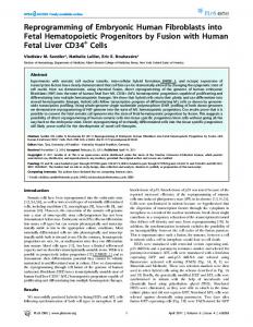
FIGURE 1. Schematic approach to generate T cells from hESC. The H1 hESC line is kept in an undifferentiated state on MEFs (Step 1). An image of two hESC colonies growing on MEFs is shown in A. In Step 2, hESC colonies are fragmented and incubated on OP9 cells to allow hematopoietic development, which is confined to HZs. An image of a small HZs at day 12 is shown in B (arrow). Immunohistochemistry analysis for CD34 expression in a HZs shows central CD34⫹ round cells (arrowhead) surrounded by flat CD34⫹ cells (arrow) C. After 10 –12 days of culture on OP9, the HZs are picked up and transferred in toto onto an OP9-DL1 monolayer (Step 3). A HZs incubated for 7 days on OP9-DL1 cells is shown in D and a magnification of the inbox in D is shown in E. Numerous small round cells compacted together in a HZs are depicted in E (asterisk). After 7 days of culture on OP9-DL1 cells, the HZs are mechanically disrupted to release and incubate the HPC on fresh OP9-DL1 monolayers for another 4 to 6 wk (Step 4 and image F). Scale bars, 100 m.
Furthermore, we show that the T cells are generated from CD34highCD43low cells present in these HZs, suggesting that these cells contain multipotent HPC. Using the approach described in this study, T cell differentiation from hESC is now easily amenable to manipulation and the principles governing differentiation can be studied. In addition, generation of T cells from hESC may be clinically relevant, especially when autologous cells with similar characteristics, such as induced pluripotent stem cells (iPS cells) (12), can be used.
Materials and Methods Cell line cultures All experiments were performed in accordance with the guidelines of the Medical Ethical Commission of Ghent University Hospital (Belgium). For this study, the hESC line H1 (National Institutes of Health code: WA01) was used (Passage 30 – 60). The hESC line was kept in an undifferentiated state on MEFs as previously described (1). The H1 cell line displayed stable growth, colony morphology, karyotype, and expression of surface markers such as SSEA-3 and SSEA-4 (data not shown). OP9 cells and OP9-DL1 cells (a gift from Juan Carlos Zu´n˜igaPflu¨cker, University of Toronto, Canada) were cultured as previously described in MEM-␣ with 20% FCS and supplemented with L-glutamine and the antibiotics streptomycin and penicillin (all from Invitrogen) (13).
Generation of HZs from hESC and transfer onto OP9-DL1 To differentiate hESC into CD34⫹ cells, the protocol from Vodyanik et al. (3) was adapted. In brief, subconfluent hESC cultures were mechanically fragmented into small clumps using glass beads (SigmaAldrich) and incubated on overgrown OP9 cells in six-well plates pre-
coated with 0.1% gelatin (Sigma-Aldrich). To estimate the number of hESC incubated on OP9 cells, hESC cultures grown from the same split/passage were used to obtain single cell suspension for counting. An average of 2 ⫻ 105 hESC were incubated per well of a six-well plate. After 10 –12 days of culture on OP9 cells, HZs were picked up under microscopic inspection using a small scalpel and pipette. The pooled HZs (average of 5,000 CD34⫹CD43⫹ cells) were transferred onto subconfluent OP9-DL1 cultures in 12-well plates with MEM-␣, 20% FCS, and supplemented with fms-like tyrosine kinase 3 receptor ligand (FLT3-L: 5 ng/ml; PeproTech), rhIL-7 (5 ng/ml; R&D Systems), and 10 ng/ml stem cell factor (SCF) (Amgen). After 5–7 days of culture, the HPC within the HZs were released by gently pipetting and incubated on a fresh OP9-DL1 monolayer for up to 6 wk. Every 3 days, half of the medium was changed, and every 5–7 days, cells were transferred on a fresh OP9-DL1 monolayer. For the kinetic studies in Figs. 3K and 4B, a large number of HZs from OP9 cultures were pooled together. To estimate the number of CD34⫹ CD43⫹ within this HZs pool, half of the collected HZs were dissociated and counted with a flow cytometer. An average of 5000 CD34⫹CD43⫹ cells were incubated in each quantitative experiment. Microscopic images were recorded with a CCD camera (Moticam 480, Motic) using phasecontrast microscopy (Leica Microsystems).
Flow cytometry and cell sorting For cell analysis, the cells were mechanically dissociated without use of enzymes. Before adding Ab, FcR blocking was performed using human IgG (Miltenyi Biotec). FACS analysis was performed on a FACSCalibur and LSR II (Becton Dickinson Immunocytometry Systems). Cell sorting was performed with FacsVantage cell sorter (BD Biosciences). For cell sorting, CD34highCD43low and CD34lowCD43high cells were gated as indicated in Fig. 4A and 5000 cells of each subset were used in each experiment. For CD34highCD43low CD7⫹/⫺ or CD34highCD43low CD45⫹/⫺ subsets, 3000 cells of each subset were
The Journal of Immunology
6881
FIGURE 2. FACS analysis of differentiating hESC on OP9 and OP9-DL1 cells. A representative FACS analysis of HZs generated after 12 days on OP9 cells is shown in A (n ⫽ 4). Four cell populations are gated: R1 (13% of the cells), R2 (2%), R3 (3%), and R4 (3%). An extensive analysis of the R2 and R3 gated CD34⫹CD43⫹ fractions is shown for the indicated cell markers. A more detailed analysis of all populations gated in A is available in supplemental Table S1. The fraction of CD34highCD43low and CD34lowCD43high cells within the total CD34⫹CD43⫹ cells after 10 –12 days on OP9 cells was very constant: 30 to 40% for CD34highCD43low cells and 60 to 70% for the CD34lowCD43high cell fraction. B, Representative FACS analysis of non-hematopoietic zones (Non-HZs) generated from hESC after 12 days on OP9 cells (n ⫽ 6). In C, a representative FACS analysis on transferred HZs at day 6 of culture on OP9-DL1 cells is shown (n ⫽ 3).
sorted using stringent gates as shown in Fig. 5. The results were analyzed using CellQuest Software or FACSDiva version 6.1.1 (BD Biosciences). The following (conjugated) anti-human mAbs were used: CD1a-allophycocyanin; CD3-allophycocyanin,-PE,-FITC,-allophycocyanin-Cy7; CD4-allophycocyanin,-PE,-FITC,-PE-Cy7; CD7-allophycocyanin,-PE,-FITC,-biotinylated; CD8␣-allophycocyanin; CD8␣-PE; CD8-PE,-allophycocyanin; CD10-PE; CD10-PECy7, CD11b-PE; CD11c-PE; CD14-PE; CD15-PE; CD16-PE; CD19-PE; CD26PE; CD27-PE,-biotynilated; CD31 (PECAM-1)-PE,-FITC; CD33-PE; CD34-allophycocyanin, -PE, -FITC; CD36-PE; CD38-PE; CD41a-PE; CD43-FITC; CD45RA-allophycocyanin,-PE; CD45-allophycocyanin,PE,-FITC; CD54 (ICAM-1)-PE; CD56-allophycocyanin,-PE, FITC,PE-Cy7; CD66-PE, CD80-PE; CD90-PE; CD106 (VCAM-1)-PE, CD117-PE (c-kit),-biotinylated; CD122 (IL-2R)-PE; CD123 (IL-3R)PE; CD127 (IL-7R)-PE; CD132-PE; CD133-PE; CD135-PE; CD144 (VE-Cadherin)-PE; CD146 (MUC-18)-PE CD161-PE; CD184 (CXCR4)-PE; CD235A (GlycophorinA)-PE; CD309 (KDR/vascular endothelial growth factor receptor (VEGFR)-2)-PE; SSEA1-PE, SSEA3-PE, SSEA4-PE, and TCR-PE. The mAbs were purchased from BD Pharm-
ingen, Beckman Coulter, eBioscience, Miltenyi Biotec, and R&D Systems. The streptavidine labels (SA-PE-Cy5.5, SA-allophycocyaninCy7) were purchased from eBioscience. The mAbs V-2,-5,-7,-12,-13,17,-22 were purchased from Beckman Coulter.
Real time RT-PCR Real-time RT-PCR analysis was performed on bulk cultures in the CD4⫹CD8⫹ double positive (DP) stage (day 30 on OP9-DL1). RNA was extracted using the RNease Mini Kit (Qiagen). cDNA was synthesized using Superscript First Strand synthesis system (Invitrogen). Primers were designed using Primer Express 3.0 software (Applied Biosystems) and manufactured by Operon. Gene and cDNA specificity of the primers was checked using NCBI-BLAST and GENATLAS, available on the internet. The following primer pairs were used: GAPDH sense: TCCTCTGACT TCAACAGCGACA; GAPDH antisense: GTGGTCGTTGAGGGCAATG; RAG-1 sense: CCTGCTGAGCAAGGTACCTCA; RAG-1 antisense: ATCTGGGGCAGAACTGAGTCC; RAG-2 sense: TGGTTTAGCGG CAAAGATTCA; RAG-2 antisense: AGTTTCTGATGGTACGTAGA
6882
T CELLS GENERATED FROM hESC
FIGURE 3. Developmental kinetics of hESC into T lineage cells. A–I, Representative FACS analysis of differentiating T lineage cells on OP9-DL1 at different time points for the indicated cell markers (n ⫽ 6). The graph in J shows the relative mRNA expression levels of RAG-1, RAG-2, and pT␣ in OP9-DL1 cells, hESC-derived endothelial cells (EC), hESC-derived T lineage cells (hESC/T cells) (d 30 on OP9-DL1), and human thymus as a positive control. GAPDH is used as a housekeeping gene and is set at 100. The result shown represents three independent experiments. In K, the expansion kinetics of hESC-derived T lineage cells is shown in absolute counts in a logarithmic scale, starting from a total of 5000 CD34⫹CD43⫹ cells (day 12 on OP9) incubated on OP9-DL1 monolayers at day 0. The result shown is a representative example for three independent experiments. TTTTTGTC; pT␣ sense: GCCCATGCATCTGTCAGGAG; pT␣ antisense: ATCCACCAGCAGCATGATTG. PCR reagents and SYBR Green I mastermixes were obtained from Eurogentec and used according to the manufacturer’s instructions. The reactions were run on an ABI Prism 7300 Sequence Detection System (Applied Biosystems). The following cycling conditions were used: 10 min at 95°C, 40 cycles at 95°C for 15 s, and 60°C for 60 s. After amplification, a melting curve was generated for every PCR product and compared with melting curve of positive control cells (human thymus). Negative control cells included undifferentiated hESC, the murine OP9-DL1 cells, and CD31⫹ CD146⫹CD45⫺ EC generated in vitro from hESC. The relative expression was calculated for each gene by the ⌬Ct method.
GeneScan analysis DNA was purified according to the manufacturer’s instructions (GenElute Mammalian Genomic DNA Miniprep Kit, Sigma-Aldrich) from bulk cultures at day 30 of coculture on OP9-DL1 (n ⫽ 3). The GeneScan was performed by two multiplex PCR as previously described by the BIOMED-2 Concerted Action (14). Each analysis was performed in duplo, including hESC and adult PBMC as negative and positive control, respectively.
IMDM (Invitrogen), supplemented with 10% FCS. After 4 days, IL-2 (40 U/ml) (BD Pharmingen) was added and refreshed every 4 days. The cells were restimulated every 12 days using the same protocol. To test for cytoplasmatic IFN-␥ production, hESC-derived T cells were subsequently incubated on anti-CD3 coated 96-wells, and soluble anti-human CD28 and anti-human CD49d mAbs, according to the protocol of the supplier (BD Pharmingen). After 1 h, GolgiPlug (BD Pharmingen) was added and after an additional 5 h, cells were analyzed with FACS LSR II (BD Pharmingen). Because the H1 cell line expresses HLA-A2, a gate was set on the HLA-A2⫹ cells for analysis. Staining for cytoplasmatic IFN-␥ was performed according to the manufacturer’s instructions (Cytofix/Cytoperm kit; BD Pharmingen) with anti-human IFN-␥-FITC or FITC Mouse IgG1, Isotype Control (BD Pharmingen) and anti-human CD3-PE (BD Pharmingen).
Statistical analysis A Mann-Whitney U test was performed with SPSS 15.0 software, and a p-value ⬍0.05 was considered as statistical significant.
Results
Functional testing of T cells
hESC generate hematopoietic zones on OP9 cells containing CD34high CD43low HPC
hESC-derived T cells were expanded on a feeder mixture consisting of irradiated JY cells and adult HLA-A2 negative PBMC cells (40 and 50 Gy, respectively) and stimulated with PHA (1 g/ml; Sigma-Aldrich) in
Several stromal cell lines have been shown to support hematopoiesis from hESCs, but the OP9 cell line was shown by Vodyanik et al. (3) to be particularly well suited for the generation of CD34⫹
The Journal of Immunology
6883
FIGURE 4. hESC-derived CD34high CD43lowcells generate T lineage cells. The gated R2 (CD34highCD43low) and R3 (CD34lowCD43high) populations generated on OP9 cells in A (day 12 on OP9) are sorted on OP9-DL1 monolayers (5000 cells each) to allow T cell differentiation. After 30 days of culture on OP9-DL1, FACS analysis on both sorted populations (arrows) is performed to detect CD4⫹CD8⫹ DP and CD3⫹TCR␣⫹ or CD3⫹TCR␥␦⫹ cells. The analysis shown is representative for three independent experiments. In B, 5000 CD34⫹CD43⫹ cells (day 12 on OP9) are incubated on OP9DL1 monolayers and supplemented with SCF (10 ng/ml) (SCF⫹) or without stem cell factor (SCF⫺). The absolute cell number of each cell subset is depicted on a logarithmic scale after 30 days of culture on OP9-DL1 cells. The data shown represent three independent experiments.
hematopoietic cells. hESC colonies (Fig. 1, Step 1) were fragmented into small clumps and incubated on OP9 monolayers to allow hematopoietic development (Fig. 1, Step 2). During the hESC/OP9 coculture period, well-organized two-dimensional structures were formed that strongly mimicked blood islands, which arise during normal embryonic development (15) (Fig. 1B). These blood island-like structures consisted of a central core of mostly CD34⫹ round cells surrounded by a rim of CD34⫹ flat endothelial-like cells (Fig. 1C). Similar structures were recently described as ES-sacs and were shown to contain precursors for megakaryocytes (11). We could demonstrate by flow cytometric analysis the presence of CD34⫹CD43⫹ HPC cells within the blood island-like structures that were mechanically picked up from the hESC/OP9 cultures (Fig. 2A), but not within areas that did not display such structures (Fig. 2B). This indicates that all CD34⫹ CD43⫹ HPC cells generated on the OP9 cells were confined to these structures. Therefore, we referred to these structures as HZs (Fig. 1, B and C). Based on the expression of CD43 and CD34, distinct cell populations can be identified within the HZs at day 10 –12 on OP9 cells (Fig. 2A, gate R1-R4). An extensive flow cytometric analysis of the gated populations is shown in supplemental Table S1.4 We could confirm, as was previously described (6), that the R1 CD34highCD43⫺ population depicted in Fig. 2A comprises CD31⫹CD146⫹CD45⫺ endothelial cells, whereas the CD34⫺CD43⫹ cells (R4 in Fig. 2A) represents CD41⫹ GlycA(CD235A)⫹ erythro-megakaryocytic lineage cells (supplemental Table S1 and data not shown). We consistently detected two major CD34⫹CD43⫹ cell populations: a CD34highCD43low cell population (R2 in Fig. 2A) that expressed variable levels of CD45 and c-kit (CD117), and a CD34lowCD43high cell subset (R3 in Fig. 2A) which was mostly devoid from CD45 and c-kit expression. The kinetics of appear4
The online version of this article contains supplementary material.
ance of the R2 population in the hESC OP9 coculture system is shown in supplemental Fig. 1A. B cell and myeloid lineage markers were absent (supplemental Table S1). Although CD56 was expressed on the majority of CD34highCD43low cells, we do not consider this evidence for NK commitment but rather as a remnant of CD56 expression by undifferentiated hESC. The fact that CD34⫹CD43⫹ cells were negative for the early NK lineage marker CD161 supports this interpretation (supplemental Table S1). Both CD34⫹CD43⫹ cell subsets expressed the lymphoid lineage marker CD7, albeit at a different frequency and intensity, but expression of cytoplasmic CD3 (cyCD3) within these cell populations was never detected (Fig. 2A). These phenotypical data are compatible with the CD34highCD43low population being an uncommitted HPC population.
Transfer of HZs on OP9-DL1 cells generates early T lineage precursors The crucial role of Notch signaling in hematopoietic cell fate decisions and T lymphocyte development in particular has been well established (13, 16, 17). Therefore, we picked up the HZs that contain CD34highCD43low HPC and transferred them in toto onto OP9-DL1 monolayers to allow further HPC development and T lineage commitment (Fig. 1, Step 3) (n ⫽ 9). Approximately 5–7 days following transfer onto the OP9-DL1 cells, the HZs/OP9DL1 cocultures (Fig. 1, D and E) were disrupted mechanically to release the hESC-derived cells. Flow cytometric analysis at that stage showed the typical CD34highCD45⫹ HPC phenotype. The cells no longer expressed CD43, but coexpressed c-kit, a marker known to be present on HPC and early T cell precursors (Fig. 2C). Expression of cyCD3 was detected within CD34highCD45⫹ c-kit⫹CD7⫹ cells (R2 in Fig. 2C), but not in CD7⫹CD34⫺ (CD45⫺) cells (R3 in Fig. 2C), suggesting T/NK lineage commitment of these CD34⫹ cells on OP9-DL1 monolayers (18). Importantly, prolonged culture of the HZs on the initial OP9 cells
6884
T CELLS GENERATED FROM hESC
FIGURE 5. Expression of CD7 and CD45 on T lineage precursors within the hESC-derived CD34highCD43lowcells. Day 12 HZ were labeled and CD43⫹ cells were sorted using the gates indicated in the upper dot plot. A total of 3000 sorted cells of each population were subsequently cultured on OP9-DL1 cells as described in material and methods and 30 –35 days later cells were analyzed for T cell lineage markers. A, Cells were sorted for CD7⫹ and CD7⫺ CD34highCD43low cells. B, Cells were sorted for CD45⫹ and CD45⫺ CD34highCD43low cells. The data shown represent three independent experiments.
or following their transfer onto fresh OP9 monolayers did not sustain (or generate) CD34highCD45⫹CD7⫹ cells, and CD45⫹CD15⫹ myelo-monocytic cells developed instead (supplemental Fig. S1, B and C). These findings indicate that DL1 triggers HPC within the HZs and specifically promotes the generation of early CD34high CD45⫹CD7⫹c-kit⫹cyCD3⫹ T/NK progenitors. Furthermore, incubation of “Non-HZs” on OP9-DL1 did not yield CD45⫹ cells, again confirming that HPC were confined to the HZs (supplemental Fig. S1D). hESC-derived HZs give rise to CD4⫹CD8⫹ DP and TCR␣⫹ and TCR␥␦⫹ cells After 14 days of coculture on OP9-DL1, most of the CD7⫹CD45⫹cyCD3⫹ cells had down-regulated CD34 expression. These CD7⫹ cells consisted of cyCD3⫹CD56⫹CD45⫹ NK lineage cells (supplemental Fig. S1E) and a significant cyCD3⫹CD5⫹ subpopulation (Fig. 3, A–D). The first CD4 single positive cells and CD4⫹CD8␣␣⫹ DP T lineage cells were detected within the cyCD3⫹CD5⫹ cell population after 14 days, suggesting that this CD5⫹ population contains T-committed precursors (Fig. 3E). After 21 days, CD4⫹CD8␣⫹ DP cells emerged in the cultures and on day 28, DP cells were prominent (Fig. 3, F and G).
Past day 30, the proportion of CD3⫹TCR␣⫹ cells in these cultures ranged between 15 and 50% of all human cells (Fig. 3, H and K). In addition to CD3⫹TCR␣⫹ cells, CD3⫹TCR␥␦⫹ appeared in all experiments (Fig. 3, I and K) (n ⫽ 9). In Fig. 3K, it is shown that 5000 HPC expand to about one hundred times that number of DP cells. At the mRNA level, pT␣, RAG-1, and RAG-2 became detectable with the appearance of DP cells (Fig. 3J), supporting the notion that T cells were generated de novo in these cultures. These data clearly show that hESC can differentiate to T lineage cells in vitro. Furthermore, the developmental pathway of these cells appears to be similar to thymic development (19). A hESC-derived CD34highCD43low cell population generates T lineage cells Next, we investigated which CD34⫹ subset in the HZs contains the T lineage precursors. Because the CD34highCD43low cell population described above has a phenotype reminiscent of adult HPC, we hypothesized that the T lineage cells generated in our system may develop from this population. Indeed, cell sorting experiments clearly demonstrated that the CD34highCD43low cells differentiated into T lineage cells when transferred on OP9-DL1 monolayers,
The Journal of Immunology
6885
FIGURE 6. hESC-derived T cells are polyclonal and functionally mature. A, Representative GeneScan analysis of TCR gene rearrangements (D-J and V-J rearrangements at the TCR locus) in hESC-derived T lineage cells (hESC/T cells) (day 30 on OP9-DL1 cells). hESC served as a negative control, whereas PBMC served as a positive control (n ⫽ 3). The pattern obtained with the primers for the J1 cluster and that obtained with the primers for the J2 cluster are shown in green and blue, respectively. As shown, hESC-derived T lineage cells and PBMC show similar polyclonal patterns whereas hESC do not display TCR gene rearrangements. B, Representative FACS analysis of the V repertoire, gated on hESC derived CD3⫹TCR␥␦⫺ cells (R1), using family specific TCR V Abs. C, Representative dot plot gated on CD3⫹ hESC-derived T cells after 40 days of coculture on OP9-DL1 cells. D and E, Representative histogram analysis for cytoplasmatic IFN-␥ (E) and isotype control (D) on anti-CD3 stimulated hESC-derived T cells (E) (n ⫽ 3).
whereas the CD34lowCD43high cells did not yield T lineage cells (Fig. 4A) (n ⫽ 3). Because day 10 –12 HZs not only contain CD43⫹ cells, but also CD43⫺ cells, we investigated whether the CD43⫺ cells also have T lymphoid potential. Cell sorting experiments did not reveal T lineage potential of CD43⫺ cells (data not shown), confirming that CD43⫹, but not CD43⫺ cells bear hematopoietic potential in the hESC/OP9 system at that developmental stage. Because the CD34highCD43low cell population is endowed with T cell potential and coexpresses the receptor c-kit, we reasoned that adding the c-kit ligand SCF might improve the generation of T lineage cells from hESC in our system. When adding SCF (10 ng/ml) to the OP9-DL1 cocultures, a dramatic increase in the absolute (⬎80-fold) and relative number (⬎14-fold) of CD4⫹ CD8⫹ DP cells was observed compared with cultures without SCF (Fig. 4B; p ⬍ 0.05). Moreover, a ⬎180-fold and 15-fold absolute increase of hESC-derived CD3⫹TCR␣⫹ and CD3⫹ TCR␥␦⫹ cells, respectively, was observed in the SCF-supplemented cultures (Fig. 4B; p ⬍ 0.05) (n ⫽ 3). CD10 was reported to mark early lymphoid committed cells present in bone marrow, blood, and thymus (20, 21). We therefore analyzed whether CD10⫹ cells could be detected in HZ. However, no CD10 expression could be observed on CD34highCD43low precursor in days 10 –12 HZ (supplemental Table S1, supplemental Fig. S2A). We subsequently analyzed whether such CD10⫹ cells
were generated from CD34highCD43low cells on OP9-DL1. Transferred cells remained consistently negative for CD10 expression (supplemental Fig. S2A), suggesting that the induction of CD10 on CD34⫹ cells requires signals that are not present in OP9-DL1 cultures. Acquisition of the surface marker CD7 on CD34⫹ HPC has been shown to be one of the earliest steps in T cell ontogeny (19, 21). To address whether the T lineage cells in our system are derived from CD34highCD43lowCD7⫹ subsets generated on OP9 cells, we compared the fate of cell sorted CD34highCD43lowCD7⫹ and CD34highCD43lowCD7⫺ cells following their transfer on OP9DL1 cells. These experiments (n ⫽ 3) clearly demonstrate that T lineage cells develop from hESC-derived CD34highCD43lowCD7⫺ cells, but not from CD34highCD43lowCD7⫹ cells generated on OP9 cells (Fig. 5A). However, as shown in Fig. 3, A–D, after transfer of CD34highCD43low cells on OP9-DL1 feeders, cells rapidly acquire CD7 and become CD7highcyCD3⫹ cells, which give rise to the later stages of T cell development (Figs. 2C and 3). This suggests that expression of a marker by itself does not signify lymphoid commitment. In the hESC/OP9 system, it was shown that CD34⫹CD43⫹ CD45⫺ precursors represent early HPC with lympho-myeloid potential (6). Acquisition of the surface marker CD45 on these cells (yielding CD34⫹CD43⫹CD45⫹ cells) was accompanied by loss of B-lymphoid potential and progressive myeloid commitment. We
6886 therefore assessed whether acquisition of CD45 was accompanied with loss of T cell lineage potential. Both CD34highCD43low CD45⫺ and CD34highCD43lowCD45⫹ subsets were sorted (n ⫽ 3) and cultured on OP9-DL1 to generate T lineage cells. Both cell populations generated DP cells and CD3⫹ T cells (Fig. 5B). The absolute cell number of CD4⫹CD8␣⫹ DP, CD3⫹TCR␣⫹, and CD3⫹TCR␥␦⫹ cells generated from CD34highCD43low CD45⫹ cells was 4-, 3-, and 2-fold higher compared with the respective populations generated from CD34highCD43low CD45⫺ cells ( p ⬍ 0.05). hESC-derived T lineage cells are polyclonal and functionally mature In the thymus, T cells are generated in a polyclonal fashion to allow the recognition of a broad range of Ags via the TCR. The genetic loci encoding the TCR undergo sequential genomic rearrangements, with randomly chosen variable (V), diversity (D), and joining (J) segments being assembled to form a variable TCR repertoire (14). To test whether the hESC-derived T lineage cells display a polyclonal repertoire, we performed a GeneScan analysis. The data in Fig. 6A show diverse and random D-J and V-DJ rearrangements at the  locus, indicating a polyclonal T lineage repertoire at the DNA level. Furthermore, flow cytometry confirmed a diverse and random V family usage at the protein level (Fig. 6B). Therefore, these data provide strong evidence that the hESCderived T lineage cells follow a stochastic pattern of gene rearrangement and do not result from the outgrowth of a single or a few T cells. Maturation status of the CD3⫹ T cells was assessed both phenotypically and functionally. T cells develop into phenotypically mature cells as defined by the up-regulation of CD27 and downregulation of CD1a (22, 23). Similar to thymic differentiation, hESC-derived T cells developed into a mature CD27⫹CD1a⫺ phenotype (Fig. 6C) (n ⫽ 9), suggesting that functionally mature cells are present. After stimulation with PHA, hESC-derived T cells proliferated and increased a 2500-fold (supplemental Fig. S2B). All CD3⫹ cells had the mature CD27⫹CD1a⫺ phenotype and both TCR␣ and TCR␥␦ positive cells were expanded (data not shown). Moreover, when these expanded cells were restimulated, IFN-␥ production could be demonstrated by flow cytometry (Fig. 6, D and E), demonstrating that these cells have acquired functional characteristics of mature T cells.
Discussion We report in this study that hESC have the potential to differentiate in vitro into T cells. To achieve this, hESC were first differentiated on OP9 cells to CD34highCD43low HPC cells in HZs. Before these cells had lost multipotency and had “spontaneously” differentiated to myeloid cells, days 10 –12 HPC cells were transferred to a DL1 environment in the presence of SCF, fms-like tyrosine kinase 3 receptor ligand, and IL-7. Under these conditions, cells became gradually committed to the T/NK lineage and generated large numbers of T cells. T cells displayed both the TCR␣ and TCR␥␦ phenotype, and were functional and polyclonal. HPC were generated on OP9 cells exclusively in hematopoietic sites, which we referred to as HZs. For these HZs to form, it is essential that the hESC/OP9 cell cultures are, apart from medium changes, not manipulated until these HZs are fully established. Similar structures were recently described by two other groups. Ledran et al. (5) described “endothelial vessel structures” that emerged on mitotically inactivated primary or established stromal cell lines from murine aorta-gonad-mesonephros and fetal liver. The developmental kinetics of these structures were slower than reported in this study, as they were first visible on day 18. How-
T CELLS GENERATED FROM hESC ever, this coincided with the peak of CD34⫹ cell numbers and hematopoietic precursor activity. Takayama et al. (11) designated similar hematopoietic sites as ES-sacs. The ES-sacs were obtained after coincubation of hESC with OP9 or C3H101/2 stromal cells and appeared around day 15 of culture. The ES-sacs were reported to be lined by endothelial cells as suggested by their morphology and CD31⫹ VEGFR-2 (CD309)⫹ UEA-1⫹ phenotype and to contain round CD31⫹ cells with some expression of CD34, VEGFR-2 (CD309), CD41a, and CD45. Both these papers report either a much higher yield in NOD/SCID repopulating activity (5) or in platelet production (11) compared with hESC differentiation protocols that do not allow the generation of these structures, suggesting that these structures are very efficient in the differentiation of ESC to HPC and/or proliferation of HPC. Recently, Galic et al. (8, 9) reported on the generation of T cells from hESC. hESC were predifferentiated to CD34⫹ cells on OP9 cells or using a serum-free embryoid system, and it was demonstrated that after in vivo injection of CD34⫹ cells in SCID-hu thymus, CD3⫹ and CD4⫹CD8⫹ DP cells were generated. In this study, we confirm and extend this observation by showing that these HZs also efficiently generate precursors for T cells. It is unclear whether at any given time hematopoietic stem cells (HSC), defined as multipotent, self renewing cells that express certain membrane markers such as CD34, CD45, and CD133, are present in the HZs generated on OP9 cells. Cells having identical membrane markers as BM stem cells are not stably present in OP9 cultures. On the one hand, cells with hematopoietic precursor activity seem to partially retain membrane molecules of their hemogenic or hemangioblastic precursors, such as VE-cadherin and VEGFR-2 (supplemental Table S1) (15). This is different from the BM stem cell compartment, where hematopoietic stem cells are believed to be no longer continuously generated from precursors and are devoid of these markers (24, 25). In contrast, they acquire membrane markers such as CD45, which are expressed on all postnatal HSC/HPC. However, acquisition of CD45 in hESC/OP9 cultures was reported to be associated with loss of lymphoid precursor capacity, suggesting that differentiation to CD34highCD45⫹ cells may be preceded by loss of multipotency in the OP9 culture system (6). In our hands, however, cell sorting experiments showed that both CD45⫹ and CD45⫺ fractions of the CD34highCD43low population have T cell potential. Furthermore, when CD34high CD43low cells were transferred to OP9-DL1 cultures, CD34high CD45⫹ cells were generated, which seem to have retained lymphoid differentiation potential. Vodyanik et al. (6) performed an extensive functional analysis of the CD34highCD43low cell population. They showed that these cells had CFU-GEMM, CFU-M, CFU-GM and some CFU-E activity. In addition, after MS5 differentiation cocultures they could show NK generation and expression of early B lineage specific markers, suggesting that these cells also contain NK and B cell precursor activity. In this study, we show that this same population can generate functionally mature TCR␥␦ and TCR␣ cells. Therefore, it seems that precursor activity for all hematopoietic lineages is present within this CD34highCD43low population. Because to our knowledge, clonal analysis was never performed, it is not known whether a single multipotent HSC or rather a mixture of committed cells is responsible for the observed phenomena. Self renewal of human HSC, a characteristic feature of stem cells, is usually addressed by injecting these cells in NOD/SCID mice. Several reports have shown that hESC-derived HPC can repopulate NOD/ SCID mice and that cells from the BM of these reconstituted mice can be used successfully for secondary transfer, suggesting that self renewing cells are present in the hESC-derived HPC (5, 26).
The Journal of Immunology However, as discussed by the authors, expression of adult globin in red lineage cells was absent, and definitive proof of lymphoid engraftment was lacking (5). Therefore, to the best of our knowledge, formal proof of the existence of hESC-derived HSC is still lacking. Based on the above discussion, we hypothesize that multipotent hematopoietic stem cells with self renewing capacity are transiently present in the HZs or ES-sacs and that the exact phenotype of these CD34highCD43low cells may vary and/or depend on the duration of culture. We have shown in this study that ESC-derived HPC can generate T cells when transferred at the right time to an environment that expresses DL1. However, in a report of Martin et al. (7), predifferentiated hESC cells on the S17 stromal lines were devoid of T cell precursor activity tested in coculture with OP9-DL1 and in fetal thymic organ culture in vitro. Whether this relates to the specific characteristics of the hESC line used, or the developmental time window by which CD34 are harvested from the stromal cell line is unclear. Nevertheless, in our hands, hESC-derived CD34⫹ cells generated on the S17 cell line after 10 –12 days, displayed similar T cell differentiation capacity compared with hESC-derived CD34⫹ cells generated on OP9 (supplemental Fig. S3). CD7 has been used extensively for the identification of lymphoid precursors within CD34⫹ HPC. Such cells were found in fetal BM and in cord blood (27). More recently, these populations were studied in the thymus: it was found that CD34⫹CD7⫺ cells are multipotent, CD34⫹CD7int cells are lymphoid precursors including B cell precursor activity and the CD34⫹CD7high population consists of T/NK precursors (21). In contrast with these findings, we found CD7 in the HZs (step 2 cultures) not to be a good marker of lymphoid commitment: CD7 was preferentially expressed on R3 cells (Fig. 2A), which do not generate T or NK cells. In addition, sorted CD34⫹CD43⫹CD7⫹ R2 cells were unable to generate T cells. We therefore believe that CD7 expression in the HZs is “aberrant”, similar to CD56 expression. However, after transfer of CD34⫹CD43⫹ cells on OP9-DL1 stromal cells, cells rapidly acquire CD7 and become CD7high. CD7 expression is then accompanied with another T/NK lymphoid marker, i.e., cyCD3. Although unproven, it is likely that these cells are the first cells which are lymphoid lineage committed and therefore in these cultures, cyCD3 appears to be a better marker. Once the cells are T/NK committed, differentiation follows the phenotypes as described in postnatal thymus. In contrast to postnatal thymopoiesis, CD1a expression is somewhat delayed as it first appears after, rather than before, CD4 expression and is therefore not the first marker for T commitment (data not shown). Similarly, CD1a disappears before the mature cells express CD27, rather than, as is observed in postnatal thymus, after CD27 expression. Interestingly, a similar expression pattern of CD1a is seen when fetal liver cells are differentiated to T cells on OP9-DL1 (data not shown). We believe the protocol as described in this study is an important new tool for future research and for the design of immunotherapy. The hESC/OP9 system allows the direct visualization of HZs and the generation of T cells from these structures. These in vitro observations may significantly improve our insights on the early steps in human developmental hematopoiesis. Indeed, in combination with genetic cell tracing strategies, the direct visualization of the HZs may allow the identification of the cellular participants and/or crucial gene expression at these hematopoietic sites (28). This system may also provide an opportunity in the field of immunotherapy where hESC-derived T cells could be engineered to express a TCR that recognizes a specific epitope expressed by malignant tumor cells (29). These engineered hESCderived T cells could then be used to target cancer. Although it is
6887 currently possible to generate such T cells from cord blood or BM from children, BM from older adults is less efficient in generating T cells (30). If the protocol described in this study could be applied to the differentiation of iPS cells, then these iPS cells would be the best source for the generation of tumor-specific T cells in older adults.
Acknowledgments We are indebted to Christiaan De Boever for performing art work. We also thank Dr. Inge Vanhaute (Red Cross Flanders, Oost-Vlaanderen, Belgium) for providing human blood samples, and Dr. Tom Boterberg for the irradiation procedures.
Disclosures The authors have no financial conflict of interest.
References 1. Thomson, J. A., J. Itskovitz-Eldor, S. S. Shapiro, M. A. Waknitz, J. J. Swiergiel, V. S. Marshall, and J. M. Jones. 1998. Embryonic stem cell lines derived from human blastocysts. Science 282: 1145–1147. 2. Zambidis, E. T., B. Peault, T. S. Park, F. Bunz, and C. I. Civin. 2005. Hematopoietic differentiation of human embryonic stem cells progresses through sequential hematoendothelial, primitive, and definitive stages resembling human yolk sac development. Blood 106: 860 – 870. 3. Vodyanik, M. A., J. A. Bork, J. A. Thomson, and I. I. Slukvin. 2005. Human embryonic stem cell-derived CD34⫹ cells: efficient production in the coculture with OP9 stromal cells and analysis of lymphohematopoietic potential. Blood 105: 617– 626. 4. Jokubaitis, V. J., L. Sinka, R. Driessen, G. Whitty, D. N. Haylock, I. Bertoncello, I. Smith, B. Peault, M. Tavian, and P. J. Simmons. 2008. Angiotensin-converting enzyme (CD143) marks hematopoietic stem cells in human embryonic, fetal, and adult hematopoietic tissues. Blood 111: 4055– 4063. 5. Ledran, M. H., A. Krassowska, L. Armstrong, I. Dimmick, J. Renstrom, R. Lang, S. Yung, M. Santibanez-Coref, E. Dzierzak, M. Stojkovic, R. A. Oostendorp, L. Forrester, and M. Lako. 2008. Efficient hematopoietic differentiation of human embryonic stem cells on stromal cells derived from hematopoietic niches. Stem Cell 3: 85–98. 6. Vodyanik, M. A., J. A. Thomson, and I. I. Slukvin. 2006. Leukosialin (CD43) defines hematopoietic progenitors in human embryonic stem cell differentiation cultures. Blood 108: 2095–2105. 7. Martin, C. H., P. S. Woll, Z. Ni, J. C. Zuniga-Pflucker, and D. S. Kaufman. 2008. Differences in lymphocyte developmental potential between human embryonic stem cell and umbilical cord blood-derived hematopoietic progenitor cells. Blood 112: 2730 –2737. 8. Galic, Z., S. G. Kitchen, A. Kacena, A. Subramanian, B. Burke, R. Cortado, and J. A. Zack. 2006. T lineage differentiation from human embryonic stem cells. Proc. Natl. Acad. Sci. USA 103: 11742–11747. 9. Galic Z., S. G. Kitchen, A. Subramanian, G. Bristol, M. D. Marsden, A. Balamurugan, A. Kacena, O. Yang, and J. A. Zack. 2009. Generation of T lineage cells from human embryonic stem cells in a feeder free system. Stem Cells 27: 100 –109. 10. Fleming, H. E., and D. T. Scadden. 2006. Embryonic stem cells make human T cells. Proc. Natl. Acad. Sci. USA 103: 12213–12214. 11. Takayama, N., H. Nishikii, J. Usui, H. Tsukui, A. Sawaguchi, T. Hiroyama, K. Eto, and H. Nakauchi. 2008. Generation of functional platelets from human embryonic stem cells in vitro via ES-sacs, VEGF-promoted structures that concentrate hematopoietic progenitors. Blood 111: 5298 –5306. 12. Takahashi, K., K. Tanabe, M. Ohnuki, M. Narita, T. Ichisaka, K. Tomoda, and S. Yamanaka. 2007. Induction of pluripotent stem cells from adult human fibroblasts by defined factors. Cell 131: 861– 872. 13. Schmitt, T. M., and J. C. Zuniga-Pflucker. 2002. Induction of T cell development from hematopoietic progenitor cells by ␦-like-1 in vitro. Immunity 17: 749 –756. 14. Dik, W. A., K. Pike-Overzet, F. Weerkamp, D. de Ridder, E. F. de Haas, M. R. Baert, P. van der Spek, E. E. Koster, M. J. Reinders, J. J. van Dongen, A. W. Langerak, and F. J. Staal. 2005. New insights on human T cell development by quantitative T cell receptor gene rearrangement studies and gene expression profiling. J. Exp. Med. 201: 1715–1723. 15. Cumano, A., and I. Godin. 2007. Ontogeny of the hematopoietic system. Annu. Rev. Immunol. 25: 745–785. 16. De Smedt, M., I. Hoebeke, and J. Plum. 2004. Human bone marrow CD34⫹ progenitor cells mature to T cells on OP9-DL1 stromal cell line without thymus microenvironment. Blood Cells Mol. Dis. 33: 227–232. 17. La Motte-Mohs, R. N., E. Herer, and J. C. Zuniga-Pflucker. 2005. Induction of T-cell development from human cord blood hematopoietic stem cells by Deltalike 1 in vitro. Blood 105: 1431–1439. 18. De Smedt, M., T. Taghon, I. Van de Walle, G. De Smet, G. Leclercq, and J. Plum. 2007. Notch signaling induces cytoplasmic CD3 epsilon expression in human differentiating NK cells. Blood 110: 2696 –2703. 19. Spits, H. 2002. Development of ␣ T cells in the human thymus. Nat. Rev. Immunol. 2: 760 –772. 20. Six, E. M., D. Bonhomme, M. Monteiro, K. Beldjord, M. Jurkowska, C. Cordier-Garcia, A. Garrigue, L. Dal Cortivo, B. Rocha, A. Fischer,
6888
21.
22.
23.
24. 25.
M. Cavazzana-Calvo, and I. Andre´-Schmutz. 2007. A human postnatal lymphoid progenitor capable of circulating and seeding the thymus. J. Exp. Med. 204: 3085–3093. Hao, Q. L., A. A. George, J. Zhu, L. Barsky, E. Zielinska, X. Wang, M. Price, S. Ge, and G. M. Crooks. 2008. Human intrathymic lineage commitment is marked by differential CD7 expression: identification of CD7- lympho-myeloid thymic progenitors. Blood 111: 1318 –1326. Vanhecke, D., G. Leclercq, J. Plum, and B. Vandekerckhove. 1995. Characterization of distinct stages during the differentiation of human CD69⫹CD3⫹ thymocytes and identification of thymic emigrants. J. Immunol. 155: 1862–1872. Vanhecke, D., B. Verhasselt, V. Debacker, G. Leclercq, J. Plum, and B. Vandekerckhove. 1995. Differentiation to T helper cells in the thymus: gradual acquisition of T helper cell function by CD3⫹CD4⫹ cells. J. Immunol. 155: 4711– 4718. ¨ . H. Yilmaz, and S. J. Morrison. 2005. CD144 (VE-cadherin) is tranKim, I., O siently expressed by fetal liver hematopoietic stem cells. Blood 106: 903–905. Timmermans, F., F. Van Hauwermeiren, M. De Smedt, R. Raedt, F. Plasschaert, M. L. De Buyzere, T. C. Gillebert, J. Plum, and B. Vandekerckhove. 2007.
T CELLS GENERATED FROM hESC
26.
27.
28.
29.
30.
Endothelial outgrowth cells are not derived from CD133⫹ cells or CD45⫹ hematopoietic precursors. Arterioscler. Thromb. Vasc. Biol. 27: 1572–1579. Tian, X., P. S. Woll, J. K. Morris, J. L. Linehan, and D. S. Kaufman. 2006. Hematopoietic engraftment of human embryonic stem cell-derived cells is regulated by recipient innate immunity. Stem Cells 24: 1370 –1380. Hao, Q. L., J. Zhu, M. A. Price, K. J. Payne, L. W. Barsky, and G. M. Crooks. 2001. Identification of a novel, human multilymphoid progenitor in cord blood. Blood 97: 3683–3690. Davis, R. P., E. S. Ng, M. Costa, A. K. Mossman, K. Sourris, A. G. Elefanty, and E. G. Stanley. 2008. Targeting a GFP reporter gene to the MIXL1 locus of human embryonic stem cells identifies human primitive streak-like cells and enables isolation of primitive hematopoietic precursors. Blood 111: 1876 –1884. van Lent, A. U., M. Nagasawa, M. M. van Loenen, R. Schotte, T. N. Schumacher, M. H. Heemskerk, H. Spits, and N. Legrand. 2007. Functional human antigenspecific T cells produced in vitro using retroviral T cell receptor transfer into hematopoietic progenitors. J. Immunol. 179: 4959 – 4968. Offner, F., T. Kerre, M. De Smedt, and J. Plum. 1999. Bone marrow CD34 cells generate fewer T cells in vitro with increasing age and following chemotherapy. Br. J. Haematol. 104: 801– 808.