1
Geometric Variability of the Scoliotic Spine Using Statistics on Articulated Shape Models Jonathan Boisvert, Farida Cheriet, Xavier Pennec, Hubert Labelle, Nicholas Ayache
Abstract— This paper introduces a method to analyze the variability of the spine shape and of the spine shape deformations using articulated shape models. The spine shape was expressed as a vector of relative poses between local coordinate systems of neighbouring vertebrae. Spine shape deformations were then modeled by a vector of rigid transformations that transforms one spine shape into another. Because rigid transforms do not naturally belong to a vector space, conventional mean and covariance could not be applied. The Fréchet mean and a generalized covariance were used instead. The spine shapes of a group of 295 scoliotic patients were quantitatively analyzed as well as the spine shape deformations associated with the CotrelDubousset corrective surgery (33 patients), the Boston brace (39 patients) and the scoliosis progression without treatment (26 patients). The variability of inter-vertebral poses was found to be inhomogeneous (lumbar vertebrae were more variable than the thoracic ones) and anisotropic (with maximal rotational variability around the coronal axis and maximal translational variability along the axial direction). Finally, brace and surgery were found to have a significant effect on the Fréchet mean and on the generalized covariance in specific spine regions where treatments modified the spine shape. Index Terms— Statistical Shape Analysis, Anatomical Variability, Rigid Transforms, Spine, Radiograph, Orthopaedic Treatment, Scoliosis
I. I NTRODUCTION Adolescent idiopathic scoliosis is a disease that causes a three dimensional deformation of the spine. As suggested by its name, the cause of the pathology remains unknown. Furthermore, the shape of a scoliotic spine varies greatly from a patient to another. Previous statistical studies (such as [1]–[4]) investigated the outcome of different treatments in term of the variation of clinical indices used by physicians to quantity the severity of the deformation. However, the variability of the spine geometry was not extensively studied. Two important reasons explain the limited number of studies interested in the geometric variability of the scoliotic spine: the availability of significant data and the lack of statistical tools Copyright (c) 2007 IEEE. Personal use of this material is permitted. However, permission to use this material for any other purposes must be obtained from the IEEE by sending a request to
[email protected]. Manuscript received August 15, 2006; revised March 25, 2007. Jonathan Boisvert and Farida Cheriet are affiliated with the École Polytechnique de Montréal, Montréal, Canada. Jonathan Boisvert, Xavier Pennec and Nicholas Ayache are affiliated with the INRIA, Sophia-Antipolis Cedex, France. Jonathan Boisvert, Farida Cheriet and Hubert Labelle are affiliated with the Sainte-Justine Hospital, Montréal, Canada. This work was funded by the Natural Sciences and Engineering Research Council (NSERC) of Canada, the Quebec’s Technology and Nature Research Funds (Fonds de Recherche sur la Nature et les Technologies du Québec) and the Canadian Institutes of Health Research (CIHR).
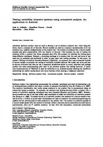
Fig. 1.
Some examples of scoliotic spine shapes from our patient database
to handle geometric primitives that are not naturally embedded in vector spaces. In the past, the vast majority of the studies analyzed the geometry of the spine using indices derived from the patient’s radiographs or from 3D reconstructions of his/her spine. Those indices were used to classify the spines’ curves [5]–[8] and also to compare the outcome of different orthopaedic treatments [1]–[4], [9]. The most popular index is certainly the Cobb angle [10], but there are several other indices such as the orientation of the plane of maximal deformity or the spine torsion [11]. Those indices have the advantage of enabling physicians to assess quickly and easily the severity of the scoliosis. However, they also present many problems. First of all, most clinical indices are global to the whole spine and thus do not provide spatial insight about the local geometry. Furthermore, most of the indices (including the Cobb angle) are computed on 2D projections, where a significant part of the curvature could be hidden (since the deformity is three–dimensional). To overcome those limitations, some authors investigated the scoliotic spine geometry or the effect of orthopaedic treatments by measuring the position and orientation of vertebrae. Those descriptors provide insights that are both local and geometric. Ghanem et al. [12] proposed a method to compute vertebrae translation and orientation during a surgery using an opto-electronic device. Unfortunately, it was a preliminary study and measures were only performed on a group of eight patients. Furthermore, translation and orientation were computed just for the vertebrae adjacent to the apex of the curvature. Sawatzky et al. [13] performed a similar study, but their goal was to find a relation between the number of hooks installed during a corrective surgery and the position and orientation of the vertebrae. This study was performed on a larger group of patients (32), which allowed the computation
2
of more advanced statistics. However, only the results for the apical vertebra were reported in their article. More recently, Petit et al. [14] compared inter–vertebral displacements in term of modifications of the center of rotation for two types of instrumentation used during corrective surgeries (Colorado and Cotrel-Dubousset). The patient sample used was larger than for previously cited studies (82 patients), which made statistically significant more subtle differences between the two groups of patients. Furthermore, results for all vertebrae were reported (not only vertebrae adjacent to the curve apex). However, the authors did not study extensively the variability of the displacement of the center of rotation or of the vertebrae rotations. One of the common limitations of those studies is that only mean values of the modifications of the positions or of the orientations of the vertebrae were extensively studied. An analysis of the mean spine shape using both orientations and positions of the vertebrae was never published. Furthermore, the variability of the spine anatomy was not studied in that context either. Unlike surgical treatments, braces effects were never analysed using the relative positions and orientations of the vertebrae. Usually, braces effects were analyzed primarily by measuring the Cobb angle (which is only a two–dimensional measure) of the major curve in the frontal plane [15], [16]. Some studies tried to take into account the three–dimensional nature of spine deformity by measuring the curve gravity in both the frontal and the sagittal plane [17]. However, repeating a 2D analysis twice is not a substitute for a true three-dimensional analysis. Finally, three-dimensional analysis of the brace effect was also conducted [18] by using a set of clinical indices extracted from three-dimensional reconstructions of the spine. However, those clinical indices were not independent, which made effect localization and analysis difficult. Moreover, brace effect variability itself was not studied. To overcome all these limitations, we propose to study the statistical variability of the spine shape and of the spine shape deformation using local features that describe both the position and the orientation of the vertebrae (i.e. rigid transformations). However, mathematical and computational tools need to be developed because conventional statistical methods usually apply under the assumption that the embedding space is a vector space where addition and scalar multiplication are defined. Unfortunately, this is not the case for rigid transformations. For example, the conventional mean is not applicable to rigid transformations since it would involve the addition of the measures followed by a division by the sample size. Recently, many researchers have been working towards the generalization of mathematical tools on Lie groups and Riemannian manifolds. A general framework for the development of probabilistic and statistical tools on Riemannian manifolds was recently proposed [19], [20]. Riemannian manifolds are more general than Lie Groups, thus findings realised on Riemannian manifolds also apply to Lie groups (and to rigid transformations by extension). Concepts such as the mean, covariance and normal distribution have been formalized for Riemannian manifolds. Many studies were realized more specifically for the tensors space, because of the develop-
ment of Diffusion Tensor Imaging (for example: [21]–[25] and references therein). The idea of computing statistics on manifolds was also used to perform anatomical shape analysis. For example, this idea can be found in our previous work [26] where an analysis of the spine shape anatomy based on Lie groups properties was proposed and in the work of Fletcher et al. [27] where a generalization of the PCA was introduced and applied to the analysis of medial axis representations of the hippocampus. However, a Riemannian approach to the study of articulated models of the spine shape was never used. To our knowledge, no previous work reported a variability analysis of the scoliotic spine shape and of the scoliotic spine shape deformations using rigid transformations as geometric descriptors. In that context, the contributions of this paper are: to introduce a new model of the variability of spine shapes and of spine shape deformations based on well posed statistics on a suitable articulated shape model, to suggest a method to compare the variability between different groups of patients, to propose a 3D visualization method of this variability and, last but not least, to present the resulting variability models computed using large groups of scoliotic patients. II. M ATERIAL AND M ETHODS This section presents the material and methods used to construct and analyze variability models of the spine shape. We will first describe the method used to create articulated models representing the spine from pairs of radiographs. The procedure used to create variability models from samples of articulated spine shapes will then be presented. Since our articulated shape models do not naturally belong to a vector space, conventional statistical methods could not be applied. However, it is still possible to define distances between articulated shape models. Therefore, some statistical notions had to be generalized based on the concept of distance between primitives. Riemannian geometry offers a good framework for this purpose. Thus, centrality and dispersion measures applied to Riemannian manifolds and their specialization to articulated models will be introduced. Finally, the visualization and the quantitative comparison of variability models built from articulated spine shapes will be discussed. A. Articulated Shape Models of the Spine from Multiple Radiographs Multi-planar radiography is a simple technique where two (or more) calibrated radiographs of a patient are taken to compute the 3D position of anatomical landmarks using a stereotriangulation algorithm. It is one of the few imaging modalities that can be used to infer the three-dimensional anatomy of the spine when the patient is standing up. Furthermore, biplanar radiography of scoliotic patients is routinely performed at Sainte-Justine hospital (Montreal, Canada). Thus, a large amount of data is available for analysis. In the case of bi-planar radiography of the spine, six anatomical landmarks are identified on each vertebra from T1 (first thoracic vertebra) to L5 (last lumbar vertebra) on a posterior-anterior and a lateral radiograph. The 3D coordinates of the landmarks are then computed and the deformation of
3
a high-resolution template using dual kriging yields 16 additional reconstructed landmarks. The accuracy of this method was previously established to 2.6 mm [28]. Once the landmarks are reconstructed in 3D, we rigidly registered registered each vertebrae to its first upper neighbour and the resulting rigid transforms were recorded. By doing so, the spine is represented by a vector of inter-vertebral rigid transformations S = [T1 , T2 , . . . , TN ] (see Fig. 2). This representation is especially well adapted to an analysis of the anatomical variability since the inter-vertebral rigid transformations describe the state of the physical links that are modified by the pathology and alter the shape of the whole spine. Most scoliotic patients are adolescents or pre-adolescents. Thus, spine length of patients afflicted by scoliosis varies considerably. In order to factor out that variability source from the statistical analysis, one could be tempted to normalize the articulated models. On the one hand, this could be desirable since the global spine size is associated primarily with patients’ growth and most physicians are more interested in analyzing the variability linked to the pathology. On the other hand, the development of many musculoskeletal pathologies, for instance adolescent idiopathic scoliosis, is tightly linked with the patient growth process. Thus, normalization could discard valuable information. Furthermore, preliminary experiments revealed that, with scoliotic patients, the only notable effect of normalization was found along the axial direction where the translational variability was almost eliminated (a reduction ranging from 3.0 mm2 in the thoracic region to 8 mm2 in the lumbar region). In summary, normalization could be desirable in certain situations, but it did not led to more probative results in our application. Thus, to preserve the clear physical interpretation of the variability models we chose not to normalize the articulated spine models. The vector S enables a local analysis of the links between the vertebrae. However, it is sometimes preferable to analyze the spine shape using absolute instead of relative transformations. For example, posture analysis is simpler when one uses absolute transformations. However, it is easy to convert S into an absolute representation Sabsolute using recursive compositions (where ◦ is the operator of composition). Sabsolute = [T1 , T1 ◦ T2 , . . . , T1 ◦ T2 ◦ . . . ◦ TN ]
(1)
The transformations are then expressed in the local coordinate system of the lowest vertebra. The choice of this reference coordinate system is arbitrary, but it can be changed easily based on the application needs. To study spine shape deformations caused by the progression of the pathology or by a treatment, we need to compute the “differences” between shape models. This can be realised once again using a vector of rigid transformations. Let S = [T1 , T2 , . . . , TN ] and S 0 = [T10 , T20 , . . . , TN0 ] be two vectors of rigid transformations extracted from two different radiological exams of the same patient (before and after a surgery, for instance), then another vector of rigid transformations can be defined with the transformations that turn the elements of S into the corresponding elements of S 0 (see Fig. 2).
... ...
Vertebra #3
T2
T2
∆T2 =T2 οT2-1 '
'
Vertebra #2
T1
T1
∆T1 =T1 ο T1-1 '
'
Vertebra #1
T0
World
T0
'
World
∆T0 =T0
'
ο T0-1
Fig. 2. Spine shape and spine shape deformation expressed using rigid transformations
The resulting vector ∆S will only depend on the difference between the two 3D spine geometries and not on the anatomy of the patient. ∆S = [∆T1 , ∆T2 , . . . ∆TN ] with ∆Ti = Ti0 ◦ Ti−1
(2)
Since this vector is still a vector of rigid transformations the analysis performed on S could also be performed on ∆S to study spine shape modifications. B. Statistics on Rigid Transforms and Articulated Models The articulated shape models that are constructed from stereo-radiographs are vectors of rigid transformations and there is no addition or scalar multiplication defined between them. Therefore, conventional statistics do not apply. However, rigid transformations belong to a Riemannian manifold and Riemannian geometry concepts can be efficiently applied to generalize statistical notions to articulated shape models of the spine. To use a Riemannian framework, we need to define a suitable distance and to find the structure of the geodesics on the manifold. To achieve this task, we introduce two representations of rigid transformations. First, a rigid transform is the combination of a rotation R and a translation t. The action of a rigid transform on a point is usually written as y = Rx + t where R ∈ SO3 and x, y, t ∈