0021-972X/98/$03.00/0 Journal of Clinical Endocrinology and Metabolism Copyright © 1998 by The Endocrine Society
Vol. 83, No. 8 Printed in U.S.A.
Gestational Changes in the Levels of Transforming Growth Factor-b1 (TGFb1) and TGFb Receptor Types I and II in the Human Myometrium* PANADDA HATTHACHOTE, JOANNA MORGAN, WILLIAM DUNLOP, G. NICHOLAS EUROPE-FINNER, AND JAMES I. GILLESPIE Departments of Surgical Sciences (J.I.G.) and Obstetrics and Gynecology, The Medical School, University of Newcastle upon Tyne, United Kingdom NE2 4HH ABSTRACT As term approaches, a number of key proteins [contraction-associated proteins (CAPs)] are expressed within the human myometrium that are essential for the activation of powerful coordinated contractions during labor. The nature of the signals that switch on the synthesis of CAPs in vivo is not known. The ryanodine-sensitive intracellular Ca21 release channel (RyR2) is a CAP whose expression in vitro is activated by transforming growth factor-b (TGFb). The present experiments were performed to determine whether TGFb and TGFb receptors are present in the human myometrium at term and to explore the idea that they might form part of a signaling system in vivo. TGFb receptor types I and II, but not III, were demonstrated in myometrial smooth muscle in tissue taken from nonpregnant, pregnant nonlaboring, and spontaneous laboring women. Western blotting was used subsequently to determine the relative expression of TGFb receptor types I and II. Using nonpregnant myometrium as a baseline control the levels of expression of receptor types I and II were
T
HE ABILITY of the myometrium to contract efficiently at term may be determined by a number of specific genes that encode for key proteins controlling the mechanisms underlying excitation contraction coupling: the contraction-associated proteins (CAPs) (1). By triggering the synthesis of these key proteins, the smooth muscle cells of the uterus can be transferred from a state of overall quiescence into one primed for activation (2). Currently, the list of CAPs includes oxytocin receptors (3), cyclooxygenase enzymes (4), gap junctions (5), ion channels (6, 7), and the ryanodinesensitive intracellular Ca21 release mechanisms, RyR2 (8). The majority of the work performed to date has focused on the role that each system might play in affecting myometrial contractility. However, the nature of the external influences and cellular processes that trigger and control the activation of these CAP genes is poorly understood. The expression of the gap junction protein connexin 43 (Cx43) has been studied in detail (9 –14). Treatment of isolated human myometrial tissue with media containing high estrogen and low progesterone concentrations resulted in an Received December 3, 1997. Revision received January 29, 1998. Rerevision received April 3, 1998. Accepted April 9, 1998. Address all correspondence and requests for reprints to: Dr. James I. Gillespie, Departments of Physiological Sciences and Obstetrics and Gynecology, The Medical School, The University, United Kingdom NE2 4HH. E-mail:
[email protected]. * This work was supported by Thai Government and the Wellcome Trust.
significantly increased by 168 6 19% (n 5 6) and 162 6 22% (n 5 7) in pregnant nonlaboring myometrium. In spontaneous laboring myometrium the levels of TGFb receptor type I and II expression were 93 6 12% (n 5 6) and 85 6 11% (n 5 7), respectively, compared to nonpregnant control values and were significantly lower than levels in pregnant nonlaboring tissues. The total TGFb1 levels in the myometrial tissues were 334 6 10, 534 6 73, and 674 6 106 pg/g tissue wet wt in nonpregnant, pregnant nonlaboring, and spontaneous laboring myometrium (n 5 3 in each group), respectively. Thus, the TGFb signaling system appears to be up-regulated in the myometrium before the onset of parturition. The apparent loss of receptors in the spontaneous laboring samples in the presence of elevated total levels of TGFb may be indicative of agonist-induced receptor downregulation. These observations support the idea that cytokines, in particular TGFb1, may play a role in the normal processes that prepare the myometrium for parturition at term. (J Clin Endocrinol Metab 83: 2987–2992, 1998)
increase in Cx43 expression (11). In addition, activation of human myometrial cells with phorbol ester increased the messenger ribonucleic acid levels of c-fos and c-jun, followed by an increased expression of Cx43 messenger ribonucleic acid, which resulted in a significant increase in Cx43 protein levels 2– 4 h later (13). This work has been interpreted to suggest that Cx43 gene expression is regulated by changes in the estrogen/progesterone ratio working through the activation of protein kinase C and the production of specific transcription factors (11–13). Less is known about the activation of other CAP genes. We have reported that the expression of the RyR2 isoform of the ryanodine-sensitive intracellular Ca21 release channel on the sarcoplasmic reticulum is up-regulated in the human myometrium as term approaches and that this increase in expression can be mimicked in vitro by the cytokine transforming growth factor-b1 (TGFb1) (8). This leads to the idea that TGFb1 could be playing a similar role in activation of the RyR2 gene in the processes of normal preparation of the myometrium in vivo. These observations raise the possibility that cytokines may form part of a signaling system, different from that activating Cx43, responsible for the activation of specific CAP genes in vivo. TGFb is a multifunctional cytokine that acts on many cells and cellular processes. The TGFbs are a family of five related peptides, three of which are found in mammalian tissues (TGFb1, TGFb2, and TGFb3) (15). They exert their actions via
2987
2988
HATTHACHOTE ET AL.
three receptors: type I, type II, and type III (16, 17). The intracellular processes linking TGFb and TGFb receptor activation to a cellular response can be complex depending on the machinery available within the cell, but ultimately this leads to the activation/inhibition of gene expression (18). Indirectly, there may be some evidence for the involvement of cytokines in the modulation of uterine function in pregnancy. In premature labors involving infection, activated leukocytes release of a number of cytokines and chemokines, including interleukin-1 (IL-1), IL-6, IL-8, IL-10, tumor necrosis factor-a, and TGFb (reviewed in Ref. 19). These agents have been reported to have a number of complex actions and interactions, including remodeling of the cervix and promotion of the synthesis of cyclooxygenase enzyme in the fetal membranes (20 –22). The resulting increased levels of PGE2 can result in myometrial activation. These strong premature contractions along with cervical softening can initiate labor and lead to delivery (20, 21). If cytokines play a role in the activation of specific CAP genes in the pregnant myometrium, it is essential to demonstrate the presence of receptors to cytokines in myometrial cells in vivo and show that the levels of these cytokines change. As there is some evidence linking RyR2 gene expression to TGFb, the present report focuses specifically on TGFb1 and TGFb receptors, types I, II, and III, in an attempt to assess their contributions to the events that prepare the myometrium for normal delivery. Materials and Methods Materials Polyclonal antibodies to TGFb receptors type I (TbRI), type II (TbRII), and type III (TbRIII) were obtained from Santa Cruz Biotechnology (Insight Biotechnology, Middlesex, UK). The TbRI antibody, R-20, corresponds to amino acids 482–501 at the carboxyl-terminus of human TbRI. The TbRII antibody, C-16, was raised against the peptide corresponding to amino acids 550 –565 within the carboxyl-terminus of human TbRII. The TbRIII antibody, C-20, corresponds to amino acids 830 – 849 within the carboxyl-terminus of human TbRIII. The cytokine TGFb1 and goat antimouse IgG were purchased from Sigma Chemical Co. (Dorset, UK). Goat antirabbit IgG-linked horseradish peroxidase was purchased from Dako (High Wycombe, UK). The protein assay kit and high grade electrophoretic reagents were purchased from Bio-Rad (Hemel Hempstead, UK). Nitro-cellulose membrane was obtained from Schleicher and Schuell (London, UK). The enhanced chemiluminescence assay and TGFb1 ELISA system were obtained from Amersham International (Buckinghamshire, UK).
Tissue collection Samples of myometrium were taken from the lower uterine segment of patients undergoing hysterectomy (nonpregnant; n 5 11), elective caesarean section (pregnant nonlaboring; n 5 11), or emergency cesarean section (spontaneous laboring; n 5 11). This investigation had the ethical approval of Newcastle Area Health Authority, and informed consent was obtained from all patients. The clinical indication for hysterectomy in nonpregnant women was benign gynecological disorder such as menorrhagia, dysmenorrhea, and myoma uteri. Cesarean section at term (38 – 40 weeks gestation) was performed due to previous section or cephalopelvic disproportion. For emergency cases where labor had progressed to the active stage with the cervix 7–10 cm dilated, the diagnosis was fetal distress. In this group amniotic membranes were intact, and no history or presence of infection was noted. The samples were excised about 5 mm away from the decidua, serosal layer, or tumor, washed in phosphate-buffered saline, cut into small pieces, and frozen at 270 C until required.
JCE & M • 1998 Vol 83 • No 8
Preparation of tissue lysates Three groups of samples (nonpregnant, pregnant nonlaboring, and spontaneous laboring samples) were used. Frozen tissues were homogenized in 3 vol cold homogenizing buffer containing 25 mmol/L Tris base (pH 7.6), 0.25 mol/L sucrose, 1 mmol/L ethylenediamine tetraacetate (23), 5 mg/mL pepstatin A, 5 mg/mL leupeptin, and 5 mg/mL aprotinin. The homogenate was then centrifuged at 1500 3 g for 30 min at 4 C. The supernatant was removed and recentrifuged to obtain a clear lysate. The protein content in tissue lysates was determined by the method of Bradford (24) using the Bio-Rad protein assay kit. Aliquots of samples were rapidly frozen and kept at 270 C until required. All procedures were carried out at 4 C.
Immunohistochemistry Paraffin-embedded sections of myometrium (4 mm thick) were mounted onto poly-l-lysine-coated slides. Sections were deparaffinized in xylene for 2 min and rehydrated in a series of ethanol and water for 1 min for each step. The following incubations were performed at room temperature. Blocking of endogenous activity was achieved by a 10-min incubation in 3% hydrogen peroxide with subsequent washing in running tap water and phosphate-buffered saline, pH 7.3, for 2 min. For TbRI and TbRII antibody incubations, sections were incubated with normal goat serum, and excess blocking serum was removed before incubation in primary antibodies at a concentration of 3 mg/mL for 2–3 h in a humidified chamber and then washed with phosphate-buffered saline; sections were incubated for 45 min with horseradish peroxidaseconjugated anti-rabbit IgG (1:100). For sections incubated with TbRIII antibody, 2% BSA was used for blocking nonspecific binding, and horseradish peroxidase-conjugated donkey antigoat IgG (1:150) was used as the secondary antibody. After two washes, sections were visualized after 5- to 10-min incubation in freshly prepared diaminobenzidine solution and subsequent washing in running tap water. Sections were counterstained with hematoxylin, washed in water, cleared in xylene, dehydrated in graded alcohol, and mounted in DPX (Merck, Leicestershire, UK). Controls were processed in an identical manner, but in the absence of primary antibody.
Western blot and immunoblot analysis Equal amounts of myometrial tissue lysates (40 mg) were solubilized in Laemmli (25) sample buffer containing 4% SDS with 10% b-mercaptoethanol and boiled for 5 min. All samples were then electrophoresed on 7.5% SDS-PAGE at 40 – 45 mA until complete elution of the dye front in a Protean II xi cell (Bio-Rad). Proteins from the gel were electrotransferred onto a nitro-cellulose membrane (0.45 mm) using a Bio-Rad semidry blotting apparatus with 25 mmol/L Tris, 0.195 mol/L glycine, and 20% (vol/vol) methanol transfer buffer, pH 8.6 (23), at 20 V for 2 h. The nitro-cellulose strips were blocked overnight in Tris-buffered saline with Tween [TBST; 10 mmol/L Tris-HCl, 150 mmol/L NaCl (pH 8), and 0.05% Tween-20] containing 5% nonfat dried milk. Thereafter, the blots were washed three times with TBST and incubated for 3 h at room temperature with primary antibodies specific to TbRI, TbRII, and TbRIII at 0.5 mg/mL (1:200 dilution) in TBST with 3% milk. After three 15-min washings, goat antirabbit and donkey antigoat horseradish peroxidase-conjugated secondary antibody in the same incubation buffer were applied to the blots at a dilution of 1:2500 for 1 h. The blots were washed three times, and the immunoreactive bands were visualized by enhanced chemiluminescence with Fuji x-ray film (Fuji, Tokyo, Japan). To ensure that the immunoreactive bands detected were specific, the blots were incubated with preneutralized receptor antibodies using the peptides to which they were raised. Antibody neutralization was performed using the protocol of Santa Cruz Technologies, and blots were then reprobed with nonneutralized antibodies to check for immunoreactive staining as a control. Immunoblottings were repeated at least twice on each tissue with similar results. The molecular weights of the immunoreactive bands were determined using molecular weight standards from Bio-Rad.
Quantification of immunoreactive bands The intensities of immunoreactive staining were measured with a GS 300 Laser Scanning Densitometer. As levels of Gb-subunit protein do not
MYOMETRIAL TGFb AND TGFb RECEPTOR EXPRESSION change during pregnancy and labor (26), this Gb-subunit was used as an internal control for the amount of protein loaded onto the gels. For each blot, normalization was performed by dividing the densitometric value of each receptor by that of the Gb-subunit protein in the same lane. Data for the expression of both receptor types in each group represent the mean and sem from six or seven independent determinations. The values are expressed as a percentage of the relative value obtained from the nonpregnant control myometrium.
TGFb1 immunoassay For assessment of the concentration of TGFb1 in the human myometrium, tissue lysates from nonpregnant, pregnant nonlaboring, and spontaneous laboring myometrium were prepared as previously described and stored at 270 C. A TGFb1 (Biotrak) ELISA system was used according to the manufacturer’s protocol. This involved activation of tissue lysates by acid hydrolysis so as to convert latent TGFb to the active form; thus, total TGFb1 levels in the tissues were determined. This assay had a sensitivity of 4 pg/mL. Samples reading as blanks were given a value of 0 pg/mL for the analysis. The concentration of cytokine was calculated from the linear regression analysis obtained from the log of TGFb1 concentrations vs. the log of the optical density standard curve. The mean total TGFb1 concentration is reported as picograms per g tissue wet wt.
Statistical analysis Data are expressed as the mean 6 sem. The levels of expression of both receptor types between groups were analyzed using ANOVA followed by the Bonferroni test. Statistic analysis for the TGFb1 immunoassay was performed using Kruskal-Wallis test. P , 0.05 was considered significant.
Results
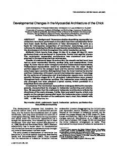
Immunohistochemistry was used to determine whether TGFb receptors were expressed in uterine smooth muscle cells in myometrial samples collected from nonpregnant, pregnant nonlaboring, and spontaneous laboring women. Figure 1, A–C, shows photomicrographs of tissue sections of human myometrium from nonpregnant, pregnant nonlaboring, and spontaneous laboring women, respectively. Each
FIG. 1. Localization of TGFb receptors in uterine smooth muscle cells in the human myometrium. Tissue sections from nonpregnant, pregnant nonlaboring, and spontaneous laboring myometrium are illustrated (A, B, and C, respectively, which are representative of several experiments). For each tissue a control section, without TGFb receptor antibody and counterstained only with hematoxylin is shown (a). The presence of TGFb receptor types I and II are illustrated in sections b and c, respectively. Positive staining is represented by the distinctive brown color within the myometrial smooth muscle cells. The calibration bar in Aa represents 100 mm.
2989
section illustrates control sections (a), sections stained for type I TGFb receptor (b), and staining for type II TGFb receptor (c). In each of the three patient groups, TGFb receptors for types I and II could be detected in the myometrial smooth muscle cells. No expression of type III TGFb receptor was detected in any of the tissues studied (data not shown). Western blotting, using the same antibodies, was subsequently employed to quantitate the relative amounts of TGFb receptors expressed in tissue lysates prepared from the different myometrial tissues. Figure 2A shows nitro-cellulose blots stained with antibodies to TGFb receptor types I, II, and III in nonpregnant, pregnant nonlaboring, and spontaneous laboring tissues, respectively. Blots were also stained with an antibody to the G protein subunit, Gb, as a loading control because the expression of this G protein subunit remains constant in the human myometrium during pregnancy and labor (26). As in Fig. 1, the receptor proteins for TGFb receptor types I and II were detected in all samples, but no protein corresponding to TGFb receptor type III was found. Incubation of nitro-cellulose blots with neutralized antibody (see Materials and Methods) resulted in the absence of any immunoreactive staining, thus indicating the specificity of the antibodies for TGFb receptor subtypes (data not shown). The relative expression of receptors in each tissue was assessed using a scanning densitometer. The data for receptor type I and II expression in the myometrial tissues after normalizing for loading (using the Gb antibody) are shown in Fig. 2B. The levels of TGFb receptor type I and II expression in nonpregnant samples were arbitrarily assigned a value of 100%. The amounts of receptors in pregnant nonlaboring tissues and spontaneous laboring myometrium are expressed relative to this value. The expression levels of TGFb receptor types I and II were significantly increased by 168 6 19% (n 5 6) and 162 6 22% (n 5 7) in pregnant nonlaboring tissues (P , 0.05) compared to those in nonpregnant myo-
2990
JCE & M • 1998 Vol 83 • No 8
HATTHACHOTE ET AL.
FIG. 3. TGFb1 in human myometrium. Measurements of the total concentration of TGFb1 in nonpregnant, pregnant nonlaboring, and spontaneous laboring myometrium. Concentrations are expressed as picograms per g tissue wet wt. Values are the mean 6 SEM (n 5 3 for each patient group).
of the tissue concentrations of the total TGFb1 (latent plus active) in nonpregnant, pregnant nonlaboring, and spontaneous laboring myometrium using ELISA. From Fig. 3, the amount of TGFb1 was increased from 334 6 10 pg/g tissue wet wt in nonpregnant myometrium to 534 6 73 and 674 6 106 pg/g tissue wet wt in pregnant nonlaboring and spontaneous laboring myometrium (n 5 3 in each group), respectively. However, these values were just outside the range of statistically significant difference between groups because of the small sample size (P 5 0.06). FIG. 2. Western immunodetection of TGFb receptor types I, II, and III in human myometrium. A, Western blots illustrating the expression of TGFb receptor types I, II, and III in tissue lysates prepared from nonpregnant (NP), pregnant nonlaboring (PNL), and spontaneous laboring myometrium (SL). The expression of the G protein Gb is also shown. This protein does not change during pregnancy and can be used as a loading control. Blotting with neutralized antibodies (see Materials and Methods) resulted in no detectable bands (data not shown). Molecular mass markers are expressed as kilodaltons. B illustrates analysis of six or seven separate myometrial lysate preparations from the different patient groups. Relative TGFb receptor levels in each tissue were determined as the ratio of TGFb receptor protein/Gb protein measured by densitometry. The values shown are the mean 6 SEM and expressed relative to the receptor levels of nonpregnant myometrium (100%). *, P , 0.05; **, P , 0.01.
metrium. The expressions of both receptor types obtained from spontaneous laboring myometrium were 93 6 12% (n 5 6) and 85 6 11% (n 5 7), respectively. These values were significantly lower than those in pregnant nonlaboring tissues (P , 0.01), which suggests that there is less protein for both receptor types I and II in spontaneously laboring myometrium. As TGFb receptors were detected in the myometrium at the end of pregnancy and in spontaneous laboring myometrium, it was important to establish whether the cytokine TGFb itself could be measured in these tissues. The presence of both cytokine and receptor is essential if this system is to have a physiological role. Figure 3 illustrates measurements
Discussion
The data reported in this investigation demonstrate the presence of TGFb receptor types I and II together with the cytokine TGFb1 in nonpregnant, term pregnant nonlaboring, and spontaneous laboring human myometrium. TGFb receptor type III was not detected in any myometrial tissue sample. The major observation reported here is that the levels of TGFb receptor types I and II were increased in pregnant nonlaboring myometrium compared to those in nonpregnant and spontaneous laboring samples. In addition, the concentration of TGFb1 in pregnant nonlaboring and spontaneous laboring samples was increased at this time relative to that in nonpregnant tissues. Taken together, these observations support the idea that TGFb may operate as an autocrine signal in the myometrium. A rise in the levels of both TGFb receptors and TGFb1 may function within a cascade of events to facilitate the activation of specific myometrial genes before term. In this context, the numbers of oxytocin receptors and endothelin receptors are also increased in the myometrium at term (27, 28). Conversely, the number of angiotensin receptors is decreased as term approaches (29). Thus, it would appear that a common mechanism that could influence myometrial function would be to up- or down-regulate the expression of key surface membrane receptors. In this way, the myometrium can alter its function from a quiescent weakly
MYOMETRIAL TGFb AND TGFb RECEPTOR EXPRESSION
contractile and poorly excitable tissue to one capable of powerful coordinated contractions. Human myometrial cells in vitro have been shown to synthesize and release TGFb (30, 31). In turn, it has been shown that the amount of TGFb synthesized can be increased by GnRH, 17b-estradiol, and medroxyprogesterone acetate (30). Taken together, these observations indicate that the TGFb signaling system is under hormonal regulation. This may also be occurring in late pregnancy. TGFb receptors are unlikely to have a role in the direct activation of contractions. It is more likely that they are associated with activation of myometrial cell growth and differentiation in preparation for parturition. Thus, it can be speculated that the myometrium is prepared for term as a result of a cascade of events involving the sequential expression of signals, receptors, and CAPs. Growth factors and cytokines have been postulated as being potential regulators of many functions in the nonpregnant uterus and in early pregnancy. TGFb has been detected in amniotic fluid isolated from the late first trimester, where it may be involved in immune responses (32, 33), and has also been detected during the second trimester, when it may stimulate the secretion of other bioactive substances. It has been reported that TGFb receptors and TGFb are present in the nonpregnant human uterus and that the levels of expression of both change during the menstrual cycle (34). TGFb has also been reported in the murine uterus at implantation, where it may be involved in synchronizing embryonic development (35, 36). In the nonpregnant ovariectomized mouse uterus, the expression of TGFb can be transiently increased by the injection of estrogen (35). Roles for estrogen and progesterone have been suggested to account for the variations in TGFb receptors and TGFb isoforms in the human uterus during the menstrual cycle (34). In other cell types, the expression of TGFb and TGFb receptors has been reported to be influenced by several hormones (37, 38). For example, ACTH up-regulates TGFb receptors in bovine adrenocortical cells (37), and androgens affect receptor expression in rat ventral prostate (38). A rise in the concentrations of other cytokines has been noted in amniotic fluid during normal and preterm deliveries. These include IL-6, IL-8, and granulocyte colony-stimulating factor (39). The source of the cytokines in late pregnancy is controversial, and the myometrium itself may be one site of production (4, 40). In addition, leukocytes invading the decidua and the decidual cells themselves have been implicated (39, 41, 42). Interestingly, the expression of IL-8 is enhanced by pretreatment with IL-1 and progestin (41), suggesting that a cascade of events may be acting to prepare the uterus for parturition. As the levels of TGFb receptor types I and II were significantly reduced compared to those in pregnant nonlaboring tissues and the levels of total TGFb1 remained elevated at parturition, this may indicate the down-regulation of TGFb receptors after exposure to elevated levels of TGFb1. If this is the case, then, the decrease in TGFb receptor expression may occur due to increased levels of active TGFb1. However, this can only be surmised, as only total levels of TGFb1 (latent plus active) were determined in this investigation. A recent report indicates that the IL-8 receptor is down-regulated
2991
upon prolonged exposure to its ligand (43). There are also data showing that a number of other receptor types are down-regulated when exposed to their ligands, e.g. the angiotensin (44), somatostatin (45), and opioid receptors (46). A related phenomenon has been noted in the human myometrium with respect to the oxytocin receptor (47). In human myometrial cells, oxytocin receptors appear to be downregulated upon prolonged exposure to oxytocin, with the effect that the myometrial cells become less sensitive to the hormone (48, 49). Thus, receptor down-regulation may be a common feature in the human myometrium to limit that action of specific hormones and agents. Several investigations have shown that the activation of the TGFb signaling pathway involves binding of TGFb to the type II receptor, resulting in the formation of a complex between type I and type II receptors with the consequent phosphorylation of the type I receptor (reviewed in Refs. 17 and 18). However, in some instances TGFb can activate cells that lack detectable levels of type II receptor (50) via activation of type I receptors alone (51). It has also been shown that a group of vertebrate homologs of Sma and Mad (mother against dpp), designated SMADs, in the cytoplasm is involved in the TGFb signaling pathway from the receptor to the nucleus (18, 52). In addition to SMADs, other proteins using the type I receptor, such as FK506-binding immunophilin FKBP12, may also modulate receptor signaling (17, 18). Members of the small GTP-binding protein family, Ras or Rac, have also been implicated in the signaling cascade (18). However, at the present time, it is not known how TGFb exerts its action on the human myometrium. References 1. Lye SJ. 1996 Initiation of parturition. Anim Reprod Sci. 42:495–503. 2. Lopez Bernal A, Rivera J, EuropeFinner GN, Phaneuf S, Asboth G. 1995 Parturition: activation of stimulatory pathways or loss of uterine quiescence? In: Ivell R, Russel J, eds. Oxytocin. New York: Plenum Press; 435– 451. 3. Kimura T, Takemura M, Nomura S, et al. 1996 Expression of oxytocin receptor in human pregnant myometrium. Endocrinology. 137:780 –785. 4. Faber BM, Metz SA, Chegini N. 1996 Immunolocalization of eicosanoid enzymes and growth factors in human myometrium and fetal placental tissues in failed labor inductions. Obstet Gynecol. 88:174 –179. 5. Chow L, Lye SJ. 1994 Expression of the gap junction protein connexin-43 is increased in the human myometrium toward term and with the onset of labor. Am J Obstet Gynecol. 170:788 –795. 6. Mershon JL, Mikala G, Schwartz A. 1994 Changes in the expression of the L-type voltage-dependent calcium channel during pregnancy and parturition in the rat. Biol Reprod. 51:993–999. 7. Boyle MB, Heslip LA. 1994 Voltage-dependent Na1 channel mRNA expression in pregnant myometrium. Receptors Channels. 2:249 –253. 8. Awad SS, Lamb HK, Morgan JM, Dunlop W, Gillespie JI. 1997 Differential expression of ryanodine receptor RyR2 mRNA in the non-pregnant and pregnant human myometrium. Biochem J. 322:777–783. 9. Hendrix EM, Mao SJ, Everson W, Larsen WJ. 1992 Myometrial connexin 43 trafficking and gap junction assembly at term and in preterm labor. Mol Reprod Dev. 33:27–38. 10. Hendrix EM, Myatt L, Sellers S, Russell PT, Larsen WJ. 1995 Steroid hormone regulation of rat myometrial gap junction formation: effects on cx43 levels and trafficking. Biol Reprod. 52:547–560. 11. Andersen J, Grine E, Eng CL, et al. 1993 Expression of connexin-43 in human myometrium and leiomyoma. Am J Obstet Gynecol. 169:1266 –1276. 12. Lefebvre DL, Piersanti M, Bai XH, Chen ZQ, Lye SJ. 1995 Myometrial transcriptional regulation of the gap junction gene, connexin-43. Reprod Fertil Dev. 7:603– 611. 13. Geimonen E, Jiang W, Ali M, Fishman GI, Garfield RE, Andersen J. 1996 Activation of protein kinase C in human uterine smooth muscle induces connexin-43 gene transcription through an AP-1 site in the promoter sequence. J Biol Chem. 271:23667–23674. 14. Kilarski WM, Fu X, Backstrom T, Roomans GM, Ulmsten U. 1996 Progesterone, oestradiol and oxytocin and their in vitro effect on maintaining the
2992
15. 16. 17. 18. 19. 20. 21.
22. 23. 24. 25. 26. 27. 28. 29. 30.
31.
32.
33. 34.
HATTHACHOTE ET AL.
number of gap junction plaques in human myometrium at term. Acta Physiol Scand. 157:461– 469. Massague J, Attisano L, Wrana JL. 1994 The TGF-b family and its composite receptors. Trends Cell Biol. 4:172–178. Massague J. 1992 Receptors for the TGF-b family. Cell. 69:1067–1070. Massague J, WeisGarcia F. 1996 Serine/threonine kinase receptors: mediators of transforming growth factor beta family signals. Cancer Surv. 27:41– 64. Heldin CH, Miyazono K, ten Dijke P. 1997 TGFb signalling from cell membrane to nucleus through SMAD proteins. Nature. 390:465– 471. Mazor M, Cohen J, Romero R, Ghezzi F, Tolosa JE, Gomez R. 1995 Cytokines and preterm labour. Fetal Matern Med Rev. 7:207–233. Norwitz ER, Lopez Bernal A, Starkey PM. 1992 Tumor necrosis factor-a selectively stimulates prostaglandin F2a production by macrophages in human term decidua. Am J Obstet Gynecol. 167:815– 820. Pollard JK, Thai D, Mitchell MD. 1993 Evidence for a common mechanism of action of interleukin-1b, tumor necrosis factor-a, and epidermal growth factor on prostaglandin production in human chorion cells. Am J Reprod Immunol. 30:146 –153. Edwin SS, LaMarche SL, Thai D, Branch DW, Mitchell MD. 1993 5-Hydroxyeicosatetraenoic acid biosynthesis by gestational tissues. Am J Obstet Gynecol. 169:1467–1471. EuropeFinner GN, Phaneuf S, Watson SP, Lopez Bernal A. 1993 Identification and expression of G-proteins in human myometrium: up-regulation of Gas in pregnancy. Endocrinology. 132:2484 –2490. Bradford MM. 1976 A rapid and sensitive method for the quantitation of microgram quantities of protein utilizing the principle of protein-dye binding. Anal Biochem. 72:248 –254. Laemmli UK. 1970 Cleavage of structural proteins during the assembly of the head of bacteriophage T4. Nature. 227:680 – 685. EuropeFinner GN, Phaneuf S, Tolkovsky AM, Watson SP, Lopez Bernal A. 1994 Down-regulation of Gas in human myometrium in term and preterm labor: a mechanism for parturition. J Clin Endocrinol Metab. 79:1835–1839. Kimura T, Tanizawa O, Mori K, Brownstein MJ, Okayama H. 1992 Structure and expression of a human oxytocin receptor. Nature. 356:526 –529. Wolff K, Faxen M, Lunell NO, Nisell H, Lindblom B. 1996 Endothelin receptor type A and B gene expression in human nonpregnant, term pregnant and preeclamptic uterus. Am J Obstet Gynecol. 175:1295–1300. Matsumoto T, Sagawa N, Mukoyama M, et al. 1996 Type 2 angiotensin II receptor is expressed in human myometrium and uterine leiomyoma and is down-regulated during pregnancy. J Clin Endocrinol Metab. 81:4366 – 4372. Chegini N, Rong H, Dou Q, Kipersztok S, Williams RS. 1996 Gonadotropinreleasing hormone (GnRH) and GnRH receptor gene expression in human myometrium and leiomyomata and the direct action of GnRH analogs on myometrial smooth muscle cells and interaction with ovarian steroids in vitro. J Clin Endocrinol Metab. 81:3215–3221. Tang XM, Dou Q, Zhao Y, McLean F, Davis J, Chegini N. 1997 The expression of the transforming growth factor-bs and TGF-b receptor mRNA and protein and the effect of TGF-bs on human myometrial smooth muscle cells in vitro. Mol Hum Reprod. 3:233–240. Altman DJ, Schneider SL, Thompson DL, Cheng HL, Tomasi TB. 1990 A transforming growth factor beta 2 (TGF-beta 2)-like immunosuppressive factor in amniotic fluid and localization of TGF-beta 2 mRNA in the pregnant uterus. J Exp Med. 172:1391–1401. Lang AK, Searle RF. 1994 The immunomodulatory activity of human amniotic fluid can be correlated with transforming growth factor-beta1 (TGF-b1) and b2 activity. Clin Exp Immunol. 97:158 –163. Chegini N, Zhao Y, Williams RS, Flanders KC. 1994 Human uterine tissue throughout the menstrual cycle expresses transforming growth factor-b1
35.
36. 37. 38. 39. 40. 41. 42.
43. 44. 45. 46.
47. 48.
49. 50. 51. 52.
JCE & M • 1998 Vol 83 • No 8
(TGFb1), TGFb2, TGFb3, and TGFb type II receptor messenger ribonucleic acid and protein and contains [125I]TGFb1-binding sites. Endocrinology. 135:439 – 449. Das SK, Flanders KC, Andrews GK, Dey SK. 1992 Expression of transforming growth factor-b isoforms (b2 and b3) in the mouse uterus: analysis of the periimplantation period and effects of ovarian steroids. Endocrinology. 130:3459 –3466. Tamada H, McMaster MT, Flanders KC, Andrews GK, Dey SK. 1990 Cell type-specific expression of transforming growth factor-b1 in the mouse uterus during the periimplantation period. Mol Endocrinol. 4:965–972. Cochet C, Feige JJ, Chambaz EM. 1988 Bovine adrenocortical cells exhibit high affinity transforming growth factor-beta receptors which are regulated by adrenocorticotropin. J Biol Chem. 263:5707–5713. Kim IY, Ahn HJ, Zelner DJ, Park L, Sensibar JA, Lee C. 1996 Expression and localization of transforming growth factor-beta receptors type I and type II in the rat ventral prostate during regression. Mol Endocrinol. 10:107–115. Saito S, Kasahara T, Kato Y, Ishihara Y, Ichijo M. 1993 Elevation of amniotic fluid interleukin 6 (IL-6), IL-8 and granulocyte colony stimulating factor (GCSF) in term and preterm parturition. Cytokine. 5:81– 88. Osmers RG, Blaser J, Kuhn W, Tschesche H. 1995 Interleukin-8 synthesis and the onset of labour. Obstet Gynecol. 86:223–229. Arici A, MacDonald PC, Casey ML. 1996 Progestin regulation of interleukin-8 mRNA levels and protein synthesis in human endometrial stromal cells. J Steroid Biochem Mol Biol. 58:71–76. Arici A, MacDonald PC, Casey ML. 1996 Modulation of the levels of interleukin-8 messenger ribonucleic acid and interleukin-8 protein synthesis in human endometrial stromal cells by transforming growth factor-b1. J Clin Endocrinol Metab. 81:3004 –3009. Ray E, Samanta AK. 1996 Dansyl cadaverine regulates ligand-induced endocytosis of interleukin-8 receptor in human polymorphonuclear neutrophils. FEBS Lett. 378:235–239. Vinson GP, Ho MM, Puddefoot JR, Teja R, Barker S. 1994 Internalization of the type-I angiotensin-II receptor (AT1) and angiotensin II function in the rat adrenal zona glomerulosa cell. J Endocrinol. 141:R5–R9. Koenig JA, Edwardson JM, Humphrey PP. 1997 Somatostatin receptors in neuro2A neuroblastoma cells: operational characteristics. Br J Pharmacol. 120:45–51. Chakrabarti S, Yang W, Law PY, Loh HH. 1997 The mu-opioid receptor down-regulates differently from the delta opioid receptor: requirement of a high affinity receptor G protein complex formation. Mol Pharmacol. 52:105–113. Bossmar T, Akerlund M, Fantoni G, Szamatowicz J, Merlin P, Maggi M. 1994 Receptors for and myometrial responses to oxytocin and vasopressin in preterm and term human pregnancy. Am J Obstet Gynecol. 171:1634 –1642. Phaneuf S, Asboth G, MacKenzie IZ, Merlin P, Lopez Bernal A. 1994 Effect of oxytocin antagonists on the activation of human myometrium in vitro: atosiban prevents oxytocin-induced desensitisation. Am J Obstet Gynecol. 171:1627–1634. Phaneuf S, Asboth G, Carrasco MP, et al. 1997 The desensitisation of oxytocin receptors in human myometrial cells is accompanied by down-regulation of oxytocin receptor messenger RNA. J Endocrinol. 154:7–18. Chen RH, Ebner R, Derynck R. 1993 Inactivation of the type II receptor reveals two receptor pathways for the diverse TGF-b activities. Science. 260:1335–1338. Weiser R, Wrana JL, Massague J. 1995 GS domain mutations that constitutively activate TbR-I, the downstream signalling component in the TGF-b receptor complex. EMBO J. 14:2199 –2208. Wrana J, Pawson T. 1997 Mad about SMADs. Nature. 388:28 –29.